Fang Zhaoa,
Jenny Perez Holmbergb,
Zareen Abbasb,
Rickard Frost
a,
Tora Sirkkaa,
Bengt Kasemoa,
Martin Hassellövb and
Sofia Svedhem*a
aDept. of Applied Physics, Chalmers University of Technology, SE-412 96 Göteborg, Sweden. E-mail: sofia.svedhem@chalmers.se
bDept. of Chemistry and Molecular Biology, University of Gothenburg, SE-412 96 Göteborg, Sweden
First published on 8th September 2016
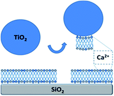
There is a need for different levels of model systems for effect studies of engineered nanoparticles and the development of nanoparticle structure–activity relationships in biological systems. Descriptors for nanoparticles based on their interactions in molecular model systems may become useful to predict toxicological responses of the nanoparticles in cells. Towards this end, we report on nanoparticle-induced formation of holes in supported model membranes. Specifically, TiO2 nanoparticle – lipid membrane interactions were studied under low ionic strength, basic conditions (pH 8), using different membrane compositions and several surface-sensitive analytical techniques. It was found that for mixed POPC/POPG (PG fractions ≥ 35%) membranes on silica supports, under conditions where electrostatic repulsion was expected, the addition of TiO2 nanoparticles resulted in transient interaction curves, consistent with the removal of part of the lipid membrane. The formation of holes was inferred from quartz crystal microbalance with dissipation (QCM-D) monitoring, as well as from optical measurements by reflectometry, and also verified by atomic force microscopy (AFM) imaging. The interaction between the TiO2 nanoparticles and the PG-containing membranes was dependent on the presence of Ca2+ ions. A mechanism is suggested where TiO2 nanoparticles act as scavengers of Ca2+ ions associated with the supported membrane, leading to weakening of the interaction between the membrane and the support and subsequent removal of lipid mass as TiO2 nanoparticles spontaneously leave the surface. This mechanism is consistent with the observed formation of holes in the supported lipid membranes.
Titanium oxide is a common material, often in the form of a nanomaterial, which is used in various applications due to its many attractive properties such as biocompatibility, photocatalysis, whiteness in paints, and charge carrier is solar cells. TiO2 exists in three crystal structures, brookite, anatase and rutile. The rutile structure, or sometimes also amorphous TiO2, is the more biocompatible form of TiO2, whereas the anatase structure is the photo(catalytically) active one.6,7 Titanium implants are widely applied in medical implants owing to their outstanding ability for osseointegration.8–11 A thin oxide layer is spontaneously formed on the titanium surface in air, and it is the titanium dioxide (TiO2) layer which is responsible for the favorable interaction of titanium implants with tissues and bones.10 Titanium oxide is also a promising material for novel photovoltaic solar cells,7,12 especially for so called dye sensitized solar cells. By controlling the geometry of titanium oxide particles, especially in the nanodimension, their properties can be tuned. While rutile is thermodynamically favored for large particle sizes, anatase and brookite are energetically favored for small nanoparticle sizes.13
Motivated by the great value and variety of applications of TiO2 NPs in biomedical and consumer products, both theoretical and experimental studies have been carried out to improve synthesis procedures and colloidal stability of particle suspensions.14–16 It is well known that the surface charge of TiO2 NPs can be controlled through varying parameters such as size, shape, and the dispersing intermediate (importantly pH).17–20 Despite the fact that the surface properties of TiO2 NPs constitute an important research topic, only few studies have been performed addressing the interfacial interaction between TiO2 NPs and lipid membranes. For example, the presence of calcium ions has been found to be important for the formation of some supported lipid bilayers on planar TiO2 substrates.21,22 One important motivation for similar studies is to understand the role of TiO2 layers for the biocompatibility of titanium implants, as well as the potential toxicity of those metal oxide NPs to cells.23–26 Such results could also be important to understand effects of nanosized wear products which may form around implants.
Surface-supported lipid membranes are extensively studied as simplified model systems for the basic functions of biological membranes, such as lipid flip-flop,27–29 membrane pore formation,30,31 functions of membrane proteins,32,33 lipid transfer,34 membrane fusion,35 or viral infection.36,37 These model membrane systems can be designed to address specific questions by varying the composition of lipid components and the choice of surface-analytical technique, to allow the investigation of the relation between structural properties and functional aspects of lipid membrane interactions. The dynamics of the supported lipid membrane platform is especially useful, e.g. in studies of tethered particles.38
In the present study, we use supported lipid membranes as model systems to probe the interaction between TiO2 NPs and lipid membranes by the quartz crystal microbalance with dissipation (QCM-D) technique. Similarly, QCM-D has previously been used to study structural rearrangements of drug carriers upon adsorption to lipid membrane surfaces.39–41
QCM-D is an acoustic sensing method which measures mass uptake and structural properties of the mass adsorbed to the sensor surface. Here, the QCM-D results are validated by two other surface analytical techniques; reflectometry and atomic force microscopy (AFM). In particular the combination of QCM-D and AFM is a powerful means to study these systems due to the different sensing principles. Phosphatidylglycerol (PG)-containing supported membranes have been used as a model for bacterial,42,43 as well as mammalian membranes.31 We focus on a comparison of the interaction of TiO2 NPs with lipid membranes of different compositions, for a given buffer condition (low ionic strength, pH 8). Our study shows the expected accumulation of TiO2 NPs on membranes under conditions for electrostatic attraction, but it also results in a mechanistic model for Ca2+-dependent removal of lipid mass from supported lipid membranes with a large enough fraction of PG lipids. This model is a contribution towards increased understanding of the interaction of TiO2 NPs with cells, especially at the nanoscale, and the mechanisms behind effects on cells which might be associated with these interactions. The results lead us to discuss the relevance of supported lipid membranes for the development of structure–activity relationships.
| Sample | Size (nm) | Polydispersity index | Zeta potential (mV) |
|---|---|---|---|
| TiO2 NPs (Tris buffer) | 57 ± 1 | 0.2 | −27 ± 2 |
| POPC liposomes (Tris buffer) | 84 ± 3 | 0.1 | −2 ± 1 |
POPC/POEPC 1 : 1 liposomes (Tris buffer) |
94 ± 1 | +59 ± 1 | |
POPC/POPG 1 : 1 liposomes (Tris buffer) |
82 ± 1 | −47 ± 1 | |
POPC/POPG 1 : 1 liposomes (Tris–CaCl2 buffer; 1 mM CaCl2) |
79 ± 1 | −27 ± 2 | |
POPC/POPG 1 : 1 liposomes (Tris–CaCl2 buffer; 2 mM CaCl2) |
77 ± 2 | −22 ± 1 | |
POPC/POPG 1 : 1 liposomes (Tris–CaCl2 buffer; 4 mM CaCl2) |
80 ± 1 | −19 ± 1 | |
POPC/POPG 1 : 1 liposomes (Tris–CaCl2 buffer; 8 mM CaCl2) |
185 ± 10 | 0.5 | −14 ± 1 |
The synthesis and stabilization of nanoparticles free from organic molecules are challenging tasks. In this particular study, additional experiments performed at a much later occasion showed similar but not identical results with respect to the particle size distribution. The particles synthesized at later stage had average particle diameter between 16 and 17 nm. Since the synthesis method is based on hydrolysis of TiCl4 a slight temperature variation can affect particle size. However, the particle suspensions at pH 2.5 were stable. Such a small variation in particle size does not have any effect on the particle surface charge density as shown in our previous study.18
In the following, we will assume that the composition of each prepared supported lipid membrane formed is similar to that of the liposomes used for their formation (suggesting a symmetric distribution of lipids between the two leaflets, for details see discussion). Thus, the zeta potential of a particular supported lipid bilayer is assumed to be represented by the zeta potential of the corresponding liposomes. The lipid membranes formed (details on the lipid membrane formation process have been reported elsewhere46,48), resulted in QCM-D frequency and dissipation shifts of Δf ∼ −26 Hz and ΔD < 0.5 × 10⁻⁶, respectively, which are characteristic values for high quality lipid membranes formed by this method. An important feature of the QCM-D technique is its ability to characterize the viscoelastic properties of an adlayer formed on the sensor surface.49 In particular, low ΔD values indicate the presence of only few or no remaining, intact liposomes on the surface.
:
1). The frequency shift (Δf) decreases (corresponding to mass uptake) and the dissipation shift (ΔD) increases (corresponding to the formation of a more dissipative layer) upon addition of TiO2 NPs. Under the present conditions, the frequency and dissipation shifts rapidly reach equilibrium (95% of the saturation value within minutes), and then stay constant upon rinsing, that is the adsorption is irreversible on the time scale of the experiments. These data are consistent with the expected, electrostatically driven, adsorption of TiO2 NPs to the membrane under the given conditions. On lipid membranes containing lower fractions of POEPC, the frequency shift (Δf) and the dissipation shift (ΔD) were both smaller, and decreased as a function of zeta potential for the corresponding liposome. These results are also expected. In the absence of the establishment of additional interactions in contact with the membrane, the adsorbed amount of TiO2 NPs at equilibrium is expected to scale with the net charge of the lipid bilayer, since upon adsorption there is a competition between the attractive NP – lipid membrane and the NP–NP repulsive electrostatic interactions. No adsorption of TiO2 NPs was observed onto lipid membranes with a fraction of POEPC below 10%. This indicates either that some force was needed to keep the NP bound to the membrane (e.g. due to thermal motions), or that EPC lipids are asymmetrically distributed between the two leaflets.
Additional experiments show that larger TiO2 NPs (∼200 nm, −34 mV) induce much higher dissipation shifts than smaller NPs for approximately the same frequency shift. This is a consequence of that larger NPs form more dissipative structures when adsorbed to the lipid membrane. Large dissipation shifts have also been observed when larger size lipid vesicles50 or liposomes51 were adsorbed on TiO2 or SiO2 surfaces compared to smaller sized vesicles and liposomes.
The affinity of the NPs to the EPC-containing lipid membranes is in all cases strong, as seen by the non-reversibility of the QCM-D frequency and dissipation shifts upon rinsing. This was further confirmed in additional experiments where the NPs were added at lower concentration. These experiments showed that the lower the NP concentration, the longer time was needed to reach equilibration, whereas the final frequency and dissipations shifts did not differ a lot, and the NPs were irreversibly adsorbed while rinsing. Thus, the equilibrium of the interaction between the NP and the positively charged lipid membrane is strongly shifted towards adsorption of NPs for fractions of POEPC > 10% in the liposomes. A likely explanation for the high affinity of the TiO2 NPs for the positively charged membrane, in view of the high mobility of lipids in the bilayer, is rearrangement of the charged lipids in the membrane, leading to accumulation of the oppositely charged lipid molecule in the membrane close to and underneath the NPs.46
:
1). In these experiments, the QCM-D frequency shift (Δf) first decreased (indicating mass uptake), then increased (indicating mass loss), and gradually returned close to its original value, while the corresponding dissipation shift (ΔD) exhibited an opposite behavior with a maximum in the signal, in time close to the frequency minimum. The interaction between the membrane and the TiO2 NPs was studied in Tris buffer, and upon changing the buffer back to Tris–NaCl–CaCl2 (which was used to form the supported membrane prior to the addition of NPs), the ΔD value could be compared to the baseline value before the addition of the TiO2 NPs, but with a small increase of the Δf value (from −27 Hz to −23 Hz). This buffer change was performed to compensate for the (unusual and) large dissipation shifts for PG-containing supported lipid membranes in response to buffer changes when switching between a Ca2+ containing and a non-Ca2+ containing buffer.47 Thus, when taking the buffer effect into consideration, the net result of the transient interaction curve obtained when the TiO2 NPs were added to the POPC/POPG 1
:
1 membrane is a small decrease in mass compared to the mass of the lipid membrane before addition of the NPs. As shown in Fig. 2, additional mass loss was observed when repeating again the TiO2 NP addition and the same sequence of buffer changes. For each cycle, more mass was lost until two thirds of the lipid membrane mass was removed from the sensor surface (Fig. 3). We associate this saturation effect with removal of all or most of the PG lipids from the membrane (see below). At this point, more POPC/POPG vesicles could be added to restore the membrane mass to its initial value (i.e. frequency and dissipations shifts corresponding the initial lipid membrane were regained, i.e. Δf ∼ −26 Hz and ΔD < 0.5 × 10⁻⁶). The restored membrane again interacted transiently with TiO2 NPs (Fig. 2, last part), causing mass loss.
![]() | ||
| Fig. 3 Lipid mass on the sensor surface at the different stages of the experiment shown in Fig. 2, as modeled from the QCM-D data in Fig. 2 using the Sauerbrey relationship. The changes in bar heights represent loss or gain of membrane mass. The bars 1–8 represent repeated exposures of the lipid membrane to TiO2 NPs, causing a monotonous loss of lipid mass. Next, the lipid membrane mass increases due to re-addition of liposomes. The last bar represents the mass after the freshly repaired lipid membrane was once again exposed to TiO2 NPs. | ||
The remaining lipid membrane mass after each transient interaction with TiO2 NPs is shown in Fig. 3. The mass loss after each addition of TiO2 NPs gradually decreases and eventually levels off after 8 additions of NPs and buffer changes. In other words TiO2 NPs appear to remove, in each cycle, lipid material from the substrate, leaving holes in the membrane. Note that the maximum mass uptake of TiO2 NPs also decreases for each successive cycle (Fig. 2, peak value of Δf), indicating that the fraction of POPG in remaining lipid patches decreases for each cycle, until the attraction between the membrane and the TiO2 NPs can no longer be restored by supplying Ca2+ ions. In other words, it appears that the TiO2 NP exposure cause selective removal of PG lipids. Mass loss is consistent with complementary combined QCM-D and reflectometry measurements where the QCM-D experiment and the optical measurement are performed at the same time and on the same lipid membrane. These experiments show the removal of lipid material for each additional cycle of addition of TiO2 NP and Ca2+ ion addition to the PG-containing membranes (ESI Fig. S1†).
:
POPG (1
:
1) supported membranes which had been exposed to TiO2 NPs revealed holes in the membrane. After two exposures to NPs, membrane defects of various sizes (hundreds of nm to several μm) were observed and holes were sparsely distributed. In Fig. 4A, one large hole is shown. The surrounding membrane was detected by force spectroscopy where the presence of a lipid membrane is revealed by a step in the resulting force spectra during the approach (Fig. 4B). This step was absent in the areas without a lipid membrane, i.e. in the holes. The height profile across a hole revealed a step of about 5 nm, a result that is in good agreement with the thickness of a lipid membrane (Fig. 4C).
It would have been interesting to image as well the structure of a layer of TiO2 NPs bound to POPC/POEPC supported membranes. However, it was not possible to image the adsorbed NPs in contact mode.
Under repulsive conditions, NPs did not bind to the lipid membrane (i.e. <30% of POPS or POPG), except for a high enough fraction (>35%) of POPG. This latter result was unexpected (see also the DLVO calculations in the ESI†). One explanation to this unexpected result would be that, in this case, the zeta potential that we measure for the POPC/POPG liposomes does not serve as an appropriate estimate for the zeta potential of the POPC/POPG membranes formed onto the silica support. Unfortunately, we have not been able to experimentally measure the zeta potential of the supported membranes due to instrumental limitations. It is perhaps not unlikely that Ca2+ ions (included in the buffer during the formation of the supported lipid membrane) associate to the PG head groups in the upper lipid leaflet such that the zeta potential is reversed. This would then explain why an attractive interaction potential is obtained (se also the DLVO calculations in the ESI†). Alternatively, Ca2+ ions are present at the interface between the silica substrate and the lower leaflet of the POPC/POPG membrane, and the displacement of the ions to the upper leaflet occurs only as the TiO2 NPs approach. Meanwhile, the repulsion between SiO2 crystal and POPG lipids become stronger in the absence of Ca2+ ions.47 This may explain why TiO2 NPs remove lipid patches rather than remain immobilized onto the lipid membrane.
:
1) showed a similar behavior as TiO2 NPs (ESI, Fig. S2†), although without any removal of lipid material, which supports the role of the TiO2 NPs as Ca2+ ion scavengers in our system. Repeated supply of calcium ions is necessary for the repeated removal of lipid material from the surface mediated by TiO2 NPs (Fig. 2 and 3), further indicating the role of Ca2+ ions. Thus, based on our results, we propose the following mechanistic model for the formation of holes in POPC/POPG (>35% PG) model membranes upon addition of TiO2 NPs (Fig. 5): (i) in the first step, a good quality POPC/POPG (1
:
1) supported membrane is formed in Tris–NaCl–CaCl2 buffer, followed by (ii) rinsing of the supported lipid membrane with the low ionic strength Tris buffer. During this rinsing step, Ca2+ ions are retained by the lipid head groups in the lower lipid leaflet, due to a strong interaction between the lipid membrane and the silica surface (this will not happen in a high ionic strength buffer, where the electrostatic attraction is screened47). We suggest that the retained Ca2+ ions are removed when (iii) adding TiO2 NPs, due to the high affinity between the TiO2 NPs and the Ca2+ ions.53 During this process, some lipids bind to the NPs and are removed by as the NP leave the surface under flow mode. After the removal of Ca2+ ions the interaction between the SiO2 support and the remaining membrane is much weakened, until (iv) upon changing the buffer back to Tris–NaCl–CaCl2 which re-establish the tight binding of the lipid membrane to the silica surface. At this point the loss of lipid material can be observed by AFM as holes in the membrane.
We cannot distinguish between the scenario where the substrate has an important role for the retention of Ca2+ ions, and a situation where the binding of Ca2+ ions to the lipid membrane head groups alone will explain the observed interactions. This issue can be further addressed in molecular dynamics (MD) simulations. MD simulations between PS-containing membranes and TiO2 substrates have previously suggested direct coordination of the phosphate or carbonyl oxygen atoms of PS and PC lipids with titanium sites.55 In particular, we foresee that it will be possible to elaborate on the role of hydrogen bonding between the lipid head groups and the Ca2+ ions by comparing MD and further experimental results. We note that previous studies of thermodynamic driving forces for lipid flip-flop in supported membranes have revealed large opposing enthalpic and entropic contributions to the free energy, thus providing new perspectives on the energetics of membrane processes.56,57
:
1) supported membranes may serve to experimentally probe for nanoparticles which act as scavengers of cations, a property which is likely governed by the pKa of functional groups at the surface of the nanoparticle along with the number of charged sites per surface area. For the case of TiO2, the density of charged sites on the surface of the oxide is much higher compared to the bilayer on average, which drives the preferential binding of Ca2+ to the TiO2 surface,24 leading to, in our model system, removal of lipid mass and the formation of holes in the supported membrane. Since TiO2 is biocompatible, this may be a physicochemical function which can be correlated to beneficial biological responses.
The role of Ca2+ ions in biomolecular adsorption to metal oxides has also been observed for adsorption of human albumin on TiO2 surface.58 Under physiologic conditions (i.e. pH 7), negatively charged albumin can bind to negatively charged TiO2 surface via Ca2+ ions, which is in analogous with our studies of POPG lipids. The way nanoparticles interact with selected proteins (or other biomolecules) is likely a good complement to the experiments with supported membranes in the search for useful structure–activity relationships.
:
POPG (1
:
1) supported lipid membrane was formed on the crystal in Tris–NaCl–CaCl2. After formation of the lipid membrane, the buffer was exchanged to Tris. As a next step, the TiO2 NPs were added and were left to incubate for 5 min. Subsequently, the surface was rinsed with Tris before changing the buffer to Tris–NaCl–CaCl2. Before imaging the surface, two cycles of NP addition were made and the buffer was changed back to Tris. Images were treated and analyzed using the SPIP software version 3.0.0.9 (Image Metrology Inc., Ljungby, Denmark). Force spectroscopy experiments were performed with a constant approach/retraction speed of v = 280–560 nm s−1, corresponding to a force loading rate vk = 8.40–16.8 nN s−1. Deflection-position raw data were converted to force–distance curves with SPIP software using specified values for the cantilever spring constant.
Footnote |
| † Electronic supplementary information (ESI) available Additional QCM-D, reflectometry and DLVO results are available as ESI. See DOI: 10.1039/c6ra05693h |
| This journal is © The Royal Society of Chemistry 2016 |