Chitosan-coated liposomes encapsulating curcumin: study of lipid–polysaccharide interactions and nanovesicle behavior

M. Hasana, G. Ben Messaouda, F. Michauxa, A. Tamayolbcd, C. J. F. Kahne, N. Belhajf, M. Lindera and E. Arab-Tehrany*a
aUniversité de Lorraine, LIBio, ENSAIA, 2 avenue de la Forêt de Haye, TSA 40602, F-54505 Vandoeuvre-lès-Nancy, France. E-mail: elmira.arab-tehrany@univ-lorraine.fr
bCenter for Biomedical Engineering, Department of Medicine, Brigham and Women's Hospital, Harvard Medical School, Boston, MA 02139, USA
cHarvard-MIT Division of Health Sciences and Technology, Massachusetts Institute of Technology, Cambridge, MA 02139, USA
dWyss Institute for Biologically Inspired Engineering, Harvard University, Boston, MA 02115, USA
eAix-Marseille Université, IFSTTAR, LBA UMR_T24, F-13016 Marseille, France
fLucasmeyer Cosmetics, ZA les Belles Fontaines, 99 route de Versailles, 91160 Champlan, France

Received 2nd March 2016 , Accepted 22nd April 2016

First published on 25th April 2016


Abstract

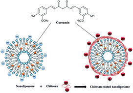
Despite various spectacular therapeutic properties, curcumin has low bioavailability mainly due to its poor solubility in water. In this paper, we encapsulated curcumin by nanoliposomes prepared from salmon purified phospholipid and coated with chitosan. Various techniques were used in order to study the interactions among phospholipid, chitosan and curcumin. FTIR results showed both electrostatic and hydrophobic interactions as well as hydrogen bonding between chitosan and phospholipid, while hydrophobic forces and hydrogen bonding dominated the interactions between curcumin and phospholipid as well as between curcumin and chitosan. Shear viscosity measurements demonstrated a flow behavior change from Newtonian to shear thinning after liposome coating. The increase/decrease stress ramp showed that the addition of chitosan layer decreased significantly the hysteresis loop area (thixotropic behavior) and therefore increased significantly the liposomal dispersion stability. The viscoelastic properties investigated by small-amplitude oscillatory shear rheology demonstrated improvement of mechanical stability after chitosan addition. Small-angle X-ray scattering experiments revealed that the liposome membrane structure was not affected by the chitosan layer or the encapsulated curcumin.


1. Introduction

Nanotechnologies supply innovations in various domains of medicine, namely therapy, diagnostics, imaging and drug delivery.1–3 In drug delivery, colloidal carriers have been suggested for effective administration of drugs having difficulties, such as toxicity, low bioavailability or poor water solubility. For these purposes, different colloidal drug delivery systems have been fabricated such as liposomes, micelles, nanoemulsions and nanoparticles.

Curcumin (diferuloyl methane) is a natural polyphenolic phytochemical molecule extracted from the powdered rhizomes of turmeric (Curcuma longa) spice. It has been widely used as a traditional medicine in many Asian countries, since it was reported that curcumin has antioxidant, anti-inflammatory, antimicrobial, and anticancer features, as well as wound healing characteristics.4–8

Although curcumin has numerous advantages, the therapeutic application of curcumin is limited because of its low bioavailability,9,10 due to its poor aqueous solubility, low stability against alkaline pH conditions, extensive first-pass metabolism and rapid systemic elimination.11,12 Therefore, a carefully designed carrier could significantly facilitate curcumin delivery and broaden the range of its possible pharmaceutical applications.

Liposomes are widely used as delivery vehicles for stabilizing drugs and overcoming barriers to cellular and tissue uptake.13 Nanoliposomes have become very versatile tools in biology, biochemistry and medicine because of their enormous diversity of structure and composition. Liposomes are microscopic vesicles formed essentially by phospholipids dispersed in water, which are amphiphilic molecules containing polar heads and hydrophobic hydrocarbon tails, which can associate spontaneously to form bilayer vesicles. This property of phospholipids gives liposomes unique properties, such as self-sealing, in aqueous media and makes them an ideal carrier system with applications in different fields including food, cosmetics, pharmaceutics, and tissue engineering.14 Liposomes can carry both hydrophilic and hydrophobic components by encapsulation in the water phase and intercalating into the hydrophobic domains, respectively. Even hydrophobic components can be formed in a stable state in an aqueous environment by high dispersion in a liposome system. In addition, the components entrapped in liposomes can effectively penetrate and overcome biological barriers to cellular and tissue uptake because of their structure being similar to that of cell membranes.15,16 Lecithin, which has two long hydrocarbon chains, is a major component of lipid bilayers of cell membranes and a natural, biological amphiphile. Furthermore, it is in many respects regarded as an ideal biological surfactant because it is biodegradable. It may be used for various purposes.17 Numerous studies, both in humans and in animals,18,19 have demonstrated that polyunsaturated fatty acids (PUFAs) of the n-3 series, in particular eicosapentaenoic acid (EPA, 20:5 n-3) and docosahexaenoic acid (DHA, 22:6 n-3), are known to have positive effects on human health by preventing cardiovascular diseases, cognitive decline, inflammation, and hypotriglyceridemic effect and improving mental capacity. Marine lecithin from salmon head (Salmo salar) contains a high percentage of PUFAs, especially EPA and DHA, so the originality of this work is the use of these salmon phospholipids to carry biomolecules.20–22

Polymer coating is a promising way to modify the surface characteristics of liposomes in order to improve their applicability. This can be achieved by simply mixing a liposome suspension with a polymer solution without chemically linking the two components.23 Among various biopolymers, chitosan, which is an hydrophilic, biocompatible and biodegradable polymer, has been widely studied as a mucoadhesive material that enhances the penetration of macromolecules across the intestinal and nasal barriers.24

Interestingly, the coating of lipid-based nanostructures with chitosan has been found to increase their stability,25 and to provide them with mucoadhesive properties.26 Because of its bioadhesive and permeation-enhancing properties, chitosan has received much attention for novel bioadhesive drug delivery systems, aimed at improving the bioavailability of drugs by prolonging their residence time at the site of absorption.27 Chitosan is predominantly composed of β-(1,4)-linked D-glucosamine units and is obtained by deacetylation of chitin, which is the primary component of the cell walls of crustaceans, fungi, and insects.28 By combining chitosan and liposomal characteristics, specific, prolonged, and controlled release may be achieved.29 Takeuchi and others showed that chitosan-coated liposomes were formed via ionic interaction between the positively charged chitosan and negatively charged diacetyl phosphate on the surface of the liposomes (Fig. 1).


image file: c6ra05574e-f1.tif
Fig. 1 Schematic illustration of curcumin-loaded nanoliposome and chitosan-coated curcumin-loaded nanoliposome.

Polymer-vesicle systems constitute a type of mixed colloidal system of particular interest. In fact, mixed biopolymer–liposome systems are good model systems for living cells since these are composed of lipidic membranes which could interact with several biopolymers.30 The control of the rheological properties of liposome dispersions is of great industrial importance.31 The rheological behavior of this type of dispersion depends on the interaction between vesicles and vesicle deformability. The interactive forces consist of van der Waals attraction, electrostatic repulsion, and “long-range” entropic repulsion as a result of thermal undulations.32

Considering that the rheological response is directly related to microstructural changes, a better understanding of the changes in microstructure under shear could provide significant opportunities for enhancing the processing of liposome dispersions.33

The present study focused primarily on the preparation of nanoliposomes from salmon purified phospholipid and coating them with chitosan in order to encapsulate curcumin. The physicochemical characteristics of the samples and their structural properties were investigated using X-ray scattering, transmission electron microscopy and infrared spectroscopic techniques; in addition, the rheological properties of systems were investigated.

2. Materials and methods

Chitosan sample (prepared from shrimp shells, practical grade) of deacetylation degree up to 75% was supplied by Sigma-Aldrich (ref. 417963). Salmon lecithin from Salmo salar was obtained by enzymatic hydrolysis. The lipids were extracted by use of an enzymatic process without any organic solvent as described by Linder et al.34 Curcumin was purchased from Sigma-Aldrich and acetic acid (100%) was supplied by Prolabo-VWR. BF3 (boron trifluoride)/methanol, chloroform, hexane, acetonitrile, methanol and other chemicals were obtained from Sigma-Aldrich (France) and Fisher (France). All organic solvents were analytical grade reagents.

2.1. Purification of marine phospholipid by acetone precipitation

Salmon phospholipid was isolated from salmon lecithin by using an acetone precipitation method as described by Lu et al.35 and Schneider and Lovaas36 with some modifications. According to Schneider and Lovaas, a total weight of 13 g salmon lecithin was dissolved in approximately 20 mL chloroform. This solution was then emptied into 100 mL of acetone (approximate ratio of 1[thin space (1/6-em)]:[thin space (1/6-em)]7.7) under vigorous stirring at ambient temperature. The ratio of lipids to solvent was according to Schneider and Lovaas.36 The mixed solution was kept at −18 °C overnight to allow phospholipid precipitation. The acetone was decanted by soft centrifugation at 1000 rpm, the precipitates were redissolved in chloroform, and the purification procedure was repeated once again. The final precipitates (purified phospholipid) were dried under nitrogen for 1 h. The residues of acetone and chloroform were further removed under vacuum at 40 °C.

2.2. Fatty acid composition

Fatty acid methyl esters (FAMEs) from salmon lecithin and phospholipid were prepared as described by Ackman.37 Then, the FAMEs were analyzed using a Shimadzu 2010 gas chromatography system (Shimadzu, France) equipped with a flame-ionization detector. Separation of FAME was accomplished on a fused silica capillary column (60 m, 0.25 mm i.d. × 0.20 μm film thicknesses, SPTM2380 Supelco, Bellefonte, PA, USA). Injector and detector temperatures were settled at 250 °C. The column temperature was fixed initially at 120 °C for 3 min, then raised to 180 °C at a rate of 2 °C min−1 and maintained at 220 °C for 25 min. Individual fatty acids were identified using standard mixtures (PUFA1 from a marine source and PUFA2 from a vegetable source; Supelco, Sigma-Aldrich, Bellefonte, PA, USA). The results were obtained from triplicate analyses.

2.3. Lipid classes

The lipid classes of different fractions of lipid from salmon, soya and rapeseed lecithin were determined by Iatroscan MK-5 TLC-FID (Iatron Laboratories Inc., Tokyo, Japan). The measurement was performed according to the protocol described in detail in a previous paper.38 Two migrations were done to determine the proportion of neutral and polar lipid fractions. All standards were purchased from Sigma (Sigma-Aldrich Chemie GmbH, Germany). Area percentages were presented as the mean value of three repetitions.

2.4. Preparation of chitosan-coated liposomes

In the first stage, 1.5 g of salmon phospholipid and 10 mg of curcumin in a completely dried round bottomed flask (RBF) were dissolved in a mixture of chloroform and methanol (2[thin space (1/6-em)]:[thin space (1/6-em)]1 v/v). A thin lipid film was then formed on the wall of the RBF using a Rotavapor and the organic solvent was completely evaporated under vacuum, followed by hydration with 47.5 mL ultrapure water. The suspension was mixed for 2 hours under agitation in inert atmosphere (nitrogen) for preparation of the liposome spontaneously; then we added 0.5 g of chitosan and 0.5 mL of acetic acid into the liposome solution already prepared. The suspension was mixed again for 4–5 min under the same conditions. Then, the sample droplet size was decreased initially by sonication at 40 kHz for 5 min (1 s on, 1 s stop) in an ice bath. Then the samples were subjected to homogenization using a high-pressure homogenizer (EmulsiFlex-C3), provided by Sodexim SA, France. The mixture was introduced in quantities of 50 mL under a pressure of 1500 bar for 7–8 cycles. Chitosan-coated liposome samples were stored in glass bottles in the dark at 4 and 37 °C.

2.5. Measurement of liposome size and zeta potential

The size distribution and zeta potential of the liposome dispersions were measured by dynamic light scattering (DLS) using a Malvern Zetasizer Nano ZS (Malvern Instruments Ltd, UK). The small interparticle space between globules results in multiple light scattering that may lead to false results, and hence dilution is necessary.39 Then, the samples were diluted (1[thin space (1/6-em)]:[thin space (1/6-em)]200) into ultra-filtrate distilled water to measure the particle size as well as the dispersed particle electrophoretic mobility that were carried out to evaluate the surface net charge around droplets. Samples were put in a standard capillary electrophoresis cell to measure their electrophoretic mobility. All size and zeta potential measurements were carried out at 25 °C with a fixed scattering angle of 173°, refractive index at 1.471 and absorbance at 0.01. The results are presented as a z-average mean (dz) as well as an average ζ-potential of the liposome suspension. All measurements were performed in triplicate.

2.6. Stability of chitosan-coated liposomes

The chitosan-coated liposomes containing curcumin and the control (without curcumin) were stored in a drying room at 4 and 37 °C for five weeks. Mean particle size, polydispersity index and electrophoretic mobility of all formulations were examined every 3 days. The above-mentioned protocols were used for each analysis (Section 2.5).

2.7. pH titration of chitosan-coated liposome solution

The chitosan-coated liposome suspension was titrated versus pH using a Multi-Purpose Titrator (MPT-Z, Malvern Instruments, UK) linked to a Malvern Zetasizer Nano ZS (Malvern Instruments Ltd, UK). The titrants used were 0.1 M, 1 M NaOH and 1 M HCl. The titration was run from pH 4 through pH 8, where size and zeta potential of the nanoparticles were measured accordingly. The experiments were done in triplicate.

2.8. Transmission electron microscopy (TEM)

TEM was employed to observe the structure of nanoliposomes and chitosan-coated liposomes with a negative staining method according to the protocol of Colas et al.40 Briefly, the samples were diluted 25-fold with distilled water to reduce the concentration of the particles. The same volume of the diluted solution was mixed with an aqueous solution of ammonium molybdate (2%) as a negative staining agent. Staining was followed by a 3 min wait at room temperature, and 5 min on a copper mesh coated with carbon; then the sample was examined using a Philips CM20 transmission electron microscope associated with an Olympus TEM CCD camera.

2.9. Membrane fluidity

Membrane fluidity of all samples was determined by fluorescence anisotropy measurements. TMA–DPH was used as fluorescent probe which is a compound that contains a cationic trimethylammonium (TMA) group that acts as a surface anchor to improve the localization of the fluorescent probe of membrane interiors, DPH. This measurement was carried out according to the method described by Maherani et al.41 Briefly, a solution of TMA–DPH (1 mM in ethanol) was added to the liposome suspension to reach finally a concentration of 4 μM and 0.2 mg mL−1 for the probe and the lipid, respectively. The mixture was lightly stirred for at least 1 h at ambient conditions and protected from light. Then, it was distributed into the wells of a 96-well black microplate at 180 μL per well. The fluorescent probe was vertically and horizontally oriented in the lipid bilayer. The fluorescence intensity of the samples was measured with a Tecan INFINITE 200R PRO (Austria) equipped with fluorescent polarizers. Samples were excited at 360 nm and emission was recorded at 430 nm under constant stirring at 25 °C. Magellan 7 software was used for data analysis. The polarization value (P) of TMA–DPH was calculated using the following equation:
 
image file: c6ra05574e-t1.tif(1)
where I is the intensity of fluorescence parallel to the excitation plane, I is the intensity of fluorescence perpendicular to the excitation plane, and G is a factor that accounts for transmission efficiency. Membrane fluidity is defined as 1/P. The results were obtained from triplicate analyses.

2.10. SAXS experiments

Small-angle X-ray scattering (SAXS) data were collected with a SAXSess mc2 apparatus (Anton Paar KG, Graz, Austria). This instrument is attached to an ID 3003 laboratory X-ray generator (General Electric) equipped with a sealed X-ray tube (PANalytical, λCu Kα = 0.1542 nm) operating at 40 kV and 50 mA. The liposome dispersions were introduced into a 1 mm capillary before being placed inside an evacuated chamber equipped with a temperature-controlled sample holder unit maintained at 25 °C. The 2D scattering patterns were detected by a CCD camera. Using SAXSQuant software (Anton Paar), the 2D images were integrated into 1D scattering intensities I(q) upon the magnitude of the scattering vector q (q = (4π/λ)sin[thin space (1/6-em)]θ, where 2θ is the total scattering angle). All data were collected in the q range from 0.11 to 6 nm−1. Scattering data, obtained with a slit collimation, contain instrumental smearing. Therefore, the beam profile was determined and used for the desmearing of the scattering data. All data were corrected for the background scattering from the capillary filled with solvent (water). The scattered intensities were evaluated on an absolute scale using water as a reference.

2.11. Fourier transform infrared spectroscopy (FTIR)

FTIR spectra were recorded using a Tensor 27 mid-FTIR spectrometer (Bruker, Germany) equipped with a diamond ATR (attenuated total reflectance) module and a DTGS (deuterated-triglycine sulfate) detector. Scanning rate was fixed to 20 kHz and 128 scans were performed for both reference and samples between 400 cm−1 and 4000 cm−1 at a resolution of 2 cm−1 at room temperature. An initial reference spectrum was then recorded. Next, a small amount of each sample was put on the diamond crystal of the optical cell and a minimum of three separate experiments were done for each sample. In addition, all treatments were carried out using OPUS software (Bruker, Karlsruhe, Germany). Crude absorbance spectra were smoothed using a nine-points Savitsky–Golay smoothing function. Then, spectra were centered and normalized using OPUS software.

2.12. Rheological characterization

Rheological studies were carried out using a Kinexus Pro rheometer (Malvern Instruments, Orsay, France). During the rheological experiments, the measuring system was covered with a humidity chamber to minimize water evaporation. Samples were allowed to rest for at least 300 s prior to analysis.
2.12.1. Shear viscosimetry. Steady-shear viscosity and shear stress measurements were determined using a cone-and-plate geometry (50 mm, 1°). For the steady-shear viscosity measurement, the shear rate was increased from 10−3 to 103 s−1.

In order to confirm the presence of dynamic hysteresis, shear stress was increased from 10−3 to 0.5 Pa and then decreased in the same shear stress range.

2.12.2. Oscillatory rheometry. To determine the mechanical properties of the liposome dispersions, an amplitude sweep was first conducted at a frequency of 0.5 Hz by changing the shear strain from 0.1% to 1000% in order to determine the linear viscoelastic region.

On the basis of this test, a value of strain within the linear regime was then used in the subsequent frequency sweep with the change of frequency between 0.01 and 10 Hz.

For these experiments, both plate-and-plate (20 mm) and cone-and-plate (2°, 60 mm) geometries were used.

3. Results and discussion

3.1. Fatty acid analyses

The main fatty acid compositions are shown in Table 1. The first observations allow us to highlight that the total PUFAs in salmon lecithin and salmon phospholipid were predominant with 46.28% and 45.11%, respectively. The percentage of the fraction of PUFAs is followed directly by the percentage of saturated fatty acids in salmon lecithin (33.40%) and salmon phospholipid (35.06%).
Table 1 Fatty acid compositions of salmon lecithin and salmon phospholipid purified by acetone precipitation
Fatty acids Salmon lecithin Salmon phospholipid
% SD % SD
C14 2.72 0.04 2.62 0.02
C15 1.13 0.01 1.16 0.04
C16 20.67 0.15 22.22 0.15
C17 0.88 0.05 0.83 0.00
C18 6.10 0.05 6.72 0.05
C20 0.35 0.03 0.36 0.15
C22 1.55 0.06 1.15 0.21
SFA 33.40   35.06  
C14:1n9 0.33 0.02 0.33 0.03
C16:1n7 3.23 0.07 2.87 0.14
C18:1n9 13.87 0.13 14.13 0.06
C20:1n9 2.11 0.06 1.91 0.01
C22:1n9 0.79 0.08 0.59 0.09
MUFA 20.32   19.83  
C18:2n6 1.20 0.04 1.05 0.07
C18:3n3 0.35 0.01 0.34 0.09
C20:4n6 3.52 0.02 3.59 0.03
C20:5n3 (EPA) 11.03 0.13 10.13 0.08
C22:5n3 3.93 0.33 4.46 0.22
C22:6n3 (DHA) 26.26 0.09 25.55 0.17
PUFA 46.28   45.11  
n-3/n-6 8.88   8.73  
DHA/EPA 2.38   2.52  


In general, the fatty acid composition of the phospholipid fraction and lecithin was not very different. The main difference between them was the slightly lower content of EPA and DHA. In purified phospholipid, the percentage of palmitic acid (C16:0) increased compared to salmon lecithin, because of the oxidation phenomenon during acetone precipitation.

On the one hand, we found that the amount of EPA and DHA decreased from 11.03% and 26.26% in lecithin fraction to 10.13% and 25.55% in purified phospholipid. On the other hand, the amount of palmitic acid increased from 20.67% in lecithin to 22.22% in purified phospholipid.

This composition was in agreement with the results presented in another study made by Lu et al.35

3.2. Lipid classes

In this study, salmon lecithin was purified through acetone precipitation with the simultaneous purpose of removing TAGs and other nonpolar lipids and thus to increase the percentage of phospholipid. The phospholipid percentage increased from 67.7% to 100%, whereas all triglycerides and cholesterol, representing respectively amounts of 31.2% and 1.2%, were removed after acetone precipitation (Table 2).
Table 2 Lipid classes and fraction of polar lipids constituting salmon lecithin and salmon phospholipid purified by acetone precipitationa
Name Salmon lecithin Salmon phospholipid
a ND: not determined.
Total phospholipids (%) 67.7 ± 1.1 100 ± 0.0
Phosphatidylcholine, PC (%) 42.4 ± 0.5 40.5 ± 0.2
Phosphatidylethanolamine, PE (%) 7.7 ± 0.1 3.6 ± 0.1
Phosphatidylserine, PS (%) 9.1 ± 0.1 8.0 ± 0.2
Phosphatidylinositol, PI (%) 13.0 ± 0.3 15.3 ± 0.4
Sphingomyelin, SPM (%) 1.5 ± 0.1 1.3 ± 0.1
Lysophosphatidylcholine, LPC (%) 2.8 ± 0.1 3.2 ± 0.2
Other phospholipids (%) 23.5 ± 0.2 28.2 ± 0.3
Triglycerides, TAGs (%) 31.2 ± 0.8 ND
Cholesterol, CHO (%) 1.2 ± 0.1 ND
Free fatty acids, FFA (%) ND ND


The results presented in Table 2 for the fraction of polar lipids in salmon lecithin and purified phospholipid showed that purified salmon phospholipid had lower contents of phosphatidylcholine (PC), phosphatidylethanolamine (PE), phosphatidylserine (PS) and sphingomyelin (SPM) than untreated marine lecithin. However, purified phospholipid had a higher level of lysophosphatidylcholine (LPC), phosphatidylinositol (PI) and other unidentified phospholipids, this probably due to hydrolysis of phospholipid during acetone precipitation.

3.3. Measurement of liposome size and zeta potential

The particle sizes of nanoliposomes were measured immediately after preparation. The minimum size that can be achieved depends on lipid composition as well as liposome preparation method such as the sonication and homogenization parameters. The particle mean size of uncoated (typical) liposomes was found to be 89.0 nm, whereas after coating with chitosan solution, it increased to 221.8 nm (Table 3). This increase can be explained by the interaction between lipid and chitosan, which includes the lipid–chitosan electrostatic interactions between lipid head groups and the specific functional groups of chitosan NH3+. Hydrophobic interactions between the lipid tails and chitosan may take place allowing chitosan to come up to the bilayer and fill the empty volume.42 The size of the curcumin-loaded liposomes was found to be smaller than that of the unloaded curcumin liposomes. Therefore, the hydrodynamic diameter of nanoliposomes decreased from 89.0 nm to 79.8 nm for curcumin-loaded liposomes. These results suggest a strong interaction between curcumin and lecithin resulting in a core compaction. These results are in good agreement with those previously reported by Mazzarino et al.43 In contrast, the size of the curcumin-loaded chitosan-coated liposomes was greater than that of the unloaded curcumin ones. Therefore, the hydrodynamic diameter of nanoparticles increased from 221.8 nm to 247.6 (Table 3). The increase in size between chitosan-coated nanoliposomes with and without curcumin encapsulation could be attributed to the loading of curcumin in the outer layer formed by chitosan around liposomal bilayer which is in good agreement with the results of Sanoj Rejinold et al.44 In addition to particle diameter, DLS also measures polydispersity index (PDI) which is a dimensionless measure of the breadth of the particle size distribution.45 As shown in Table 3, the PDIs were between 0.30 and 0.33 which indicated that particle size was well controlled with a narrow dispersity. According to mean values and PDIs, there was no significant difference between the breadths of distributions of the samples. In comparing these results with the results published previously,38 we observed that the size of liposomes prepared with purified phospholipid was smaller and the PDI was slightly greater. This difference can be explained by the presence of triglycerides in unpurified lecithin, which induce an increase in liposome flexibility, and break the large liposomes into smaller ones in order to increase the homogeneity of the population.46
Table 3 Mean particle size, polydispersity index, zeta potential and membrane fluidity of liposomes and chitosan-coated liposomes (each value represents the mean of triplicates)a
Sample Particle size (nm) Polydispersity index Zeta potential (mV) Membrane fluidity
a CH-LP: chitosan-coated liposome.
Salmon liposome 89.0 ± 0.6 0.31 ± 0.00 −45.8 ± 2.4 3.24 ± 0.05
Curcumin-loaded salmon liposome 79.8 ± 1.6 0.30 ± 0.01 −46.0 ± 0.8 3.04 ± 0.01
Salmon CH-LP 221.8 ± 2.3 0.33 ± 0.01 +60.9 ± 0.2 2.81 ± 0.08
Curcumin-loaded salmon CH-LP 247.6 ± 2.0 0.33 ± 0.01 +61.5 ± 0.7 2.73 ± 0.10


Zeta potential measurements are commonly used to investigate certain physical properties of colloidal drug delivery systems. They give an indication of the surface electrical charge of the particles, which is a particularly important parameter that affects liposome behavior.47 The magnitude of the zeta potential gives an indication of the stability of colloidal systems: as the potential increases, the repulsion between particles is greater, thus leading to more stable colloidal dispersions. If all the particles in the suspension have a large negative or positive zeta potential, they will repel each other, and there will be no tendency for the particles to aggregate.48

This was attributed to the adsorption of the cationic polymer increasing the density of the positive charge around the liposomal surface. After adsorption, the strong electrostatic attraction between positively charged polyelectrolyte and oppositely charged surface groups of liposome forces them to come into close proximity, forming a thin layer around the surface.49 It is clear from Table 3 that coating the nanoliposomes with chitosan shifted the zeta potential from negative (−45.8 mV) to positive values (+61.9 mV). The increase in zeta potential was attributed to the cationic polymer groups adsorbed to the liposomal surface. Since chitosan carried a high positive charge, the adsorption of chitosan increased the density of the positive charge and led to a positive zeta potential value.46

The increased particle size and zeta potential of chitosan-coated nanoliposomes reflect several changes in the surface properties of nanoliposomes due to polymer–liposome interactions.

With respect to the stability of chitosan-coated nanoliposomes with time, we observed no significant variation in particle size after 30 days of incubation at 4 and 37 °C.

3.4. Influence of pH on z-average and zeta potential of chitosan-coated liposomes

The size and surface charge of chitosan-coated liposomes were evaluated in response to variations in pH. As shown in Fig. 2, we observe that both the z-average and zeta potential of the particles were susceptible to the pH of the media. Increasing the suspension pH from 4 to 8, the zeta potential of the particles progressively decreased from about +55.0 mV at pH 4 to near 0 mV at pH 8. The zeta potential decreased when pH increased; this can be explained by the charge neutralization of chitosan due to the addition of NaOH, during which the protons H+ of the amine group of chitosan are lost when they interact with the OH groups of NaOH. When most of the protons in chitosan-coated liposomes have been neutralized, the surface charge presents a neutral value (isoelectric point).
image file: c6ra05574e-f2.tif
Fig. 2 Effect of pH on size and zeta potential of chitosan-coated liposomes.

In parallel, the particle size of the sample is stable near pH 6.5; the size of the particles increased rapidly from pH 6.8 and the aggregation of particles appeared. These results confirmed that particle stability depends on electrostatic nature and the polysaccharide component defined the particle surface properties. In fact, the chitosan pKa value is around 6.5, where the particles are totally aggregated, but we observed the aggregation of particles begins at pH 6.5, when they are partially deprotonated. The chitosan chains retain their affinity to the negative charge of liposomes, which is due to a combination of various mechanisms such as adsorption, coagulation and bridging between them.50,51

3.5. Morphology of the liposomes

Surface morphological studies on the shape of the prepared systems using TEM indicated that the systems were almost spherical. TEM images indicated that liposomes prepared by sonication and high-pressure homogenization methods were in the form of multilamellar vesicles (Fig. 3a). The morphology of the chitosan-coated liposomes showed a contrasting band surrounding each liposome vesicle, as shown in Fig. 3b.
image file: c6ra05574e-f3.tif
Fig. 3 Transmission electron microscopic images of salmon nanoliposomes before (a) and after (b) coating with chitosan.

3.6. Membrane fluidity

Bilayer fluidity reflects the order and dynamics of phospholipid alkyl chains in the bilayer. The release of entrapped bioactive agents from nanoliposomes depends on the number of bilayers and their permeability and fluidity.52

The influence of lipid composition on vesicle membrane fluidity was explored by Coderch et al. and Calvagno et al.52,53 They determined a significant difference in the drug release profiles as a result of two factors: (i) the strength of the drug–liposomal lipid interaction and (ii) the fluidity of the bilayer. In fact, drug release to the aqueous medium is increased by increasing the membrane fluidity of the vesicle. The FA composition of the membrane bilayer tunes the membrane fluidity level. Indeed, the presence of saturated FAs increased the lipid ordering in the membrane, which excluded water in the proximity of the bilayer surface and reduced membrane fluidity. Whereas, unsaturated FAs could reduce the packing between phospholipids and preserve a higher level of membrane hydration, thus keeping membrane fluidity.54

It appears that nanoliposomes made of salmon phospholipid have slightly higher membrane fluidity, 3.24 ± 0.05, in comparison with those made of unpurified lecithin, the value of which was 3.19 ± 0.08,38 probably due to absence of cholesterol after the purification procedure. This is responsible for higher stiffness of the liposomal membrane because it enters into the lipid bilayer and it has an ordering effect on the membrane structure.55,56

To recognize the action of curcumin and chitosan on membrane fluidity, it is necessary to understand the behavior of curcumin and chitosan with respect to solution composition. Indeed, in a previous study, we showed that the presence of curcumin decreased the membrane fluidity of nanoliposomes.38 The presence of a curcumin molecule can weaken hydrophobic interactions among acyl chains of phospholipids.57 This perturbs the packing characteristics of the phospholipid bilayer and thus enhances the packing density of the hydrocarbon moieties in the lipid bilayer.

According to Table 3, we observed that chitosan reduced the membrane fluidity of nanoliposomes. It makes a new layer around the liposome, which is probably incorporated within the membrane bilayer, causing the rigidity of bilayers, and decreasing the movement of fatty acid chains of phospholipid. Consequently, the membrane bilayer fluidity decreases. The motional freedom of phosphate group was reduced in the presence of chitosan.58

The rotational motions of the probes that result in depolarization of fluorescence are tightly coupled to acyl chain orientational fluctuations and, consequently, reflect the degree of molecular packing (order) in the membrane.59

3.7. SAXS experiments

The influence of the chitosan coating on nanoliposome structure has been studied by SAXS. Furthermore, the influence of the presence of curcumin in the lipid membrane on the scattering signal has also been investigated. Small-angle scattering methods allow the investigation of the size and shape of nanoliposomes or their membrane structure depending on the employed experimental q-range. The lower the q-value is, the greater the observation window of the system. In this study, the experimental q-range (7.5 × 10⁻² to 4 nm−1) is suitable for investigating the phospholipid bilayer structure but not the overall structure of the liposome. This q-range corresponds indeed to sizes from 1.5 to 84 nm (d = 2π/q).

Several pieces of information are obtained from scattering curves presented in Fig. 4a. At small angles (low q values; q < 0.2 nm−1), a q−2 slope is detected for every sample. According to the literature, this specific regime at low q values corresponds to the form factor of the liposome lamellae.60 Nevertheless, the experimental configuration does not allow one to reach the plateau which should appear at lower q required to determine the liposomes size. Since DLS experiments have been performed to measure particle diameters, we focused the SAXS experiments on a higher q-range to study the bilayer structure.


image file: c6ra05574e-f4.tif
Fig. 4 (a) SAXS profiles (log–log representation) of uncoated nanoliposomes (without curcumin (green line, triangle), with curcumin (orange line, diamond)) and nanoliposomes coated by chitosan (without curcumin (blue line, square), with curcumin (black line, circle)). (b) Zoom of the SAXS profiles in the 0.6–1.5 nm−1 q-range (linear representation) to highlight the diffraction peak located at 1.0 nm−1. Electronic density profiles of liposome (c) and liposome coated by chitosan (d).

The phospholipid multilamellar arrangements of the liposome membrane usually lead to the presence of an intense diffraction peak between 0.6 and 1.6 nm−1 if the number of layers is sufficient. The repeat distance of the lamellar structure is deduced from the position of this peak (between 4 and 10 nm).61 In Fig. 4a, no diffraction peaks are clearly detected in this range, and only a small diffraction line at 1.0 nm−1 is observed (Fig. 4b). This tiny signal may correspond to the first reflection of the lamellar structure with a repeat distance of 6.3 nm. Nevertheless, the absence of a clear diffraction peak for every sample means that the liposome membrane is composed of only a few phospholipid bilayers. Furthermore, the presence of chitosan in the system affects the SAXS signal. The intensities of the samples coated with the polymer are indeed higher than those without chitosan. However, the curve shapes are similar which means that the bilayer structure is not affected by the presence of chitosan around the liposome. This difference of intensities may come from the modification of the contrast term between samples. To scatter the X-rays, a difference of electronic densities between different parts of the sample must exist and the scattered intensity depends on this difference (called contrast term).60 In the case of liposomes, the X-rays are scattered because water and phospholipids present different electronic densities (Fig. 4c). The presence of chitosan around the liposomes modifies this contrast which may lead to a change in the scattered intensities (Fig. 4d). Moreover, concerning the influence of curcumin on the liposome structure, no difference between samples with and without curcumin is observed regardless of the presence of chitosan or not. The related scattering curves remain identical (Fig. 4a).

3.8. FTIR spectroscopy

Fig. 5 shows FTIR spectra in the wavenumber range of 4000–400 cm−1 for salmon liposome, chitosan, and curcumin, each of them separately and with different types of formulation in order to study the interaction between them.
image file: c6ra05574e-f5.tif
Fig. 5 FTIR spectra of (a) (i) salmon liposome, (ii) curcumin encapsulated liposome, (iii) chitosan-coated liposome, and (b) (iv) curcumin, (v) chitosan, and (vi) mixture of chitosan and curcumin.

The main bands appearing in the spectrum of chitosan were due to stretching vibrations of OH groups in the range from 3700 cm−1 to 3000 cm−1, which are overlapped with the stretching vibration of N–H; and C–H bond stretching vibrations of –CH3 at 2926 cm−1 and –CH2 at 2883 cm−1. Bending vibrations of methyl and methylene groups were also visible at 1381 cm−1 and 1404 cm−1, respectively.62 The vibrations of carbonyl bonds (C[double bond, length as m-dash]O) of the amide group CONHR (secondary amide, 1635 cm−1) and the vibrations of protonated amine group (NH3+, 1541 cm−1) were also detected.63 The absorption bands at 950–1200 cm−1 were attributed to a saccharine structure. The sharp peaks at 1022 cm−1 and 1065 cm−1 are assigned to C–O stretching vibrations (ν(COC)) (Fig. 5v).64

As shown in Fig. 5i, the FTIR spectrum displays the main characteristic bands of phospholipid vesicles, especially those of the CH stretching modes with maxima of peaks at 2854 cm−1 and at 2924 cm−1, corresponding to the symmetric and antisymmetric stretching in the CH2 groups of alkyl chains, respectively, with minor contribution from the symmetric and antisymmetric stretching vibration in CH3 groups at 2893 cm−1 and 2958 cm−1, respectively. The broad band from 3750 to 3050 cm−1 represents OH. The band at 1732 cm−1 corresponds to the stretching vibrations of the ester carbonyl groups of phospholipid, and the relatively strong band centered at 1651 cm−1 corresponds to the stretching vibrations of alkene carbon–carbon double bond –C[double bond, length as m-dash]C–. The scissoring vibrations of the CH2 groups are represented by the band at 1456 cm−1, and the band at 1406 cm−1 corresponds to ([double bond, length as m-dash]C–H) bending (rocking) vibrations. While the relatively weak band at 1394 cm−1 represents the umbrella deformation vibrations of the CH3 groups of alkyl chains. In addition, the spectral bands at 1086 and 1224 cm−1 represent the symmetric and antisymmetric PO2− stretching vibration of phospholipids, and the band representing the antisymmetric N+\CH3 stretching vibrations was detected at 970 cm−1.65–67

The FTIR spectrum of curcumin (Fig. 5iv) showed a sharp peak at 3508 cm−1 indicating the phenolic O–H stretching, with a broad band in the range from 3100 to 3400 cm−1, which is due to the ν(OH) group (in enol form), C–H (methyl) at 2844 cm−1, and C–H (aryl) at 3014 cm−1. The strong peak at 1626 cm−1 has a predominantly mixed ν(C[double bond, length as m-dash]C) and ν(C[double bond, length as m-dash]O) character. Another strong band at 1601 cm−1 is attributed to the symmetric aromatic ring stretching vibrations ν(C[double bond, length as m-dash]C ring). The 1508 cm−1 peak is assigned to ν(C[double bond, length as m-dash]O), and the C–O–C stretching peak of ether appears at 1026 cm−1; and significance bands were observed at 730 and 797 cm−1 for C[double bond, length as m-dash]C–H aromatic stretching frequency.68–70

In order to determine the structure of lipid/polysaccharide nanoparticles, the interactions between the polysaccharide and the lipid have been studied by FTIR spectrometry. The FTIR spectra of nanoparticles and of the components lecithin and chitosan alone are shown in Fig. 5. FTIR was used, as a no perturbing technique, to analyze possible changes in the structure of phospholipid by analyzing the frequency of different functional groups of the lipid molecule in the presence or absence of chitosan. Formation of a complex between the liposomes and chitosan resulted in a considerable change in the absorption bands of OH group, which were broad. This is due to the presence of more hydroxyl groups and the possibility of new intermolecular hydrogen bond formation. The presence of chitosan with liposome caused also considerable changes in the absorption bands corresponding to acyl chains (3000–2800 cm−1) toward lower wavenumbers. For example, the symmetric and antisymmetric stretching in the CH2 and CH3 groups of alkyl chain absorption bands located at 2854 cm−1 and 2924 cm−1 for CH2, and at 2893 cm−1 and 2958 cm−1 for CH3 respectively in case of free liposomes, were merged into two peaks at 2852 and 2924 cm−1 easily distinguishable as narrow and intense in the case of chitosan-coated liposomes (Fig. 5iii). A significant shift from 2954 to 2952 cm−1 is also detected. We observed also a shift in the bending vibrations of acyl chain bands from 1406 cm−1 toward 1404 cm−1, whereas the peak was more intense.

The band at 1651 cm−1 corresponding to the stretching vibrations of alkene carbon–carbon double bond –C[double bond, length as m-dash]C– underwent a large shift towards 1641 cm−1 after chitosan coating. The low frequency shifts of the absorption bands in the hydrophobic region correspond to the decrease in the acyl chain mobility in liposomes, as has been demonstrated earlier.71,72 Evidently, complex formation with chitosan leads to restriction of acyl chain mobility in the bilayer structure which implies a decrease in the membrane fluidity and thereby stabilization of the system.73

The major potential binding site of anionic liposomes with amino polysaccharides (chitosan) via electrostatic interactions is the phosphate groups. Indeed, we found that the absorption band of liposomal phosphate groups shifted to high frequency (from 1224 to 1232 cm−1), which means that the interaction with polymer ligands leads to dehydration of phosphate groups. In fact, the characteristic band of chitosan at 1541 cm−1, corresponding to scissoring vibration of protonated amine group NH3+, was shifted to higher frequency at 1549 cm−1. Apparently, most hydrogen bonds of the phosphate group are broken due to electrostatic interactions with the amino group of the chitosan.

Another potential binding site of chitosan conjugate on liposomes is the carbonyl group, which carries a partial negative charge on the oxygen atom. Liposome formation with chitosan was found to result in considerable changes in the region of the carbonyl group absorption. We found that the interaction between liposomes and chitosan leads to a considerable shift in the absorption band to higher frequencies (from 1732 to 1738 cm−1). This position of the absorption band indicates a considerable decrease in the group hydration.72 This evidences destruction of some of the hydrogen bonds in which the carbonyl group is involved due to interaction with cationic groups of the polymer, as has been observed in the case of the liposome phosphate groups. The data prove that both carbonyl and phosphate groups act as chitosan binding sites.74

The characteristic band of chitosan at 1635 cm−1 of carbonyl bonds (C[double bond, length as m-dash]O) of the amide group CONHR (secondary amide) was not present. It was probably merged with the band of carbonyl groups of phospholipid resulting in more important intensity of the peak at 1738 cm−1.

Moreover, we have studied the interaction between the liposome and the encapsulated curcumin by FTIR. We found the encapsulated curcumin caused considerable change in the absorption bands of acyl chains (3000–2800 cm−1) toward lower wavenumbers as in the case of chitosan, but to a lesser degree. For example the absorption bands located at 2854 cm−1 and at 2958 cm−1 were shifted towards lower frequency at 2852 cm−1 and 2956 cm−1, respectively. We observed a new peak at 3011 cm−1, which corresponds to C–H (aryl) stretching of the molecule of curcumin, shifted also toward low frequency compared to the initial vibration at 3014 cm−1. The band at 1651 cm−1 corresponds to the stretching vibrations of alkene carbon–carbon double bond –C[double bond, length as m-dash]C– shifted towards 1647 cm−1 (Fig. 5ii). Then, the low frequency shifts of the absorption bands in the hydrophobic region of phospholipid with encapsulated curcumin indicate the hydrophobic interaction between them and this corresponds to a decrease the acyl chain mobility and an increase in the order of the bilayer.75 In respect of the bands of carbonyl and phosphate groups of phospholipid at 1732 and 1224 cm−1, respectively, we found considerable changes in the absorption of the two groups, which shifted from 1732 to 1738 cm−1 for carbonyl group, and from 1224 to 1230 cm−1 for phosphate group. Additionally, the appearance of the O–H region changed possibly due to the presence of the phenolic O–H stretching of curcumin. This was related to the possibility of the breaking of hydrogen bonds and formation of intermolecular hydrogen bonds between the phenolic OH groups of curcumin and the functional groups of phospholipid (phosphate and carboxyl groups).76,77 The curcumin molecule was demonstrated to be anchored inside the phospholipid bilayer through the hydrogen bonding of OH groups of phenolic rings of curcumin with the head group of phospholipid and the hydrophobic interactions of the aromatic rings of curcumin with phospholipid acyl chains.78

We studied the interaction between curcumin and chitosan. It was observed that the stretching vibrations of CH3 and CH2 groups of chitosan at 2922 cm−1 and 2879 cm−1, respectively, were shifted in the presence of curcumin towards 2922 cm−1 and 2875 cm−1, indicating a hydrophobic interaction between chitosan and the hydrophobic region of curcumin. The peaks at 2922 cm−1 were also sharpened which could be attributed to the presence of a greater number of CH groups.

We observed that the peak at 1541 cm−1 related to the protonated amine group NH3+ of chitosan was shifted in the presence of curcumin to 1539 cm−1, and the peak at 1635 cm−1 related to carbonyl bonds (C[double bond, length as m-dash]O) of the amide group CONHR was also shifted to 1633 cm−1 (Fig. 5vi). These results suggest the formation of intermolecular hydrogen bonds between chitosan and curcumin. Then the results indicate the formation of hydrophobic interactions and hydrogen bond formation between curcumin and chitosan.79,80

In fact, these interactions between molecules of curcumin and the vector, whether liposome alone or chitosan-coated liposome, can help in reducing the release rate of curcumin thereby helping controlled release.

3.9. Rheological study

3.9.1. Steady-state shear viscosity. Fig. 6a shows the flow curves of the different liposomal samples. The chitosan coating had an impact on the fluid rheological behavior. However there was no remarkable difference between unloaded and curcumin-loaded lipid vesicles.
image file: c6ra05574e-f6.tif
Fig. 6 (a) Flow curves of the liposomal samples. The solid lines are the fits to a power law model. (b) Casson plots of the square root of shear stress versus the square root of shear rate. (c) Shear viscosity evolution at low shear rates. (d) The shear stress was increased from 10−3 to 0.5 Pa and decreased in the same shear stress range.

The flow curves of the liposomal samples are plotted in Fig. 6a, where the solid lines represent fits to the power law model:

 
σ = n (2)
where σ is the shear stress, k is the consistency index, γ is the shear rate, and n is the flow behavior index.

The consistency k is numerically equal to the viscosity at 1 s−1 and could be useful as a general measure of viscosity for comparative purposes.

In order to determine the plastic viscosity and the yield stress of the different liposomal samples, the flow curves of the square root of shear stress were plotted against the square roots of shear rate (Fig. 6b).81

In fact, the Casson model (eqn (3)) is a structure-based model:

 
(σ)0.5 = K0c + Kc(γ)0.5 (3)
where the Casson yield stress is calculated as the square of the intercept, σ0c = (K0c)2, and the Casson plastic viscosity as the square of the slope, ηCasson = (Kc)2.

Eqn (2) and (3) were used to fit the experimental data of different samples, and the rheological parameters are shown in Table 4.

Table 4 Rheological properties of the different liposomal samples
Sample Power law model Casson model
K n R2 Plastic viscosity (mPa s) Yield stress (mPa) R2
Salmon LP 0.002 ± 0.0004 1.057 ± 0.0683 0.9879 2.64 ± 0.53 1.79 ± 0.10 0.9668
Curcumin-loaded salmon LP 0.004 ± 0.0006 0.9441 ± 0.0524 0.9903 2.59 ± 0.35 1.70 ± 0.12 0.9650
Salmon CH-LP 0.038 ± 0.002 0.8389 ± 0.0082 0.9983 14.09 ± 2.26 3.11 ± 0.24 0.9927
Curcumin-loaded salmon CH-LP 0.047 ± 0.004 0.8505 ± 0.0128 0.9987 17.85 ± 2.47 3.33 ± 0.14 0.9968


The n and k values ranged from 0.83 to 1.05 and from 2 × 10⁻³ to 47 × 10−3, respectively. Uncoated liposome suspension exhibited a nearly Newtonian flow behavior (n ≈ 1). The chitosan coating caused a decrease of the flow behavior index (n < 1) which resulted in shear-thinning behavior.48

In fact, shear-thinning behavior is generally desirable for pharmaceutical product formulation, providing a dispersion with a low viscosity when sheared, whereas at rest, the system has a certain solid-like consistency.33

The yield stress (the minimum stress needed to cause a Bingham plastic to flow) and the plastic viscosity increased, respectively, from 1.70 mPa and 2.64 mPa s for uncoated liposomes to 3.33 Pa and 17.85 mPa s after lipid vesicle coating with chitosan. Therefore, the addition of chitosan layer allowed a better stability of the lipid vesicle dispersion at rest.

It is interesting to note that the shear viscosity evolution as a function of shear rates did not show a Newtonian plateau at small shear (Fig. 6c) which represents an infinite resistance to flow below the critical shear stress (yield stress).

The zero shear viscosity of the salmon liposomes increased from 23 to 123 Pa s after lipid vesicle coating. The difference between the coated and uncoated lipid vesicles could be related to surface charge and size.

From Fig. 6a–c, we can conclude that the inclusion of curcumin into the phospholipid bilayer did not affect the rheological flow behavior of lipid vesicles.

In order to study the time-dependent flow behavior of the liposomal samples, the shear stress was increased from 10−3 to 0.5 Pa during 5 minutes and then decreased in the same stress range. The plots show a thixotropic behavior for all liposomal dispersions. Nevertheless, chitosan addition decreased significantly the hysteresis loop area which increases significantly the liposomal dispersion stability. In fact, chitosan liposomes are less thixotropic and their structure rebuilds faster than uncoated liposomes after shearing, which could be very useful for several applications including cosmetics where liposomal dispersions have to recover immediately to their original structure after extrusion or applying to the skin.

This observation is in a good agreement with a previous study focused on lecithin/chitosan dispersion which demonstrated that chitosan induced a change in the time-dependent flow of the lecithin dispersion from rheopexic to thixotropic by allowing the transition of lecithin dispersion from planar sheets to closed structures such as vesicles.82

The hysteresis loop area corresponding to the thixotropic behavior is related to the separation of vesicles from vesicle aggregates. The chitosan layer enhanced therefore the stability of the lipid vesicle dispersion and decreased liposome deformation under shearing.

3.9.2. Oscillatory rheometry. In order to study the mechanical stability of the liposomal samples, oscillatory rheology was performed at a fixed strain within the linear viscoelastic region. The mechanical spectra of the liposome dispersions are reported in Fig. 7.
image file: c6ra05574e-f7.tif
Fig. 7 Frequency sweep test of liposomal samples. (a) Complex modulus G* (Pa) evolution with frequency at a shear strain of 2% and (b) elastic modulus (G′) and viscous modulus (G′′) at a shear strain of 10%.

For these experiments, two geometries were used: plate-and-plate (20 mm) with a gap of 500 μm (Fig. 7a) and cone-and-plate (2°, 60 mm) with a gap of 70 μm (Fig. 7b).

The complex modulus G* (G*(w) = G′(w) + iG′′(w)) of coated liposomes is more than one order of magnitude higher than that of uncoated lipid vesicles. Moreover, coated liposomes showed a low frequency dependence of G* which demonstrates a better mechanical stability due to the additional chitosan layer (Fig. 7a).

However, the encapsulated curcumin did not affect the general mechanical spectra of coated and uncoated lipid vesicles. In general, the viscoelastic properties of lipid vesicles are related mainly to temperature and to the volume fraction. In fact, for high volume fractions near the maximum packing fraction, the average repulsive force is affected.32

Several factors could be the origin of the better stability of coated liposomes which could be related to the nanoparticle size and to the inner mechanical properties of the chitosan layer which could also act as a protective coating by reducing phospholipid layer deformation. The interactive forces could also participate in improving the mechanical stability of the coated liposomal dispersions.

In general, the mechanical stability of a liposomal dispersion is mediated by lipid vesicle interaction types such as van der Waals attraction (w), electrostatic repulsion (e), and “long-range” entropic repulsion (u). The total forces are assumed to be additive:83

 
ϕ = ϕw + ϕe + ϕu (4)

In our case, the rheological experiments were performed at 25 °C < T of the phase transition of liposomal dispersion.84 When the lipid bilayers are in the gel state, the entropic repulsion (u) and van der Waals attraction (w) are of the same order of magnitude.32 Therefore, the increase of mechanical stability could be as a result of the increase of electrostatic repulsion because the addition of the chitosan layer resulted in an increase of the absolute value of the zeta potential (from 45 to 60 mV after lipid vesicle coating) (Table 4).

Fig. 7b shows the frequency dependence of the elastic modulus (G′) and viscous modulus (G′′) using the cone-and-plate geometry at a gap of 70 μm. These experiments should be carefully monitored since the gap and dilute state of the liposomal samples could lead to very large phase angle (δ) and therefore the rheological test will be dominated by inertial effects.

The results showed that in the studied frequency range (0.1–10 Hz), uncoated lipid vesicles showed several crossover points between G′ and G′′ and therefore indicating several relaxation processes related to diffusive behavior and to the deformation of the vesicles.32

The coating of liposomes with chitosan resulted in a crossover point at a higher frequency (1 Hz) than uncoated vesicles which reflected a better resistance to shear and twist.

4. Conclusion

Our present studies give information about the physicochemical properties of nanoliposomes and chitosan-coated nanoliposomes before and after encapsulation of curcumin.

The increase of particle size and zeta potential of chitosan-coated nanoliposomes reflect several changes in the surface properties of nanoliposomes due to polymer–liposome interactions.

The coating of liposomes by a chitosan layer was confirmed by electron microscope images of liposome dispersions.

Coating with chitosan decreased the membrane fluidity of nanoliposomes causing the rigidity of bilayers, and decreasing the movement of fatty acid chains of phospholipid as well as the presence of curcumin decreased membrane fluidity.

FTIR results indicate there are electrostatic interactions between positive ammonium groups in the chitosan chain and negatively charged liposomes, and hydrophobic interactions as well as hydrogen bonding between chitosan and phospholipid, while hydrophobic forces and hydrogen bonding dominated the interactions between curcumin and phospholipid as well as between curcumin and chitosan.

Small-angle X-ray scattering experiments revealed that the liposome membrane structure was not affected by the chitosan coating even if the membrane fluidity is modified. Only a variation of the scattered intensity has been reported corresponding to the modification of the electronic contrast of the system induced by the presence of the polymer around the liposomes. Furthermore, with or without chitosan, curcumin did not affect the bilayer arrangement.

The flow curves of the different liposomal samples showed that the chitosan coating had an impact on the fluid rheological behavior. However, there was no remarkable difference between unloaded and curcumin-loaded lipid vesicles.

Concerning the flow behavior, uncoated liposome suspension exhibited a nearly Newtonian behavior (n ≈ 1); however, the chitosan coating resulted in a shear-thinning behavior. On the other hand, the addition of chitosan decreased the thixotropic behavior of liposomal dispersions suggesting a significant increase in the stability of the liposomal dispersions.

The mechanical properties of the liposomal samples investigated by small-amplitude oscillatory shear rheology showed greater resistance to shear and twist and therefore better stability after liposome coating with chitosan.

Acknowledgements

This work was supported by the Syrian Ministry of Higher Education and Aleppo University.

References

  1. G. M. Barratt, Pharm. Sci. Technol. Today, 2000, 3, 163–171 CrossRef CAS PubMed .
  2. R. A. Freitas Jr, DM, Dis.-Mon., 2005, 51, 325–341 CrossRef PubMed .
  3. F. Sonvico, C. Dubernet, P. Colombo and P. Couvreur, Curr. Pharm. Des., 2005, 11(16), 2095–2105 CrossRef PubMed .
  4. Bhawana, R. K. Basniwal, H. S. Buttar, V. K. Jain and N. Jain, J. Agric. Food Chem., 2011, 59, 2056–2061 CrossRef CAS PubMed .
  5. T. K. Biswas and B. Mukherjee, Int. J. Lower Extremity Wounds, 2003, 2(1), 25–39 CrossRef PubMed .
  6. A. B. Kunnumakkara, P. Anand and B. B. Aggarwal, Cancer Lett., 2008, 269(2), 199–225,  DOI:10.1016/j.canlet.2008.03.009 .
  7. R. C. Lantz, G. J. Chen, A. M. Solyom, S. D. Jolad and B. N. Timmermann, Phytomedicine, 2005, 12(6–7), 445–452 CrossRef CAS PubMed .
  8. A. C. Reddy and B. R. Lokesh, Mol. Cell. Biochem., 1992, 111(1–2), 117–124 CAS .
  9. K. S. Parvathy, P. S. Negi and P. Srinivas, Food Chem., 2009, 115, 265–271 CrossRef CAS .
  10. P. Wilart, J. Jinnantina, P. Uma and M. Samlee, Am. J. Agric. Biol. Sci., 2009, 4, 83–91 CrossRef .
  11. H. H. Tønnesen, Pharmazie, 2002, 57, 820–824 Search PubMed .
  12. P. Anand, A. B. Kunnumakkara, R. A. Newman and B. B. Aggarwal, Mol. Pharm., 2007, 4, 807–818 CrossRef CAS PubMed .
  13. G. Gregoriadis, Liposomes as Drug Carriers: Recent Trends and Progress, John Wiley & Sons, New York, 1988 Search PubMed .
  14. A. D. Bangham, Nature, 1961, 192, 1197–1198 CrossRef CAS PubMed .
  15. G. H. Shin, S. K. Chung, J. T. Kim, H. J. Joung and H. J. Park, J. Agric. Food Chem., 2013, 61, 11119–11126 CrossRef CAS PubMed .
  16. M. Brandl, Biotechnol. Annu. Rev., 2001, 7, 59–85 CAS .
  17. C. C. Lin, H. Y. Lin, H. C. Chen, M. W. Yu and M. H. Lee, Food Chem., 2009, 116, 923–928 CrossRef CAS .
  18. P. C. Calder and P. Yaqoob, Postgrad. Med., 2009, 121, 148–157 CrossRef PubMed .
  19. G. Van der Meerena, M. Tlusty, A. Metzlerc and T. Van der Meerend, N. Z. J. Mar. Freshwater Res., 2009, 43, 225–232 CrossRef .
  20. G. A. Gbogouri, M. Linder, J. Fanni and M. Parmentier, Eur. J. Lipid Sci. Technol., 2006, 108, 766–775 CrossRef CAS .
  21. N. Belhaj, E. Arab-Tehrany and M. Linder, Process Biochem., 2010, 45, 187–195 CrossRef CAS .
  22. N. Belhaj, F. Dupuis, E. Arab-Tehrany, F. M. Denis, C. Paris, I. Lartaud and M. Linder, Eur. J. Pharm. Sci., 2012, 47, 305–312 CrossRef CAS PubMed .
  23. H. Takeuchi, H. Yamamoto, T. Toyoda, H. Toyobuku, T. Hino and Y. Kawashima, Int. J. Pharm., 1998, 164, 103–111 CrossRef CAS .
  24. J. W. Lee, J. H. Park and J. R. Robinson, J. Pharm. Sci., 2000, 89(7), 850–866 CrossRef CAS PubMed .
  25. I. Henriksen, S. R. Vagen, S. A. Sande, G. Smistad and J. Karlsen, Int. J. Pharm., 1997, 146, 193–203 CrossRef CAS .
  26. H. Takeuchi, Y. Matsui, H. Yamamoto and Y. Kawashima, J. Controlled Release, 2003, 86, 235–242 CrossRef CAS PubMed .
  27. A. F. Kotzé, H. L. Luessen, M. Thanou, J. C. Verhoef, A. B. G. de Boer, H. E. Junginger and C.-M. Lehr, Bioadhesive Drug Delivery Systems: Fundamentals, Novel Approaches, and Development, ed. E. Mathiowitz, D. E. Chickering and C.-M. Lehr, Marcel Dekker, New York, 1999, pp. 341–386 Search PubMed .
  28. R. A. A. Muzzarelli, J. Boudrant, D. Meyer, N. Manno, M. DeMarchis and M. G. Paoletti, Carbohydr. Polym., 2012, 87, 995–1012 CrossRef CAS .
  29. H. Takeuchi, H. Yamamoto, T. Niwa, T. Hino and Y. Kawashima, Pharm. Res., 1996, 13, 896–901 CrossRef CAS .
  30. E. F. Marques, O. Regev, A. Khan, M. D. Miguel and B. Lindman, Macromolecules, 1999, 32, 6626–6637 CrossRef CAS .
  31. J. Kevelam, A. C. Hoffmann, J. Engberts, W. Blokzijl, J. van de Pas and P. Versluis, Langmuir, 1999, 15, 5002–5013 CrossRef CAS .
  32. K. H. deHaas, C. Blom, D. vandenEnde, M. H. G. Duits, B. Haveman and J. Mellema, Langmuir, 1997, 13, 6658–6668 CrossRef CAS .
  33. M. G. Berni, C. J. Lawrence and D. Machin, Adv. Colloid Interface Sci., 2002, 98, 217–243 CrossRef CAS PubMed .
  34. M. Linder, E. Matouba, J. Fanni and M. Parmentier, Eur. J. Lipid Sci. Technol., 2002, 104, 455–462 CrossRef CAS .
  35. F. S. H. Lu, N. S. Nielsen, C. P. Baron, B. W. K. Diehl and C. Jacobsen, J. Agric. Food Chem., 2012, 60, 12388–12396 CrossRef CAS PubMed .
  36. M. Schneider and E. Lovaas, US pat., 2009/0028989, 2009 .
  37. R. G. Ackman, J. Am. Oil Chem. Soc., 1998, 541–545 CrossRef CAS .
  38. M. Hasan, N. Belhaj, H. Benachour, M. Barberi-Heyob, C. J. F. Kahn, E. Jabbari, M. Linder and E. Arab-Tehrany, Int. J. Pharm., 2014, 461, 519–528 CrossRef CAS PubMed .
  39. S. Sood, K. Jain and K. Gowthamarajan, Colloids Surf., B, 2014, 113, 330–337 CrossRef CAS PubMed .
  40. J. C. Colas, W. L. Shi, V. Rao, A. Omri, M. R. Mozafari and H. Singh, Micron, 2007, 38, 841–847 CrossRef CAS PubMed .
  41. B. Maherani, E. Arab-Tehrany, A. Kheirolomoom, V. Reshetov, M. J. Stebe and M. Linder, Analyst, 2012, 137, 773–786 RSC .
  42. P. Wydro, B. Krajewska and K. Hac-Wydro, Biomacromolecules, 2007, 8, 2611–2617 CrossRef CAS PubMed .
  43. L. Mazzarino, C. Travelet, S. Ortega-Murillo, I. Otsuka, I. Pignot-Paintrand, E. Lemos-Senna and R. Borsali, J. Colloid Interface Sci., 2012, 370, 58–66 CrossRef CAS PubMed .
  44. N. S. Rejinold, P. R. Sreerekha, K. P. Chennazhi, S. V. Nair and R. Jayakumar, Int. J. Biol. Macromol., 2011, 49, 161–172 CrossRef PubMed .
  45. M. L. T. Zweers, D. W. Grijpma, G. H. M. Engbers and J. Feijen, J. Biomed. Mater. Res., Part B, 2003, 66, 559–566 CrossRef PubMed .
  46. J. Guo, Q. Ping, G. Jiang, L. Huang and Y. Tong, Int. J. Pharm., 2003, 260, 167–173 CrossRef CAS PubMed .
  47. Encyclopedia of medical devices and instrumentation, ed. D. Paolino, M. Fresta, D. Sinha, M. Ferrari and J. G. Webester, John Wiley and Sons, 2nd edn, 2006, pp. 437–495 Search PubMed .
  48. M. M. Mady, M. M. Darwish, S. Khalil and W. M. Khalil, Eur. Biophys. J., 2009, 38, 1127–1133 CrossRef CAS PubMed .
  49. D. Guzey and D. J. McClements, Adv. Colloid Interface Sci., 2006, 128, 227–248 CrossRef PubMed .
  50. J. Filipovic-Grcic, N. Skalko-Basnet and I. Jalsenjak, J. Microencapsulation, 2001, 18(1), 3–12 CrossRef CAS PubMed .
  51. O. Mertins and R. Dimova, Langmuir, 2013, 29, 14545–14551 CrossRef CAS PubMed .
  52. M. G. Calvagno, C. Celia, D. Paolino, D. Cosco, M. Iannone, F. Castelli, P. Doldo and M. Fresta, Curr. Drug Delivery, 2007, 4, 89–101 CrossRef CAS .
  53. L. Coderch, J. Fonollosa, M. De Pera, J. Estelrich, A. De La Maza and J. L. Parra, J. Controlled Release, 2000, 68, 85–95 CrossRef CAS PubMed .
  54. S. Leekumjorn, H. J. Cho, Y. Wu, N. T. Wright, A. K. Sum and C. Chan, Biochim. Biophys. Acta, Biomembr., 2009, 1788, 1508–1516 CrossRef CAS PubMed .
  55. A. Karewicz, D. Bielska, B. Gzyl-Malcher, M. Kepczynski, R. Lach and M. Nowakowska, Colloids Surf., B, 2011, 88, 231–239 CrossRef CAS PubMed .
  56. A. Arora, T. M. Byrem, M. G. Nair and G. M. Strasburg, Arch. Biochem. Biophys., 2000, 373, 102–109 CrossRef CAS PubMed .
  57. D. Patra, E. El Khoury, D. Ahmadieh, S. Darwish and R. M. Tafech, Photochem. Photobiol., 2012, 88, 317–327 CrossRef CAS PubMed .
  58. X.-Y. Shi, C.-M. Sun and S.-K. Wu, Polym. Int., 1999, 48, 212–216 CrossRef CAS .
  59. B. R. Lentz, Chem. Phys. Lipids, 1993, 64, 99–116 CrossRef CAS PubMed .
  60. N. Kučerka, M.-P. Nieh, J. Katsaras and I. Ales, in Advances in Planar Lipid Bilayers and Liposomes, Academic Press, 2011, vol. 12, pp. 201–235 Search PubMed .
  61. P. Sautot, M. Tarek, M. J. Stebe, C. Paris, E. Arab-Tehrany and M. Linder, Eur. J. Lipid Sci. Technol., 2011, 113, 744–755 CrossRef CAS .
  62. N. R. Pires, P. L. R. Cunha, J. S. Maciel, A. L. Angelim, V. M. M. Melo, R. C. M. de Paula and J. P. A. Feitosa, Carbohydr. Polym., 2013, 91, 92–99 CrossRef CAS PubMed .
  63. R. H. Marchessault, F. Ravenelle and X. X. Zhu, Polysaccharides for Drug Delivery and Pharmaceutical Applications, American Chemical Society, 2006 Search PubMed .
  64. H. Y. Zhang, E. Arab Tehrany, C. J. F. Kahn, M. Ponnçot, M. Linder and F. Cleymand, Carbohydr. Polym., 2012, 88, 618–627 CrossRef CAS .
  65. H. Yang and J. Irudayaraj, J. Am. Oil Chem. Soc., 2000, 77, 291–295 CrossRef CAS .
  66. B. Pawlikowska-Pawlega, L. E. Misiak, B. Zarzyka, R. Paduch, A. Gawron and W. I. Gruszecki, Biochim. Biophys. Acta, 2013, 1828(2), 518–527,  DOI:10.1016/j.bbamem.2012.10.013 .
  67. K. Shimizu, Y. Maitani, K. Takayama and T. Nagai, J. Pharm. Sci., 1996, 85, 741–744 CrossRef CAS PubMed .
  68. J. Pan Ch, J. J. Tang, Y. J. Weng, J. Wang and N. Huang, J. Controlled Release, 2006, 116(1), 42–49 CrossRef CAS PubMed .
  69. P. R. K. Mohan, G. Sreelakshmi, C. V. Muraleedharan and R. Joseph, Vib. Spectrosc., 2012, 62, 77–84 CrossRef CAS .
  70. S. Hatamie, M. Nouri, S. K. Karandikar, A. Kulkarni, S. D. Dhole, D. M. Phase and S. N. Kale, Mater. Sci. Eng., C, 2012, 32, 92–97 CrossRef CAS .
  71. D. K. Hincha, E. Zuther, E. M. Hellwege and A. G. Heyer, Glycobiology, 2002, 12, 103–110 CrossRef CAS PubMed .
  72. K. Cieslik-Boczula and A. Koll, Biophys. Chem., 2009, 140(1–3), 51–56,  DOI:10.1016/j.bpc.2008.11.009 .
  73. B. Biruss, R. Dietl and C. Valenta, Chem. Phys. Lipids, 2007, 148, 84–90 CrossRef CAS PubMed .
  74. I. M. Deygen and E. V. Kudryashova, Russ. J. Bioorg. Chem., 2014, 40, 547–557 CrossRef CAS .
  75. F. Severcan, I. Sahin and N. Kazanci, Biochim. Biophys. Acta, Biomembr., 2005, 1668, 215–222 CrossRef CAS PubMed .
  76. J. F. Zhang, Q. Tang, X. Y. Xu and N. Li, Int. J. Pharm., 2013, 448, 168–174 CrossRef CAS PubMed .
  77. K. Maiti, K. Mukherjee, A. Gantait, B. P. Saha and P. K. Mukherjee, Int. J. Pharm., 2007, 330, 155–163 CrossRef CAS PubMed .
  78. J. Barry, M. Fritz, J. R. Brender, P. E. S. Smith, D. K. Lee and A. Ramamoorthy, J. Am. Chem. Soc., 2009, 131, 4490–4498 CrossRef CAS PubMed .
  79. A. L. Parize, M. Heller, V. T. Favere, M. C. M. Laranjeira, I. M. C. Brighente, G. A. Micke and T. C. R. Souza, Lat. Am. J. Pharm., 2009, 28, 19–26 CAS .
  80. Erdawati and N. H. Fithriyah, J. Basic. Appl. Sci. Res., 2013, 3(1), 5–14 Search PubMed .
  81. H. Sakai, A. Sato, S. Takeoka and E. Tsuchida, Langmuir, 2007, 23, 8121–8128 CrossRef CAS PubMed .
  82. S. Madrigal-Carballo, D. Seyler, M. Manconi, S. Mura, A. O. Vila and F. Molina, Colloids Surf., A, 2008, 323, 149–154 CrossRef CAS .
  83. P. Versluis, J. C. van de Pas and J. Mellema, Langmuir, 2001, 17, 4825–4835 CrossRef CAS .
  84. L. Bouarab, B. Maherani, A. Kheirolomoom, M. Hasan, B. Aliakbarian, M. Linder and E. Arab-Tehrany, Colloids Surf., B, 2014, 115, 197–204 CrossRef CAS PubMed .

This journal is © The Royal Society of Chemistry 2016