Synthesis and photocatalytic activity of g-C3N4/BiOI/BiOBr ternary composites

Ding Yuana, Liying Huanga, Yeping Li*b, Yuanguo Xua, Hui Xuc, Shuquan Huanga, Jia Yana, Minqiang He*a and Huaming Lic
aSchool of Chemistry and Chemical Engineering, Jiangsu University, Zhenjiang 212013, P. R. China. E-mail: ypli@ujs.edu.cn; jbmwgkc@126.com; Tel: +86-511-85038170
bSchool of Pharmacy, Jiangsu University, Zhenjiang 212013, P. R. China
cInstitute for Energy Research, Jiangsu University, Zhenjiang 212013, P. R. China

Received 2nd March 2016 , Accepted 8th April 2016

First published on 11th April 2016


Abstract

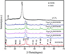
A novel ternary composite photocatalyst (g-C3N4/BiOI/BiOBr) was prepared via a facile solvothermal method. The samples were characterized by powder X-ray diffraction, transmission electron microscopy, UV-visible diffuse reflection spectrometry, X-ray photoelectron spectrometry and photoluminescence measurements. Under irradiation with visible light, the g-C3N4/BiOI/BiOBr photocatalyst showed a higher photocatalytic activity than pure g-C3N4 and BiOI/BiOBr for the degradation of methylene blue. Among the hybrid photocatalysts, 3% g-C3N4/BiOI/BiOBr showed the highest photocatalytic activity for the degradation of MB. These results suggest that the heterostructure combination of g-C3N4, BiOI and BiOBr provides a synergistic effect through an efficient charge transfer process.


1. Introduction

Contamination by organic pollutants has become a major environmental concern with industrial development and increases in the human population.1 Photocatalytic techniques are often used to eliminate hazardous pollutants from the atmosphere and the aqueous environment.2 TiO2 has attracted much attention as a photocatalyst for the decomposition of organic pollutants as a result of its high oxidative power, photostability and non-toxicity.3 However, because of its wide band gap (3.20 eV) and rapid recombination of photoinduced electrons–holes, TiO2 has a low efficiency for the utilization of solar energy, which limits its applications. Therefore there is a need to develop new photocatalysts with high activity under irradiation with visible light.4

Polymeric graphitic carbon nitride (g-C3N4) is a novel metal-free visible light induced semiconductor with a narrow band gap of 2.7 eV.5 It has been used in many applications, such as environmental purification,6 H2 production7 and CO2 reduction.8 However, its low quantum efficiency and fast charge recombination limits its applications.9 As composite photocatalysts have been shown to have a better photocatalytic performance than individual photocatalysts,10,11 g-C3N4 has been used to make composite photocatalysts with narrow or wide band gap semiconductors, such as ZnO,12 Co3O4,13 WO3,14 In2O3,15 AgX (X = Br, I),16 Ag3PO4,17 SmVO4,18 CdS,19 Pt/ZnO,20 Au/Pt,21 Ag3VO4,22 BiOCl,23 BiOI24 and Ag2O.25 Some ternary photocatalysts have also been synthesized, such as TiO2–In2O3@g-C3N4 (ref. 26) and g-C3N4/Bi2O3/TiO2.27 By combining semiconductors with g-C3N4, the ternary composites obtained, such as TiO2–In2O3@g-C3N4, showed a much higher photocatalytic performance than individual photocatalysts for the degradation of Rhodamine B under irradiation with visible light and in the production of hydrogen. The g-C3N4/Bi2O3/TiO2 composite showed enhanced activity under visible light irradiation and improved photoelectrochemical activity.

The V–VI–VII ternary bismuth compounds (BiOX; X = Cl, Br, I) have attracted considerable attention as a result of their excellent electrical and optical properties resulting from the internal static electric fields between the [Bi2O2]2+ slabs and interleaved halogen ion layers.28 Coupling BiOX with other types of semiconductors to construct heterostructures is considered to be an effective strategy to improve their photocatalytic reactivity.29 BiOX heterojunctions, including BiOBr/Bi2O3,30 BiOBr/BiOI,31 BiOCl/Bi2S3 (ref. 32) and g-C3N4/BiOClxBr1−x (ref. 33) have been reported. The coupling of BiOX with appropriate semiconductors has resulted in enhanced photocatalytic activities because the other semiconductor helps to increase the photocatalytic response of BiOX by providing a suitable band gap, efficient electron transfer and a low recombination rate for the photogenerated charge carriers.

Based on above investigations, binary BiOX composites have been synthesized for the enhanced photocatalytic activity, whereas there have been few reports on ternary composites based on BiOX binary composites. In this work, g-C3N4 was introduced to a BiOI/BiOBr binary composite to form the ternary composite g-C3N4/BiOI/BiOBr. The photocatalysts were characterized by XRD, SEM, EDS, HRTEM and diffuse reflectance spectrometry. The photocatalytic performance of the photocatalysts in the degradation of methylene blue (MB) was tested under irradiation with visible light. A possible photocatalytic mechanism is proposed based on the relative band positions in the three semiconductors.

2. Experimental section

2.1. Synthesis of photocatalysts

All the starting reagents (analytical-reagent grade purity) were purchased from Sinopharm and used without further purification.
2.1.1. Synthesis of g-C3N4. The g-C3N4 powder was prepared by heating dicyandiamide powder to 520 °C for 4 h at a heating rate of 20 °C min−1 in a muffle furnace according to a previously reported method.14 The yellow product was collected and ground into a powder.
2.1.2. Synthesis of BiOI/BiOBr composite. In a typical process, 1 mmol of Bi(NO3)3·5H2O was dissolved in 10 mL of ethylene glycol with magnetic stirring for about 30 min (solution A). Then 0.6 mmol of cetyltriethylammonium bromide (CTAB) and 0.4 mmol of KI were dissolved in 10 mL of water with ultrasonic treatment for 30 min (solution B). Solution A was then added into solution B with magnetic stirring and a yellow precipitate formed immediately (solution C). Solution C was stirred for 10 min and then transferred into a 25 mL Teflon-lined autoclave to 80% of the volume, which was then sealed and heated at 140 °C for 24 h.34 After cooling to room temperature naturally, the products was collected by filtration and washed five times with distilled water before drying at 80 °C for 12 h.
2.1.3. Synthesis of g-C3N4/BiOI/BiOBr composites. In a typical synthesis of 1% g-C3N4/BiOI/BiOBr composite, 7.7 mg of g-C3N4 were added to solution C (total mass of Bi(NO3)3·5H2O + CTAB + KI = 770 mg) and the suspension was stirred for 10 min and then transferred into a 25 mL Teflon-lined autoclave up to 80% of the volume, which was then sealed and heated at 140 °C for 24 h. After cooling to room temperature naturally, the products were collected by filtration and washed five times with distilled water and then dried at 80 °C for 12 h before being used in the photocatalytic reactions and for further characterization.

In suspension C, taking the mass of g-C3N4 as m and the total mass of Bi(NO3)3·5H2O + CTAB + KI as M, based on the result of (m/M) × 100% = 1%, the obtained product was named as the 1% g-C3N4/BiOI/BiOBr composite. According to this method, different mass ratios of the g-C3N4/BiOI/BiOBr composites were synthesized and labeled as 3% g-C3N4/BiOI/BiOBr, 5% g-C3N4/BiOI/BiOBr and 7% g-C3N4/BiOI/BiOBr.

2.2. Characterization

The g-C3N4/BiOI/BiOBr nanocomposites were analyzed by XRD using a Bruker D8 diffractometer with Cu Kα radiation (λ = 1.5418 Å) in the 2θ range 10–80°. The morphology and structure of the obtained samples were examined with transmission electron microscopy (TEM; Hitachi H-600-II). Ultraviolet-visible (UV-visible) diffuse reflectance spectra were measured using a UV-visible spectrophotometer (Shimadzu UV-2450) in the range 200–800 nm. BaSO4 was used as the standard reflectance material. Fourier-transform infrared (FTIR) spectra of the samples were recorded on a Nicolet Avatar-370 spectrometer at room temperature. Photoluminescence (PL) spectra of the catalysts were measured on a QuantaMaster 40 spectrofluorometer (Photon Technology International) with an excitation wavelength of 420 nm. X-ray photoemission spectrometry (XPS) was carried out on a PHI5300 instrument with a monochromatic Mg Kα source.

2.3. Photocurrent measurements

Photocurrent tests and electrochemical impedance spectroscopy (EIS) measurements were conducted using an electrochemical analyzer with a standard three-electrode configuration. A 500 W xenon lamp was used as the photo source. The working electrodes were ITO glass (0.5 × 1.5 cm2) coated with the as-prepared samples (0.1 mg) and the counter electrode was a platinum wire. The reference electrode was a saturated Ag–AgCl electrode and an aqueous solution of 0.1 M phosphate-buffered saline (pH 7.0) was used as the electrolyte. EIS was performed in the dark using a 0.1 M KCl solution containing 5 mM Fe(CN)63−/Fe(CN)64−.

2.4. Photocatalytic activity

The photocatalytic activities of the as-prepared samples were evaluated by the degradation of MB (20 mg L−1) under visible light irradiation at room temperature. A 75 mg mass of the photocatalyst was suspended in 75 mL of aqueous MB. Before irradiation, the suspension was magnetically stirred in the dark for about 30 min to establish an adsorption–desorption equilibrium between the photocatalysts and the MB dye and the solution was also magnetically stirred during the reaction. Under visible light illumination, 4 mL aqueous sample was withdrawn at certain time intervals and centrifuged at 10[thin space (1/6-em)]000 rpm before analysis. The MB concentration was determined by spectrometry by recording the intensity of the maximum band at 280 nm in the UV-visible absorption spectrum.

3. Results and discussion

3.1. XRD analysis

Fig. 1 shows the XRD patterns of the g-C3N4, BiOI/BiOBr and g-C3N4/BiOI/BiOBr composites. Two broad peaks were observed in the XRD pattern of g-C3N4. The strongest XRD peak at 27.4° originates from the (002) interlayer reflection of a graphite-like structure and the other pronounced XRD peak indexed as (100) at about 13.1° arises from the in-plane ordering of tris-triazine units.35 For BiOI/BiOBr, the diffraction peaks can be readily indexed to the tetragonal phases of BiOBr (JCPDS file no.73-2061)36 and BiOI (JCPDS file no. 10-0445).37 In the case of the g-C3N4/BiOI/BiOBr composites, the diffraction peaks can be indexed to the tetragonal phases of BiOBr and BiOI. However, no typical diffraction peak of g-C3N4 appeared in the g-C3N4/BiOI/BiOBr composite. This is a result of the low contents of g-C3N4 in the g-C3N4/BiOI/BiOBr composites and the fact that g-C3N4 was fully mixed with BiOI/BiOBr. This result is similar to that reported previously.38 The existence of g-C3N4 was confirmed by FTIR spectrometry and XPS.
image file: c6ra05565f-f1.tif
Fig. 1 XRD patterns of g-C3N4, BiOI/BiOBr, 1% g-C3N4/BiOI/BiOBr, 3% g-C3N4/BiOI/BiOBr, 5% g-C3N4/BiOI/BiOBr and 7% g-C3N4/BiOI/BiOBr.

3.2. SEM and TEM analyses

The dispersion state and the structure of the g-C3N4, BiOI, BiOBr, BiOI/BiOBr, 3% g-C3N4/BiOI/BiOBr and 7% g-C3N4/BiOI/BiOBr composites were obtained by TEM (Fig. 2). The TEM image of the prepared g-C3N4 sample showed a lamellar structure (Fig. 2A), which is in agreement with previous work.39 TEM images of the prepared BiOI and BiOBr samples are shown in Fig. 2B and C. BiOI formed rounded sheets with a size of about 50–150 nm and BiOBr formed quadrate sheets of about 200–500 nm. For BiOI/BiOBr, it could be clearly seen that the quadrate BiOBr substrates overlapped with rounded thin pieces of the BiOI nanosheets (Fig. 2D), which confirmed the presence of BiOI/BiOBr.29 In the TEM images of the 3% g-C3N4/BiOI/BiOBr (Fig. 2E) and 7% g-C3N4/BiOI/BiOBr (Fig. 2F) composites, the composites showed a much darker color than BiOI/BiOBr, which is a result of the introduction of g-C3N4. Although having undergone ultrasonic treatment before the TEM observations, 7% g-C3N4/BiOI/BiOBr showed a firm connection between g-C3N4, BiOI and BiOBr (Fig. 2F), in which it can be clearly seen that curved g-C3N4 was attached to the surface of the composite, which is favorable for the formation of heterojunctions. The morphologies of the 3% g-C3N4/BiOI/BiOBr composite were examined by SEM (Fig. 2G); the as-prepared 3% g-C3N4/BiOI/BiOBr showed a sheet-like structure. The chemical composition of the 3% g-C3N4/BiOI/BiOBr composite was further confirmed by EDS analysis. Fig. 2H shows that Bi, O, C, N, Br and I peaks were present in the spectrum; the Au peak is attributed to the gold plating.
image file: c6ra05565f-f2.tif
Fig. 2 TEM images of (A) g-C3N4, (B) BiOI, (C) BiOBr, (D) BiOI/BiOBr, (E) 3% g-C3N4/BiOI/BiOBr and (F) 7% g-C3N4/BiOI/BiOBr. (G) SEM image and (H) EDS spectrum of 3% g-C3N4/BiOI/BiOBr.

3.3. XPS analysis

XPS was carried out to further analyze the elemental composition and valence bond structure of the g-C3N4, BiOI/BiOBr and g-C3N4/BiOI/BiOBr composites (Fig. 3). The survey XPS spectrum (Fig. 3A) clearly showed that the 3% g-C3N4/BiOI/BiOBr composite was composed of C, N, Bi, O, I and Br. High-resolution spectra of Bi4f, Br3d, I3d, O1s, C1s and N1s are shown in Fig. 3B–G. The Bi4f spectra of BiOI/BiOBr and 3% g-C3N4/BiOI/BiOBr (Fig. 3B) could be resolved into two spin orbit components at binding energies of 158.7 and 163.9 eV, which were assigned to the Bi4f7/2 and Bi4f5/2 of Bi3+ in BiOBr, respectively.40 In the BiOI/BiOBr composite, the typical peaks at binding energies of about 67.6 and 68.4 eV (Fig. 3C) belonged to Br3d5/2 and Br3d3/2.41,42 The I3d peaks of BiOI/BiOBr were located at 618.4 and 629.6 eV (Fig. 3D), which correspond to the I3d5/2 and I3d3/2 binding energies.43 However, in the case of the 3% g-C3N4/BiOI/BiOBr composite, the binding energy of Br3d3/2 (68.5 eV) and I3d3/2 (629.8 eV) showed a slight shift compared with that of the BiOI/BiOBr composite. The shift of Br3d3/2 and I3d5 confirmed the interaction between g-C3N4 and BiOI/BiOBr.
image file: c6ra05565f-f3.tif
Fig. 3 (A) XPS survey spectrum and high-resolution XPS spectra of (B) Bi4f, (C) Br3d, (D) I3d, (E) O1s, (F) C1s and (G) N1s regions for 3% g-C3N4/BiOI/BiOBr.

The XPS spectra of O1s for BiOI/BiOBr and 3% g-C3N4/BiOI/BiOBr are shown in Fig. 3E. For BiOI/BiOBr, the O1s profile is asymmetrical and can be fitted to three symmetrical peaks located at 529.5, 530.8 and 532.5 eV, respectively, indicating three different O species in the sample. The O1s peak at 529.5 eV was attributed to crystal lattice O atoms (Bi–O). The peak at 530.8 eV was attributed to the Bi–O bonds in the [Bi2O2] slabs of BiOI.44 The peak at 532.5 eV was associated with the presence of the –OH group or water molecules on the surface of the BiOI/BiOBr and 3% g-C3N4/BiOI/BiOBr composites.45 For the 3% g-C3N4/BiOI/BiOBr composite, the O1s peak at 530.8 eV for BiOI/BiOBr shifted to 531.0 eV after hybridization, indicating a slight change in the oxygen environment after the introduction of g-C3N4 into BiOI/BiOBr.

The XPS spectra of C1s for g-C3N4 and the 3% g-C3N4/BiOI/BiOBr composite are shown in Fig. 3F. For g-C3N4, the peak at 284.8 eV is regarded as the carbon species from g-C3N4.46 The peak at 286.2 eV was attributed to the C–NH2 species on g-C3N4.47 The other peak at 288.3 eV was assigned to the sp2-hybridized carbon in the N[double bond, length as m-dash]C–N2 coordination,48 which is ascribed to carbon atoms that have one double and two single bonds with three N neighbors, respectively.49 After hybridization, the peak at 288.3 eV for pure g-C3N4 shifted to 287.8 eV, indicating that there were interactions among g-C3N4, BiOI and BiOBr. Compared with pure g-C3N4, the peak intensity of C1s (287.8 eV) in the 3% g-C3N4/BiOI/BiOBr composite decreased significantly, which is a result of the lower levels of g-C3N4.

The XPS results for N1s in g-C3N4 and the 3% g-C3N4/BiOI/BiOBr composite are shown in Fig. 3G. The N1s binding energy of g-C3N4 at 398.8 eV can be assigned to sp2-hybridized nitrogen (C[double bond, length as m-dash]N–C),50,51 confirming the presence of sp2-bonded graphitic carbon nitride. The binding energies at 400.1 and 401.3 eV can be assigned to tertiary nitrogen bonded to carbon atoms by C–NH and N–(C)3 bonds,49 respectively. Compared with g-C3N4, the binding energy of the N1s (398.4 eV) of the 3% g-C3N4/BiOI/BiOBr composite had a negative shift and the intensity decreased significantly. This shift further indicates that there are interactions among g-C3N4, BiOI and BiOBr. As a result of the low content of g-C3N4 in the 3% g-C3N4/BiOI/BiOBr composite, the peak intensity of N1s is very weak. This has also been found in similar systems.38

Based on the results of the XRD, TEM, EDS and XPS analyses, the interaction among g-C3N4, BiOI and BiOBr was confirmed, which may be favorable for charge transfer and the separation of photogenerated electron–hole pairs, which could promote the photocatalytic performance.

3.4. FTIR analysis

Fig. 4 shows the FTIR spectra for g-C3N4 and the BiOI/BiOBr and g-C3N4/BiOI/BiOBr composites, with different weight ratios of g-C3N4 in the g-C3N4/BiOI/BiOBr composites. For pure g-C3N4, the band at 808 cm−1 is related to the breathing mode of the heptazine arrangement, whereas those at 1244–1639 cm−1 can be assigned to aromatic C–N breathing modes.52,53 For the BiOI/BiOBr sample, the valent symmetrical A2u-type vibrations of the Bi–O bond were observed at 508 cm−1 in BiOI/BiOBr.54 The absorption at 767 cm−1 was assigned to the asymmetrical stretching vibration of the Bi–O bond.55,56 The sharp and strong absorption at 1626 cm−1 was assigned to the bending vibrations of O–H resulting from the adsorption of free water molecules on the photocatalyst surface. The characteristic peaks of g-C3N4 and BiOI/BiOBr are present in all the g-C3N4/BiOI/BiOBr composites.
image file: c6ra05565f-f4.tif
Fig. 4 FTIR spectra of g-C3N4, BiOI/BiOBr, 1% g-C3N4/BiOI/BiOBr, 3% g-C3N4/BiOI/BiOBr, 5% g-C3N4/BiOI/BiOBr and 7% g-C3N4/BiOI/BiOBr.

3.5. UV-visible diffuse reflectance spectrometry

The optical properties of the g-C3N4, BiOI/BiOBr and g-C3N4/BiOI/BiOBr composites were investigated by UV-visible diffuse reflectance spectrometry. As shown in Fig. 5, the g-C3N4 and BiOI/BiOBr samples absorb UV to visible light, which signifies their visible-light-driven photocatalytic activities. g-C3N4 can absorb light with a wavelength <450 nm,57 and the BiOI/BiOBr sample showed strong absorption over the whole UV-visible range of 200–600 nm.58 The absorption of the g-C3N4/BiOI/BiOBr composites decreased with the increasing g-C3N4 mass ratios, whereas they still showed an absorption edge in the visible region. The result implies that the fabrication of the composites provides the potential in the following visible-light photocatalytic test.
image file: c6ra05565f-f5.tif
Fig. 5 UV-visible diffuse reflectance spectra of g-C3N4, BiOI/BiOBr, 1% g-C3N4/BiOI/BiOBr, 3% g-C3N4/BiOI/BiOBr, 5% g-C3N4/BiOI/BiOBr and 7% g-C3N4/BiOI/BiOBr.

3.6. Photocatalytic activity and kinetics

The photocatalytic capability of the g-C3N4, BiOI/BiOBr and g-C3N4/BiOI/BiOBr composites was evaluated by decomposing MB under visible light. Fig. 6A shows that MB self-degradation was almost negligible in the absence of a photocatalyst. g-C3N4 and BiOI/BiOBr could degrade MB up to 50% and 40%, respectively, within 2.5 h and all the g-C3N4/BiOI/BiOBr composites showed a higher photocatalytic activity than both g-C3N4 and BiOI/BiOBr under visible light irradiation. Significantly, the 3% g-C3N4/BiOI/BiOBr composite had the best activity for the decomposition of MB, resulting in 80% degradation within 2.5 h. However, on further increasing the proportion of g-C3N4, the degradation rate showed a slight decrease, although it remained higher than that of g-C3N4 and BiOI/BiOBr. The decrease in the activity of the samples with a heavy loading of g-C3N4 may be a result of the shading effect,59 which can block the absorption of the incident light by BiOI/BiOBr.
image file: c6ra05565f-f6.tif
Fig. 6 (A) Photodegradation of MB on g-C3N4/BiOI/BiOBr composites. (B) Kinetic fit for the degradation of MB with g-C3N4, BiOI/BiOBr and g-C3N4/BiOI/BiOBr composites.

To quantitatively investigate the reaction kinetics of the degradation of MB, the experimental data were fitted by a first-order model as expressed by the formula:

ln(C0/C) = kt
where C0 and C are the dye concentrations in solution at times 0 and t, respectively, and k is a first-order rate constant.

Fig. 6B shows that the pseudo-first-order rate constants (k) for the degradation of MB with the g-C3N4, BiOI/BiOBr and g-C3N4/BiOI/BiOBr composites (1, 3, 5 and 7%) were about 0.27, 0.21, 0.46, 0.68, 0.54 and 0.35 h−1, respectively. The rate constant of the 3% g-C3N4/BiOI/BiOBr composite was 2.5 times as high as that of g-C3N4 and 3.2 times as high as that of BiOI/BiOBr. As a result, the 3% g-C3N4/BiOI/BiOBr composite had the best performance and was selected for the subsequent recycling experiment.

3.7. PL analysis

To investigate the effect of the separation efficiency of the generated electrons and holes in the semiconductors, the PL spectra for the BiOI/BiOBr and 3% g-C3N4/BiOI/BiOBr composites were determined at an excitation wavelength of 420 nm (Fig. 7); the results were similar to previous reports.41 The BiOI/BiOBr and 3% g-C3N4/BiOI/BiOBr composites both showed a similar peak shape and peak position. Compared with the BiOI/BiOBr composite, the intensity of the peaks for the 3% g-C3N4/BiOI/BiOBr composite was much lower, suggesting that the composite had a much lower recombination rate for the photogenerated charge carriers.
image file: c6ra05565f-f7.tif
Fig. 7 Photoluminescence spectra of BiOI/BiOBr and 3% g-C3N4/BiOI/BiOBr.

3.8. Photocurrent measurements

Photocurrent measurements were performed to further investigate the photoelectric performance because a stronger photocurrent intensity often suggests a higher separation efficiency of holes and electrons.60 Hence the higher the photocurrent, the better the electron and hole separation efficiency and therefore the higher the photocatalytic activity. Fig. 8 shows the transient photocurrent response curves of the g-C3N4, BiOI/BiOBr and 3% g-C3N4/BiOI/BiOBr composites. Compared with g-C3N4 and BiOI/BiOBr, the 3% g-C3N4/BiOI/BiOBr composite showed an enhanced photocurrent response, which was 5 and 1.7 times the intensity of the g-C3N4 and BiOI/BiOBr composites, respectively. This result indicates that the formation of heterojunctions is favorable for charge separation.61
image file: c6ra05565f-f8.tif
Fig. 8 Transient photocurrent response for pure g-C3N4, BiOI/BiOBr and 3% g-C3N4/BiOI/BiOBr.

3.9. EIS analysis

The charge transfer efficiency at the interface of semiconductor photoelectrodes can be indicated by EIS. The EIS Nyquist plots of the g-C3N4, BiOI/BiOBr and 3% g-C3N4/BiOI/BiOBr composites were measured (Fig. 9). The results show that the BiOI/BiOBr and 3% g-C3N4/BiOI/BiOBr composites both had smaller diameter Nyquist circles than g-C3N4. In addition, the 3% g-C3N4/BiOI/BiOBr composite had a smaller diameter than BiOI/BiOBr. Compared with pure g-C3N4 and BiOI/BiOBr, 3% g-C3N4/BiOI/BiOBr composite had the lowest resistance, which is favorable for the interfacial charge transfer process. The results of the EIS analysis suggested that the combination of g-C3N4 and BiOI/BiOBr can improve the separation and charge transfer efficiency of photogenerated electron–hole pairs and enhance the photocatalytic activity.62
image file: c6ra05565f-f9.tif
Fig. 9 EIS profiles for pure g-C3N4, BiOI/BiOBr and 3% g-C3N4/BiOI/BiOBr.

3.10. Stability evaluation

The stability and recyclability of photocatalysts are important in practical applications. Additional experiments were carried out to degrade MB under visible light irradiation over three cycles with the 3% g-C3N4/BiOI/BiOBr sample. Fig. 10A shows the relationship between the removal ratio for MB and the cycle time. After three repeated runs, the MB removal ratio decreased to 55%, which may be a result of the loss of the photocatalysts during the cycling experiment. The sample was collected after the cycling runs and analyzed by XRD (Fig. 10B). The XRD peaks of the used 3% g-C3N4/BiOI/BiOBr sample were identical to those of the fresh sample, which implied that the sample was stable during degradation.
image file: c6ra05565f-f10.tif
Fig. 10 (A) Cycling runs of 3% g-C3N4/BiOI/BiOBr composite under visible light irradiation for 2.5 h. (B) XRD patterns of 3% g-C3N4/BiOI/BiOBr before and after the cycling photocatalytic experiments.

3.11. Detection of reactive species

Photoinduced holes, superoxide radicals and hydroxyl radicals are the main active species in the degradation of organic pollutants.63 The roles of active species in the photodegradation process were tested by adding individual scavengers to the photodegradation suspensions containing 3% g-C3N4/BiOI/BiOBr. The scavengers used were isopropanol for hydroxyl radicals (˙OH), N2 for superoxide radicals (˙O2) and triethanolamine for holes (h+). Fig. 11 shows that the addition of isopropanol, N2 and triethanolamine caused a decrease in the photodegradation efficiency of 3% g-C3N4/BiOI/BiOBr, indicating that ˙OH, ˙O2 and h+ are the active species in this photocatalytic process. A significant suppression of photocatalytic performance was observed when triethanolamine was added, which indicates that h+ is the main active species. These experiments suggest that ˙OH, ˙O2 and h+ affect the degradation of MB, but that h+ is the main reactive species in the degradation of MB.64,65
image file: c6ra05565f-f11.tif
Fig. 11 Reactive species trapping experiments over 3% g-C3N4/BiOI/BiOBr.

3.12. Possible photocatalytic mechanism of the ternary hybrid composites

Two possible photocatalytic mechanisms based on the band structures and microstructures of the catalysts are proposed for g-C3N4/BiOI/BiOBr (Fig. 12). The valence band (VB) and conduction (CB) levels for g-C3N4 were 1.57 and −1.12 eV,66,67 respectively, and those for BiOI were 2.31 and −0.63 eV;68 the VB and CB edge potentials of BiOBr were 0.24 and 3.05 eV.34 Based on the band gap structures of g-C3N4, BiOBr and BiOBr, the separation processes for the photoexcited electrons–holes are shown in Fig. 12A and B. If the charge carriers of the g-C3N4/BiOI/BiOBr composite transfer as shown in Fig. 12A, then the generic electron–hole separation process seen in a great number of composite photocatalysts takes place. g-C3N4, BiOI and BiOBr all absorbed visible light and became excited. Because the CB edge of g-C3N4 is more negative than that of both BiOI and BiOBr, electrons from g-C3N4 were injected into the less negative CB of the surrounding BiOI. Similarly, the electrons of BiOI jumped toward BiOBr and the holes in the VB of BiOBr migrated to the VB of BiOI and g-C3N4. As a result, the accumulated electrons in the CB of BiOBr cannot reduce O2 to ˙O2 and the holes in the VB of g-C3N4 cannot oxidize OH to ˙OH. Therefore when the charge carriers of the photocatalyst transfer in accordance with the traditional model, the formation of the active species ˙O2and ˙OH is not favorable. Therefore the mechanism in Fig. 12A does not match the results of the reactive species experiment.
image file: c6ra05565f-f12.tif
Fig. 12 Proposed mechanisms for the photodegradation of MB on g-C3N4/BiOI/BiOBr composites (see text for discussion).

However, if the charge carriers of the g-C3N4/BiOI/BiOBr photocatalyst transfer according to Fig. 12B, which involves a direct Z-scheme,69,70 then the photoexcited electrons in the CB of BiOBr and the photoexcited holes in the VB of BiOI are rapidly combined. At the same time, the electrons in the CB of BiOI, which have a more negative potential, reduce molecular oxygen to yield ˙O2. The holes in the VB of BiOBr, which have a more positive potential, generate abundant active ˙OH radicals. The holes in the VB of BiOBr and g-C3N4 are then available to degrade organic pollutants. Thus a model based on the Z-scheme between BiOBr and BiOI is favorable for the production of the ˙O2, ˙OH and h+ reactive species.

4. Conclusion

Novel g-C3N4/BiOI/BiOBr composite photocatalysts were successfully prepared via a facile solvothermal method. The structure, surface morphology and chemical states of the elements were investigated and the interaction between g-C3N4 and BiOI/BiOBr was determined. Compared with the g-C3N4 and BiOI/BiOBr composites, the g-C3N4/BiOI/BiOBr composites showed a remarkable improvement in photocatalytic activity under irradiation with visible light. The degradation rate constant of the optimized 3% g-C3N4/BiOI/BiOBr composite was 0.68 h−1, 3.2 times that of BiOI/BiOBr. A possible photocatalytic mechanism is proposed based on the experimental results. The matched band positions of g-C3N4, BiOI and BiOBr allowed the development of a model containing the Z-scheme, which is favorable for the separation and transfer of photogenerated charge carriers and the production of ˙O2, ˙OH and h+ reactive species.

Acknowledgements

The authors genuinely appreciate the financial support of this work from the National Nature Science Foundation of China (21406094, 21476097 and 21416078), Postdoctoral Foundation of China (2015M571693) and the Foundation of Jiangsu University (14JDG184).

References

  1. M. Chong, B. Jin, C. Chow and C. Saint, Water Res., 2010, 44, 2997–3027 CrossRef CAS PubMed.
  2. C. Chen, W. Ma and J. Zhao, Chem. Soc. Rev., 2010, 39, 4206–4219 RSC.
  3. X. Chen and S. Mao, Chem. Rev., 2007, 107, 2891–2959 CrossRef CAS PubMed.
  4. Y. Chen, W. Huang, D. He, Y. Situ and H. Huang, ACS Appl. Mater. Interfaces, 2014, 6, 14405–14414 CAS.
  5. Y. Wang, X. Wang and M. Antonietti, Angew. Chem., Int. Ed., 2012, 51, 68–89 CrossRef CAS PubMed.
  6. J. Xia, J. Di, S. Yin, H. Li, H. Xu, L. Xu, H. Shu and M. He, Mater. Sci. Semicond. Process., 2014, 24, 96–103 CrossRef CAS.
  7. H. Liu, Z. Xu, Z. Zhang and D. Ao, Appl. Catal., B, 2016, 192, 234–241 CrossRef CAS.
  8. J. Wang, H. Yao, Z. Fan, L. Zhang, J. Wang, S. Zang and Z. Li, ACS Appl. Mater. Interfaces, 2016, 8, 3765–3775 CAS.
  9. H. Shi, G. Chen, C. Zhang and Z. Zou, ACS Catal., 2014, 4, 3637–3643 CrossRef CAS.
  10. N. Zhang, M. Q. Yang, S. Liu, Y. Sun and Y. J. Xu, Chem. Rev., 2015, 115, 10307–10377 CrossRef CAS PubMed.
  11. S. Liu, Z. R. Tang, Y. Sun, J. Colmenares and Y. Xu, Chem. Soc. Rev., 2015, 44, 5053–5075 RSC.
  12. Y. He, Y. Wang, L. Zhang, B. Teng and M. Fan, Appl. Catal., B, 2015, 168, 1–8 Search PubMed.
  13. C. Han, L. Ge, C. Chen, Y. Li, X. Xiao, Y. Zhang and L. Guo, Appl. Catal., B, 2014, 147, 546–553 CrossRef CAS.
  14. L. Huang, H. Xu, Y. Li, H. Li, X. Cheng, J. Xia, Y. Xu and G. Cai, Dalton Trans., 2013, 42, 8606–8616 RSC.
  15. S. W. Cao, X. F. Liu, Y. P. Yuan, Z. Y. Zhang, Y.-S. Liao, J. Fang, S. C. J. Loo, T. C. Sum and C. Xue, Appl. Catal., B, 2014, 147, 940–946 CrossRef CAS.
  16. H. Xu, J. Yan, Y. Xu, Y. Song, H. Li, J. Xia, C. Huang and H. Wan, Appl. Catal., B, 2013, 129, 182–193 CrossRef CAS.
  17. Y. He, L. Zhang, B. Teng and M. Fan, Environ. Sci. Technol., 2015, 49, 649–656 CrossRef CAS PubMed.
  18. T. Li, L. Zhao, Y. He, J. Cai, M. Luo and J. Lin, Appl. Catal., B, 2013, 129, 255–263 CrossRef CAS.
  19. J. Fu, B. Chang, Y. Tian, F. Xi and X. Dong, J. Mater. Chem., 2013, 1, 3083–3090 RSC.
  20. J. Xiao, X. Zhang and Y. Li, Int. J. Hydrogen Energy, 2015, 40, 9080–9087 CrossRef CAS.
  21. J. Xue, S. Ma, Y. Zhou, Z. Zhang and M. He, ACS Appl. Mater. Interfaces, 2015, 7, 9630–9637 CAS.
  22. S. Wang, D. Li, C. Sun, S. Yang, Y. Guan and H. He, Appl. Catal., B, 2014, 144, 885–892 CrossRef CAS.
  23. S. Shi, M. A. Gondal, A. A. Al-Saadi, R. Fajgar, J. Kupcik, X. Chang, K. Shen, Q. Xu and Z. S. Seddigi, J. Colloid Interface Sci., 2014, 416, 212–219 CrossRef CAS PubMed.
  24. C. Chang, L. Zhu, S. Wang, X. Chu and L. Yue, ACS Appl. Mater. Interfaces, 2014, 6, 5083–5093 CAS.
  25. H. T. Ren, S. Y. Jia, Y. Wu, S. H. Wu, T. H. Zhang and X. Han, Ind. Eng. Chem. Res., 2014, 53, 17645–17653 CrossRef CAS.
  26. Z. Jiang, D. Jiang, Z. Yan, D. Liu, K. Qian and J. Xie, Appl. Catal., B, 2015, 170–171, 195–205 CrossRef CAS.
  27. Y. Zhang, J. Lu, M. Hoffmann, Q. Wang, Y. Cong, Q. Wang and H. Jin, RSC Adv., 2015, 5, 48983–48991 RSC.
  28. J. C. Ahern, R. Fairchild, J. S. Thomas, J. Carr and H. H. Patterson, Appl. Catal., B, 2015, 179, 229–238 CrossRef CAS.
  29. H. Huang, X. Han, X. Li, S. Wang, P. K. Chu and Y. Zhang, ACS Appl. Mater. Interfaces, 2015, 7, 482–492 CAS.
  30. Q. Wang, D. Jiao, J. Lian, Q. Ma, J. Yu, H. Huang, J. Zhong and J. Li, J. Alloys Compd., 2015, 649, 474–482 CrossRef CAS.
  31. L. Lin, M. Huang, L. Long, Z. Sun, W. Zheng and D. Chen, Ceram. Int., 2014, 40, 11493–11501 CrossRef CAS.
  32. C. F. Guo, S. Cao, J. Zhang, H. Tang, S. Guo, Y. Tian and Q. Liu, J. Am. Chem. Soc., 2011, 133, 8211–8215 CrossRef CAS PubMed.
  33. S. Shi, M. A. Gondal, S. G. Rashid, Q. Qi, A. A. Al-Saadi, Z. H. Yamani, Y. Sui, Q. Xu and K. Shen, Colloids Surf., A, 2014, 461, 202–211 CrossRef CAS.
  34. Z. Liu, H. Ran, B. Wu, P. Feng and Y. Zhu, Colloids Surf., A, 2014, 452, 109–114 CrossRef CAS.
  35. Z. Tong, D. Yang, J. Shi, Y. Nan, Y. Sun and Z. Jiang, ACS Appl. Mater. Interfaces, 2015, 7, 25693–25701 CAS.
  36. J. Zhang, F. Shi, J. Lin, D. Chen, J. Gao, Z. Huang, X. Ding and C. Tang, Chem. Mater., 2008, 20, 2937–2941 CrossRef CAS.
  37. L. Ye, J. Chen, L. Tian, J. Liu, T. Peng, K. Deng and L. Zan, Appl. Catal., B, 2013, 130–131, 1–7 CAS.
  38. J. Di, J. Xia, S. Yin, H. Xu, M. He, H. Li, L. Xu and Y. Jiang, RSC Adv., 2013, 3, 19624–19631 RSC.
  39. M. Xu, L. Han and S. Dong, ACS Appl. Mater. Interfaces, 2013, 5, 12533–12540 CAS.
  40. L. Ye, J. Liu, C. Gong, L. Tian, T. Peng and L. Zan, ACS Catal., 2012, 2, 1677–1683 CrossRef CAS.
  41. J. Di, J. Xia, M. Ji, B. Wang, X. Li, Q. Zhang, Z. Chen and H. Li, ACS Sustainable Chem. Eng., 2016, 4, 136–146 CrossRef CAS.
  42. W. Li, X. Jia, P. Li, B. Zhang, H. Zhang, W. Geng and Q. Zhang, ACS Sustainable Chem. Eng., 2015, 3, 1101–1110 CrossRef CAS.
  43. X. Zhang, L. Zhang, T. Xie and D. Wang, J. Phys. Chem. C, 2009, 113, 7371–7378 CAS.
  44. C. Chang, L. Zhu, S. Wang, X. Chu and L. Yue, ACS Appl. Mater. Interfaces, 2014, 6, 5083–5093 CAS.
  45. Y. Wang, B. Li, C. Zhang, L. Cui, S. Kang, X. Li and L. Zhou, Appl. Catal., B, 2013, 130–131, 277–284 CrossRef CAS.
  46. D. Jiang, J. Li, C. Xing, Z. Zhang, S. Meng and M. Chen, ACS Appl. Mater. Interfaces, 2015, 7, 19234–19242 CAS.
  47. F. He, G. Chen, Y. Yu, S. Hao, Y. Zhou and Y. Zheng, ACS Appl. Mater. Interfaces, 2014, 6, 7171–7179 CAS.
  48. Y. Li, R. Wang, H. Li, X. Wei, J. Feng, K. Liu, Y. Dang and A. Zhou, J. Phys. Chem. C, 2015, 119, 20283–20292 CAS.
  49. M. Rong, L. Lin, X. Song, T. Zhao, Y. Zhong, J. Yan, Y. Wang and X. Chen, Anal. Chem., 2015, 87, 1288–1296 CrossRef CAS PubMed.
  50. X. F. Li, J. Zhang, L. H. Shen, Y. M. Ma, W. W. Lei, Q. L. Cui and G. T. Zou, Appl. Phys. A: Mater. Sci. Process., 2009, 94, 387–392 CrossRef CAS.
  51. S. C. Yan, Z. S. Li and Z. G. Zou, Langmuir, 2010, 26, 3894–3901 CrossRef CAS PubMed.
  52. L. Liu, D. Ma, H. Zheng, X. Li, M. Cheng and X. Bao, Microporous Mesoporous Mater., 2008, 110, 216–222 CrossRef CAS.
  53. S. C. Yan, Z. S. Li and Z. G. Zou, Langmuir, 2009, 25, 10397–10401 CrossRef CAS PubMed.
  54. Y. Wang, K. Deng and L. Zhang, J. Phys. Chem. C, 2011, 115, 14300–14308 CAS.
  55. K. Gurunathan, Int. J. Hydrogen Energy, 2004, 29, 933–940 CrossRef CAS.
  56. Z. Liu, X. Xu, J. Fang, X. Zhu, J. Chu and B. Li, Appl. Surf. Sci., 2012, 258, 3771–3778 CrossRef CAS.
  57. X. Fan, L. Zhang, M. Wang, W. Huang, Y. Zhou, M. Li, R. Cheng and J. Shi, Appl. Catal., B, 2016, 182, 68–73 CrossRef CAS.
  58. H. Lin, H. Ye, X. Li, J. Cao and S. Chen, Ceram. Int., 2014, 40, 9743–9750 CrossRef CAS.
  59. Y. Li, J. Zhan, L. Huang, H. Xu, H. Li, R. Zhang and S. Wu, RSC Adv., 2014, 4, 11831–11839 RSC.
  60. Z. Zhu, Z. Lu, D. Wang, X. Tang, Y. Yan, W. Shi, Y. Wang, N. Gao, X. Yao and H. Dong, Appl. Catal., B, 2016, 182, 115–122 CrossRef CAS.
  61. B. Long, Y. Huang, H. Li, F. Zhao, Z. Rui, Z. Liu, Y. Tong and H. Ji, Ind. Eng. Chem. Res., 2015, 54, 12788–12794 CrossRef CAS.
  62. J. Di, J. Xia, M. Ji, L. Xu, S. Yin, Q. Zhang, Z. Chen and H. Li, Carbon, 2016, 98, 613–623 CrossRef CAS.
  63. F. Dong, Z. Zhao, Y. Sun, Y. Zhang, S. Yan and Z. Wu, Environ. Sci. Technol., 2015, 49, 12432–12440 CrossRef CAS PubMed.
  64. H. Wang, Y. Su, H. Zhao, H. Yu, S. Chen, Y. Zhang and X. Quan, Environ. Sci. Technol., 2014, 48, 11984–11990 CrossRef CAS PubMed.
  65. X. Wang, W. Yang, F. Li, Y. Xue, R. Liu and Y. Hao, Ind. Eng. Chem. Res., 2013, 52, 17140–17150 CrossRef CAS.
  66. K. Li, S. Gao, Q. Wang, H. Xu, Z. Wang, B. Huang, Y. Dai and J. Lu, ACS Appl. Mater. Interfaces, 2015, 7, 9023–9030 CAS.
  67. K. Maeda, ACS Catal., 2013, 3, 1486–1503 CrossRef CAS.
  68. F. Chen, C. Niu, Q. Yang, X. Li and G. Zeng, Ceram. Int., 2016, 42, 2515–2525 CrossRef CAS.
  69. X. Yang, Z. Chen, J. Xu, H. Tang, K. Chen and Y. Jiang, ACS Appl. Mater. Interfaces, 2015, 7, 15285–15293 CAS.
  70. Y. He, L. Zhang, X. Wang, Y. Wu, H. Lin, L. Zhao, W. Weng, H. Wan and M. Fan, RSC Adv., 2014, 4, 13610–13619 RSC.

This journal is © The Royal Society of Chemistry 2016