A diarylethene-based fluorescent chemosensor for the sequential recognition of Fe3+ and cysteine

Guanming Liao, Chunhong Zheng*, Dandan Xue, Congbin Fan, Gang Liu and Shouzhi Pu*
Jiangxi Key Laboratory of Organic Chemistry, Jiangxi Science and Technology Normal University, Nanchang, Jiangxi 330013, PR China. E-mail: articlechem@163.com; pushouzhi@tsinghua.org.cn; Fax: +86-791-83831996; Tel: +86-791-83831996

Received 2nd March 2016 , Accepted 28th March 2016

First published on 30th March 2016


Abstract

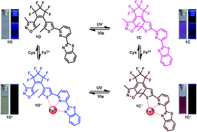
A new fluorescent chemosensor for the sequential recognition of Fe3+ and cysteine has been constructed based on photochromic diarylethene. It exhibits sequential recognition of Fe3+ and cysteine via an “on–off–on” molecular switch. The title compound can selectively and sensitively recognize Fe3+ in methanol, causing quenching of the fluorescence. Then, the formed complex was found to be a fluorescent chemosensor for cysteine with an increase in the fluorescence intensity. A detection limit as low as 4.09 × 10⁻⁸ mol L−1 for Fe3+ and 2.89 × 10⁻⁸ mol L−1 for cysteine was obtained. Moreover, a two input INHIBIT logic gate was fabricated by using Fe3+ and cysteine as inputs and taking I439 as the output.


Introduction

The design of fluorescent chemosensors for various metal ions has attracted considerable interest from researchers because they play critical roles in fundamental processes, including osmotic regulation, catalysis, metabolism, biomineralization, and signaling.1–3 Iron is one of the most important elements in metabolic processes, being indispensable for all living systems. It is extensively distributed in environmental and biological materials.4,5 A lack of iron in the human body can lead to anemia,6 liver and kidney damages,7 diabetes8 and heart diseases,9 whereas excessive iron may result in some types of cancers and a deterioration of organic functions such as Parkinson's, Alzheimer's disease.10,11 Moreover, excess iron can result in some environmental problems.12 In consideration of the wide role of iron(III) (Fe3+), designing a highly selective chemosensor for Fe3+ is still a challenge.

In recent years, study of fluorescent chemosensors for detection of thiol-containing amino acids has attracted significant attention due to their crucial roles in a wide range of biological processes.13–15 Cysteine (Cys), one of the most abundant intracellular biothiols, possesses various important biochemical functions including protein synthesis, detoxification, metabolism and metal binding.16–18 Cys deficiency may cause health problems such as slow growth of children, hair depigmentation, edema, lethargy, liver damage, muscle and fat loss, skin lesions, and weakness.19 Therefore, the development of fluorescent chemosensors for detection of Cys has become quite desirable. In view of the significance of Fe3+ and Cys, highly selective fluorimetric methods for their detection have been developed.20,21

The research on photochromic functional materials has been becoming increasingly popular owing to their potential applications as sensors, photoresponsive self-assemblies, optical memory storage, live cell imaging, molecular logic gates, and multifunctional switches.22–27 Among diverse photochromic compounds, diarylethene derivatives are the most promising candidate fluorescent switch systems because of their fast response, excellent thermal stability and outstanding fatigue resistance.28–30 Due to the distinct feature, an increasing number of diarylethene-based fluorescence chemosensors for selective detection of metal ions such as Hg2+, Cu2+, Al3+, Fe3+, and Zn2+ were designed and synthesized.31–35 However, the reported derivatives can only recognize of metal ions, and their fluorescent modulation efficiency and selectivity need to be further enhanced. It is still a challenge to develop new dual functional fluorescent chemosensors based on diarylethenes that can sequentially detect of cations and amino acids. This would reduce analytic time, increase analysis speed and cut cost.36–40 Moreover, chemosensors with benzothiazole moiety have received much attention due to their potential use as fluorescent sensors. Herein, a fluorescent chemosensor with benzothiazole moiety was designed and synthesized. Its sequential sensing properties towards various metal ions and amino acids were studied. The structure and its photochromic process of 1O are shown in Scheme 1.


image file: c6ra05558c-s1.tif
Scheme 1 Photochromism of 1O.

Experimental

General methods

All solvents were spectroscopic grade and purified by distillation before use. Other reagents were obtained from J&K Scientific LTD and used without further purification. The solutions of metal ions (0.1 mol L−1) were prepared by dissolution of their respective metal nitrates in distilled water, except for K+, Hg2+, Fe2+, Mn2+ and Ba2+ (all of their counter ions were chloride ions). Amino acids (cysteine (Cys), glutathione (GSH), tryptophan (Trp), histidine (His), isoleucine (Ile), phenylalanine (Phe), lysine (Lys), methionine (Met), alanine (Ala), leucine (Leu), valine (Val), glycine (Gly), arginine (Arg), serine (Ser), glutamine (Gln), aspartic acid (Asp), proline (Pro), threonine (Thr), glutamic acid (Glu), and tyrosine (Tyr)) (0.1 mol L−1) were also freshly prepared in distilled water. NMR spectra were recorded on a Bruker AV400 (400 MHz) spectrometer with CDCl3 as solvent and tetramethylsilane as internal standard. Infrared spectra (IR) were recorded on a Bruker Vertex-70 spectrometer. Melting point was measured on a WRS-1B melting point apparatus. Absorption spectra were measured using an Agilent 8453 UV/vis spectrophotometer. Photoirradiation was carried out using a MUΛ-165 UV lamp and a MVL-210 visible lamp. The required wavelength was isolated by the use of appropriate filters. Fluorescence spectra were measured using a Hitachi F-4600 spectrophotometer. Mass spectra were obtained on a Bruker AmaZon SL spectrometer. Elemental analysis was measured with a PE CHN 2400 analyzer. The fluorescence quantum yield was measured with an Absolute PL Quantum Yield Spectrometer QY C11347-11.

Synthesis of 1-(3,5-dimethylisoxazol-4-yl)-2-[2-methyl-5-(2-pyridyl-6-benzothiazolyl)-3-thienyl]perfluorocyclopentene (1O)

To a stirred solution of thiol aniline (0.16 g, 1.3 mmol) in anhydrous methanol (10 mL), ZrCl4 (0.03 g, 0.13 mmol) was added. The mixture was stirred at room temperature for 10 min. Then, 1-(3,5-dimethylisoxazol-4-yl)-2-[2-methyl-5-(6-pyridinecarboxaldehyde)-3-thienyl]perfluoroyclopentene41 (0.61 g, 1.3 mmol) was added to the mixture, and was stirred at 353 K under reflux for 24 h. The reaction was cooled to room temperature, and quenched with NaHCO3 (3 mL, 0.5 mol L−1). The product was extracted with dichloromethane (20 mL × 3). The organic layer was dried over anhydrous Na2SO4, filtered and evaporated in vacuo. The crude product was purified by column chromatography using petroleum ether/ethyl acetate (4[thin space (1/6-em)]:[thin space (1/6-em)]1) as the eluent to obtain 0.29 g of the target compound 1O as pale yellow solid in 39% yield. Mp 503–504 K; 1H NMR (400 MHz, CDCl3): δ 2.00 (s, 3H), 2.03 (s, 3H), 2.18 (s, 3H), 7.36 (t, J = 8.0 Hz, 1H), 7.45 (t, J = 8.0 Hz, 1H), 7.52 (s, 1H), 7.62 (d, J = 8.0 Hz, 1H), 7.80 (t, J = 8.0 Hz, 1H), 7.89 (d, J = 8.0 Hz, 1H), 8.03 (d, J = 8.0 Hz, 1H), 8.17 (d, J = 8.0 Hz, 1H); 13C NMR (100 MHz, CDCl3): δ 10.9, 12.2, 14.9, 100.0, 105.0, 119.2, 119.7, 122.0, 123.7, 123.9, 125.1, 125.8, 126.4, 136.3, 138.0, 142.8, 144.7, 151.1, 154.3, 158.5, 169.1, 169.7; IR (KBr, ν, cm−1): 1446 (–C[double bond, length as m-dash]N); LRMS (ESI+): m/z 578.0 [M + H]+; calcd for C27H17F6N3OS2 (%): calcd C, 56.29; H, 2.99; N, 7.21. Found C, 56.25; H, 2.97; N, 7.28.

Results and discussion

Photochromism and fluorescence of 1O

Colorless of solution of the open-ring 1O in methanol (2.0 × 10⁻⁵ mol L−1) was observed to turn purple upon irradiation with 297 nm UV light. As shown in Fig. 1A, 1O showed an absorption band at 303 nm due to π–π* transition.25 Upon irradiation with 297 nm UV light, a new absorption band centered at 539 nm emerged, indicating the formation of closed-ring isomer 1C. Characteristic isosbestic points at 315 nm were found, which supported the two-component photochromic reaction42,43 as illustrated in Scheme 1. The purple color faded to colorless upon irradiation with visible light (>510 nm), and the absorption spectrum returned to the initial state of 1O. Using 1,2-bis(2-methyl-5-phenyl-3-thienyl)perfluorocyclopentene44 as a reference, the cyclization and cycloreversion quantum yields of 1O were determined to be 0.28 and 0.004, respectively. When excited at 298 nm light, the emission peak of 1O was observed at 415 nm. The absolute fluorescence quantum yield was determined to be 0.017. Like most reported diarylethenes,26,43 1O exhibited an evident fluorescent switch by photoirradiation in methanol. Upon irradiation with 297 nm UV light, the emission intensity of 1O rapidly decreased, accompanied with an obvious fluorescence color change from blue to medium blue. The back irradiation with appropriate visible light regenerated the open-ring isomer, and recovered the original emission intensity. As shown in Fig. 1B, the fluorescence intensity of 1O was quenched to ca. 30% when the photostationary state was reached. The residual fluorescence in the photostationary state may be attributed to the incomplete cyclization due to the existence of isomers with parallel conformation.45
image file: c6ra05558c-f1.tif
Fig. 1 Changes in the absorption and fluorescence spectra of 1O upon irradiation with UV/vis light in methanol (2.0 × 10⁻⁵ mol L−1): (A) absorption spectral changes; and (B) fluorescence spectral changes, excited at 298 nm.

Fluorescence responses of 1O toward Fe3+

To evaluate the selectivity of 1O in methanol, various metal ions such as Fe3+, Sr2+, Co2+, Ni2+, Mn2+, Fe2+, Ba2+, Pb2+, Hg2+, Ca2+, Mg2+, K+, Sn2+, Cd2+, Zn2+, Al3+, Cr3+, and Cu2+ were employed. As shown in Fig. 2, addition of Fe3+ (24 equiv.) to the solution of 1O (2.0 × 10⁻⁵ mol L−1) induced an almost complete fluorescence emission quenching. However, the addition of the other metal ions produced a negligible change in the fluorescence spectra of 1O. Therefore, the results disclosed that 1O was a Fe3+-specific turn-off fluorescent chemosensor. Achieving high selectivity toward Fe3+ over the other competitive species coexisting is a very important feature to evaluate the performance of the fluorescent chemosensor 1O. Therefore, the competition experiments were also conducted. As shown in Fig. 3A, when Fe3+ was respectively added to the solution of 1O in the presence of other ions such as Sr2+, Co2+, Ni2+, Mn2+, Fe2+, Ba2+, Pb2+, Hg2+, Ca2+, Mg2+, K+, Sn2+, Cd2+, Zn2+, Al3+, Cr3+, and Cu2+, the emission spectra of 1O were almost identical to that in the presence of Fe3+ alone. The results implied that 1O exhibited high selectivity for Fe3+ even in the presence of other competitive metal ions. The stepwise Fe3+ addition to 1O led to a decrease in the fluorescence intensity at 415 nm, where the intensity became almost zero upon addition of 24 equiv. of Fe3+ (Fig. 3B). A bathochromic shift from 415 nm to 439 nm in the emission spectrum was observed. The fluorescence quantum yield of the 1O–Fe3+ complex was determined to be 0.001. The limit of detection of 1O toward Fe3+ was calculated to be 4.09 × 10⁻⁸ mol L−1 according to the reported method (Fig. S1A).46 The results indicated that 1O was sensitive to Fe3+ and could be potentially used to quantitatively detect the Fe3+ concentration in methanol. Moreover, 1C exhibited similar changes in fluorescence induced by Fe3+ (Fig. S2).
image file: c6ra05558c-f2.tif
Fig. 2 Changes in the fluorescence of 1O induced by the addition of various metal ions (24 equiv.) in methanol (2.0 × 10⁻⁵ mol L−1): (A) emission spectral changes, excited at 298 nm; and (B) photos demonstrating changes in its fluorescence.

image file: c6ra05558c-f3.tif
Fig. 3 (A) Competitive test for the fluorescent responses of 1O to various metal ions in methanol. Gray bars represent the addition of 24 equiv. of various metal ions to the solution of 1O, red bars represent the addition of Fe3+ (24 equiv.) to the above solution, respectively; and (B) fluorescence spectral changes of 1O induced by Fe3+ in methanol (2.0 × 10⁻⁵ mol L−1), excited at 298 nm.

In order to understand the coordination ratio of 1O with Fe3+, Job's plot analysis was carried out (Fig. 4). The results indicated that 1O formed a 1[thin space (1/6-em)]:[thin space (1/6-em)]1 stoichiometrical complex with Fe3+ (1O′), which was further supported by the ESI mass spectra and IR spectra of 1O and 1O′. As shown in Fig. S3A, the free 1O displayed a characteristic peak at 578.0 m/z for [1O + H]+, when excess amounts of Fe3+ were added to 1O, a new peak at 631.6 m/z emerged due to the formation of [1O′ − H]+. In the presence of Fe3+, the absorption at 1446 cm−1 attributed to C[double bond, length as m-dash]N valence vibrations was shifted to 1425 cm−1, shown in Fig. S3B. Based on the Job's plot, ESI-mass and IR spectrometry analysis, we propose the structure of 1O′ as shown in Scheme 2. Moreover, the association constant of Fe3+ binding to 1O was found to be 1.26 × 10⁴ L mol−1 on the basis of Benesi–Hildebrand expression (Fig. S1B).47


image file: c6ra05558c-f4.tif
Fig. 4 Job's plot showing the 1[thin space (1/6-em)]:[thin space (1/6-em)]1 complex of 1O with Fe3+.

image file: c6ra05558c-s2.tif
Scheme 2 Proposed interaction mode of 1O with Fe3+ and Cys.

Fluorescence responses of 1O′ toward cysteine

Cysteine (Cys) is an important amino acid, which is the only one containing a free thiol moiety among some 20 common aminoacids. The combination of heavy metals with the thiol group, have received considerable attentions.40,48 In this study, we applied 1O for sequential detection of Cys after its coordination to Fe3+. As shown in Fig. 5, only addition of Cys to the solution of 1O′ can lead to the fluorescence enhancement, manifesting the satisfactory selectivity of 1O′ to Cys over other amino acids, such as GSH, Trp, His, Ile, Phe, Lys, Met, Ala, Leu, Val, Gly, Arg, Ser, Gln, Asp, Pro, Thr, Glu and Tyr. This indicated that Cys competitively bound with Fe3+ in the 1O′, freeing 1O from 1O′ and the fluorescence intensity underwent a regular increase. Moreover, the fluorescence response in the presence of the above mentioned amino acids was investigated. As a result, as shown in Fig. 6A, no interference was observed for the detection of Cys by 1O′. Consequently, 1O′ is suitable for analysis of Cys in the presence of other amino acids. To the best of our knowledge, this is the first Cys-selective ”off–on” fluorescent chemosensor using iron complex based on a diarylethene. The binding properties of 1O′ with Cys were further studied by fluorescence titration experiments. As shown in Fig. 6B, upon addition of Cys, the fluorescence intensity of 1O′ gradually enhanced with increasing concentrations of Cys, accompanied with the color change from colorless to light blue. The considerable increase in the fluorescence quantum yield (0.012) of 1O′ for Cys suggest this system is a proficient sensing platform for Cys. 1C′ also exhibited similar changes in fluorescence induced by Cys (Fig. S4).
image file: c6ra05558c-f5.tif
Fig. 5 Changes in fluorescence of 1O′ induced by various amino acid in methanol (2.0 × 10⁻⁵ mol L−1): (A) emission spectral changes, excited at 298 nm; and (B) photos demonstrating changes in its fluorescence.

image file: c6ra05558c-f6.tif
Fig. 6 (A) Competitive test for the fluorescent responses of 1O′ to various amino acid in methanol. Red bars represent the addition of 45 equiv. of various amino acid to the solution of 1O′, gray bars represent the addition of Cys (45 equiv.) to the above solution, respectively; and (B) fluorescence spectral changes of 1O′ induced by Cys in methanol (2.0 × 10⁻⁵ mol L−1), excited at 298 nm.

The Job's plot analysis indicated a 1[thin space (1/6-em)]:[thin space (1/6-em)]1 ratio between Cys and 1O′ (Fig. 7). The stoichiometry was confirmed by ESI-mass spectrometry analysis (Fig. S5A). The positive ion mass spectrum showed that a peak at 578.0 m/z was assignable to [1O + H]+, resulting from release from 1O′. It was noticed that the peak of [Cys–Fe3+ + H]+ was observed at 178.3 m/z, indicating the 1[thin space (1/6-em)]:[thin space (1/6-em)]1 binding mode between Fe3+ and Cys. Based on the Job's plot and ESI-mass spectrometry analysis, we proposed that 1O′ might undergo the demetallation (Scheme 2). Based on the Benesi–Hildebrand expression, the association constant between 1O′ and Cys was calculated as 8.3 × 10² L mol−1 (Fig. S5B). Moreover, the limit of detection of 1O′ toward Cys (Fig. S5C) was calculated to be 2.89 × 10⁻⁸ mol L−1, suggesting that 1O′ could be an excellent chemosensor for the detection of Cys.


image file: c6ra05558c-f7.tif
Fig. 7 Job's plot showing the 1[thin space (1/6-em)]:[thin space (1/6-em)]1 complex of 1O′ with Cys.

Logic gate application

Nowadays, an increasing number of researchers have shown considerable interest towards fluorescent chemosensors due to their broad applications in molecular logic gates.25–27 Upon addition of Fe3+ and Cys with different sequences, the fluorescence spectra of 1O changed differently. Based on the changes in the fluorescence, a logic gate can be successfully mimicked. As shown in Fig. 8, the chemical species Fe3+ (input 1) and Cys (input 2) were used as inputs, and the fluorescence intensity at 439 nm (output 1) and 415 nm (output 2) were taken as the outputs. In this concrete system, the ‘on’ state (output = 1) is defined as the fluorescence intensity was high at 439 nm, whereas the ‘off’ state (output = 0) corresponds to the weak fluorescence. The presence and absence of two chemical inputs Fe3+ and Cys can be specified as ‘1’ and ‘0’ states. The threshold value of fluorescence intensity is stated to be 439 nm as output. The emission intensity at 439 nm was distinctly low only when the inputs were in a (1, 0) sequence, while the output was high when inputs were (1, 1). Thus, a two input INHIBIT logic gate was fabricated by using Fe3+ and Cys as inputs and taking I439 as the output.
image file: c6ra05558c-f8.tif
Fig. 8 Truth table for the logic gate; and the combinatorial logic scheme of INHIBIT logic operations; and construction of an INHIBIT logic gate.

Conclusions

In summary, a new diarylethene with a pyridine-linked benzothiazole unit was designed and synthesized. It has been used for “on–off–on” detection of Fe3+ and cysteine in methanol with high sensitivity and selectivity. By adding Fe3+, the fluorescence was quenched. Then, the fluorescence can be enhanced upon addition cysteine. Importantly, this is the first dual functional fluorescent chemosensor based on photochromic diarylethene for the sequential detection of Fe3+ and cysteine. Detection limits of 4.09 × 10⁻⁸ mol L−1 for Fe3+ and 2.89 × 10⁻⁸ mol L−1 for cysteine were achieved. With the obvious change in the fluorescence induced by sequential stimuli of Fe3+ and cysteine, a two input INHIBIT logic gate was fabricated. This work would be a guidance to the development of a new type of the sequential recognition of Fe3+ and cysteine.

Acknowledgements

The authors are grateful for the financial support from the National Natural Science Foundation of China (21362013, 51373072), the Science Funds of Natural Science Foundation of Jiangxi Province (20142BAB203005, 20132BAB203005), the Project of the Science Funds of Jiangxi Education Office (KJLD12035), and the Masters' Innovative Foundation of Jiangxi Province (YC2014-S432, YC2014-S433).

Notes and references

  1. B. Valeur and I. Leray, Coord. Chem. Rev., 2000, 205, 3–40 CrossRef CAS.
  2. J. S. Wu, W. M. Liu, J. C. Ge, H. Y. Zhang and P. F. Wang, Chem. Soc. Rev., 2011, 40, 3483–3495 RSC.
  3. K. P. Carter, A. M. Young and A. E. Palmer, Chem. Rev., 2014, 114, 4564–4601 CrossRef CAS PubMed.
  4. S. K. Sahoo, D. Sharma, R. K. Bera, G. Crisponi and J. F. Callan, Chem. Soc. Rev., 2012, 41, 7195–7227 RSC.
  5. Y. M. Ma, V. Abbate and R. C. Hider, Metallomics, 2015, 7, 212–222 RSC.
  6. D. A. Weinstein, C. N. Roy, M. D. Fleming, M. F. Loda, J. I. Wolfsdorf and N. C. Andrews, Blood, 2002, 100, 3776–3781 CrossRef CAS PubMed.
  7. A. R. Nissenson and J. Strobos, Kidney Int., 1999, 55, 18–21 CrossRef.
  8. P. S. Hardikar, S. M. Joshi, D. S. Bhat, D. A. Raut, P. A. Katre, H. G. Lubree, A. Jere, A. N Pandit, C. H. D. Fall and C. S. Yajnik, Diabetes Care, 2012, 35, 797–802 CrossRef CAS PubMed.
  9. J. C. Colet, M. Lainscak, K. Dickstein, G. S. Filippatos, P. Johnson, T. F. Lüscher, C. Mori, R. Willenheimer, P. Ponikowski and S. D. Anker, Eur. Heart J., 2013, 34, 30–38 CrossRef PubMed.
  10. A. S. Dornelles, V. A. Garcia, M. N. M. de Lima, G. Vedana, L. A. Alcalde, M. R. Bogo and N. Schröder, Neurochem. Res., 2010, 35, 564–571 CrossRef CAS PubMed.
  11. S. J. Dixon and B. R. Stockwell, Nat. Chem. Biol., 2014, 10, 9–17 CrossRef CAS PubMed.
  12. R. M. Chambers, J. W. Fourqurean, S. A. Macko and R. Hoppenot, Limnol. Oceanogr., 2001, 46, 1278–1286 CrossRef CAS.
  13. Y. Zhou and J. Yoon, Chem. Soc. Rev., 2012, 41, 52–67 RSC.
  14. C. X. Yin, F. J. Huo, J. J. Zhang, R. M. Máňez, Y. T. Yang, H. G. Lv and S. D. Li, Chem. Soc. Rev., 2013, 42, 6032–6059 RSC.
  15. L. Y. Niu, Y. Z. Chen, H. R. Zheng, L. Z. Wu, C. H. Tung and Q. Z. Yang, Chem. Soc. Rev., 2015, 44, 6143–6160 RSC.
  16. K. G. Reddie and K. S. Carroll, Curr. Opin. Chem. Biol., 2008, 12, 746–754 CrossRef CAS PubMed.
  17. C. E. Paulsen and K. S. Carroll, Chem. Rev., 2013, 113, 4633–4679 CrossRef CAS PubMed.
  18. E. Weerapana, C. Wang, G. M. Simon, F. Richter, S. Khare, M. B. D. Dillon, D. A. Bachovchin, K. Mowen, D. Baker and B. F. Cravatt, Nature, 2010, 468, 790–795 CrossRef CAS PubMed.
  19. S. Shahrokhian, Anal. Chem., 2001, 73, 5972–5978 CrossRef CAS PubMed.
  20. M. Kumar, R. Kumar and V. Bhalla, Org. Lett., 2011, 13, 366–369 CrossRef CAS PubMed.
  21. M. Kumar, N. Kumar and V. Bhalla, Dalton Trans., 2013, 42, 981–986 RSC.
  22. S. Bonacchi, M. E. Garah, A. Ciesielski, M. Herder, S. Conti, M. Cecchini, S. Hecht and P. Samorì, Angew. Chem., Int. Ed., 2015, 54, 1–6 CrossRef PubMed.
  23. I. F. A. Mariz, F. Siopa, C. A. B. Rodrigues, C. A. M. Afonso, X. Chen, J. M. G. Martinho and E. M. S. Maçôas, J. Mater. Chem. C, 2015, 3, 10775–10782 RSC.
  24. K. Y. Liu, Y. Wen, T. Shi, Y. Li, F. Y. Li, Y. L. Zhao, C. H. Huang and T. Yi, Chem. Commun., 2014, 50, 9141–9144 RSC.
  25. Z. X. Li, L. Y. Liao, W. Sun, C. H. Xu, C. Zhang, C. J. Fang and C. H. Yan, J. Phys. Chem. C, 2008, 112, 5190–5196 CAS.
  26. S. Z. Pu, H. C. Ding, G. Liu, C. H. Zheng and H. Y. Xu, J. Phys. Chem. C, 2014, 118, 7010–7017 CAS.
  27. S. J. Xia, G. Liu and S. Z. Pu, J. Mater. Chem. C, 2015, 3, 4023–4029 RSC.
  28. M. Irie, Chem. Rev., 2000, 100, 1685–1716 CrossRef CAS PubMed.
  29. H. Tian and S. J. Yang, Chem. Soc. Rev., 2004, 33, 85–97 RSC.
  30. M. Irie, T. Fukaminato, K. Matsuda and S. Kobatake, Chem. Rev., 2014, 114, 12174–12277 CrossRef CAS PubMed.
  31. J. J. He, J. X. He, T. T. Wang and H. P. Zeng, J. Mater. Chem. C, 2014, 2, 7531–7540 RSC.
  32. Q. Zou, X. Li, J. J. Zhang, J. Zhou, B. B. Sun and H. Tian, Chem. Commun., 2012, 48, 2095–2097 RSC.
  33. S. Z. Pu, Z. P. Tong, G. Liu and R. J. Wang, J. Mater. Chem. C, 2013, 1, 4726–4739 RSC.
  34. S. Y. Huang, Z. Y. Li, S. S. Li, J. Yin and S. H. Liu, Dyes Pigm., 2012, 92, 961–966 CrossRef CAS.
  35. C. C. Zhang, S. Z. Pu, Z. Y. Sun, C. B. Fan and G. Liu, J. Phys. Chem. B, 2015, 119, 4673–4682 CrossRef CAS PubMed.
  36. X. J. Wang, Q. Q. Miao, T. J. Song, Q. P. Yuan, J. H. Gao and G. L. Liang, Analyst, 2014, 139, 3360–3364 RSC.
  37. Q. H. You, A. W. M. Lee, W. H. Chan, X. M. Zhu and K. C. F. Leung, Chem. Commun., 2014, 50, 6207–6210 RSC.
  38. Y. W. Choi, J. J. Lee, G. R. You and C. Kim, RSC Adv., 2015, 5, 38308–38315 RSC.
  39. H. Xu, S. S. Huang, C. Y. Liao, Y. Li, B. Z. Zheng, J. Du and D. Xiao, RSC Adv., 2015, 5, 89121–89127 RSC.
  40. Z. Li, Y. Wang, Y. N. Ni and S. Kokot, Sens. Actuators, B, 2015, 207, 490–497 CrossRef CAS.
  41. G. M. Liao, C. H. Zheng and S. Z. Pu, J. Photochem. Photobiol., A, 2016, 317, 115–124 CrossRef CAS.
  42. S. Kawai, T. Nakashima, K. Atsumi, T. Sakai, M. Harigai, Y. Imamoto, H. Kamikubo, M. Kataoka and T. Kawai, Chem. Mater., 2007, 19, 3479–3483 CrossRef CAS.
  43. M. Taguchi, T. Nakagawa, T. Nakashima and T. Kawai, J. Mater. Chem., 2011, 21, 17425–17432 RSC.
  44. M. Irie, T. Lifka, S. Kobatake and N. Kato, J. Am. Chem. Soc., 2000, 122, 4871–4876 CrossRef CAS.
  45. C. B. Fan, S. Z. Pu, G. Liu and T. S. Yang, J. Photochem. Photobiol., A, 2008, 197, 415–425 CrossRef CAS.
  46. M. Zhu, M. J. Yuan, X. F. Liu, J. L. Xu, J. Lv, C. S. Huang, H. B. Liu, Y. L. Li, S. Wang and D. B. Zhu, Org. Lett., 2008, 10, 1481–1484 CrossRef CAS PubMed.
  47. H. A. Benesi and J. H. Hildebrand, J. Am. Chem. Soc., 1949, 71, 2703–2707 CrossRef CAS.
  48. S. Chen, H. L. Gao, W. W. Shen, C. Lu and Q. P. Yuan, Sens. Actuators, B, 2014, 190, 673–678 CrossRef CAS.

Footnote

Electronic supplementary information (ESI) available. See DOI: 10.1039/c6ra05558c

This journal is © The Royal Society of Chemistry 2016