Self-assembled fibrillar networks comprised of a naturally-occurring cyclic peptide—LOB3

M. A. Rogers*a, Q. Fenga, V. Ladizhanskyb, D. B. Goodb, A. K. Smitha, M. Corridinic, D. A. S. Grahamea, B. C. Bryksaa, P. D. Jadhavd, S. Sammynaikene, L.-T. Lima, B. Guilda, Y. Y. Shimd, P.-G. Burnettd and M. J. T. Reaneydf
aDepartment of Food Science, University of Guelph, Guelph, ON N1G 2W1, Canada. E-mail: mroger09@uoguelph.ca; Tel: +1 519 824 4120 ext. 54327
bDepartment of Physics, University of Guelph, Guelph, ON N1G 2W1, Canada
cDepartment of Food Science, Rutgers University, New Brunswick, NJ 08901-8520, USA
dDepartment of Plant Sciences, University of Saskatchewan, Saskatoon, SK S7N 5A8, Canada
eSaskatchewan Structural Sciences Centre, University of Saskatchewan, Saskatoon, SK S7N 5C9, Canada
fGuangdong Saskatchewan Oilseed Joint Laboratory, Department of Food Science and Engineering, Jinan University, Guangzhou, Guangdong 510632, People's Republic of China

Received 26th February 2016 , Accepted 17th April 2016

First published on 19th April 2016


Abstract

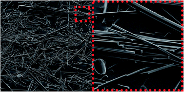
To the best of our knowledge, this is the first report of a self-assembling orbitide that is capable of forming 1D nano-fibers and ultimately 3D molecular gel networks. LOB3 (a.k.a. cyclolinopeptide A), extracted from Linum usitatissimum L. (flaxseed), forms molecular gels in acetonitrile. LOB3 molecular gels, illustrate that cyclic peptides may be comprised of more complex amino acid sequences than have been currently reported. It appears that cyclization, to form orbitides, imparts conformational aspects to the molecule facilitating self-organization into crystalline nano-fibers. These nanoscale fibers, ∼300 nm in diameter and >100 μm in length, aggregate into bundles of fibers which may exceed micron dimensions. Within the nano-fibers, the orbitides adapt an antiparallel β-sheet-like conformation with high molecular periodicity, as illustrated by CD and XRD.

Introduction

Molecular gels, comprised of low molecular mass organogelators (LMOGs), have garnered tremendous interest for their potential applications in recovery of spilled oil,1,2 in vehicles for controlled drug release,3–5 and in edible oleogels.6,7 Despite the vast body of literature devoted to these self-assembling fibrillar network (SAFiN) gels,8 new LMOGs are still, more often than not, discovered serendipitously rather than through rational design.9 Thus, it remains challenging to predict, a priori, the structure of a potential gelator or to foresee solvents that may be gelled by a known gelator.10–13 Challenges in designing these hierarchical architectures lie in their contrasting parameters that promote epitaxial growth whilst limiting solubility. One class of SAFiNs that has drawn exceptional interest recently is peptide-derived molecular gelators due to their structural simplicity, biocompatibility and versatility.14–22

Peptides, capable of acting as bottom-up building blocks for supramolecular architectures, typically are comprised of synthetic diphenylalanine motifs, which, via π–π stacking, form discrete, extraordinarily stiff nanotubes.16,22,23 Interestingly, the diphenylalanine functional motif is the core recognition motif for the Alzheimers' β-amyloid polypeptide.22 Peptides, as small as diphenylalanine,16,22–24 pentapeptides,19 larger molecular weight peptides25,26 and synthetic cyclic D,L-α-peptides,27,28 have been shown to assemble into nanotubes, fibers and/or ribbons. Numerous engineered oligopeptide based molecular gels have been shown to assemble into β-sheets.19 Ajayaghosh and George have suggest that the supramolecular ordering of molecules is driven by hydrogen-bonding, π–π stacking, solvophobic, and donor–acceptor interactions.29 Tomasini and Castellucci have recently published an excellent review on peptide and peptidomimetics gelators.30

Cyclic peptides, capable of self-assembling into crystalline hollow nano-tubes, form artificial ion channels that could be used in the construction of functional artificial transmembrane transporters.31 The structure of synthetic cyclic peptides can be modified to control aspects of their 3-dimensional (3D) supramolecular structure including their internal diameter and external surface properties, allowing for selective transport of small molecules.31

The orbitide, [1–9-NαC]-linusorb B3 (LOB3) (Scheme 1), extracted from Linum usitatissimum L. (flaxseed), is a ribosomally-synthesized homodetic plant peptide that are cyclized by N-to-C amide bonds rather than disulfide bonds.32 These orbitides are being investigated in a broad array of potential applications from cancer treatments33 to light emitting semi-conductors.34 To the best of our knowledge, this is the first orbitide or, indeed cyclic peptide of natural origin, to be reported to self-assemble into crystalline fibrillar aggregates capable of gelling specific solvents.


image file: c6ra05154e-s1.tif
Scheme 1

Methods

LOB3 [NαC-(Ile-Leu-Val-Pro-Pro-Phe-Phe-Leu-Ile), catalog number B3] was provided as an “in kind” contribution from Prairie Tide Chemicals Inc. (Saskatoon, SK, Canada). The linear LOB3 peptide analogs (Scheme 2) denoted as peptide 1 through 7 were synthesized to a minimum purity of 95% from GenicBio Limited (Shanghai, China).
image file: c6ra05154e-s2.tif
Scheme 2

It was not possible to prepare peptide 8 (Pro-Phe-Phe-Leu-Ile-Ile-Leu-Val-Pro) at a minimum purity of 95% and this compound was not included in this study. Acetone, acetonitrile (ACN), carbon tetrachloride (CCL4), dichloromethane (CH2Cl2), dimethylformamide (DMF), hexane, hexanol, methanol, tetrahydroxyfuran (THF) and toluene were purchased from Fisher Scientific (Fair Lawn, NJ, USA) and each had greater than 99% purity and were used as received.

Gelation test and Hansen space

Each peptide was dissolved in the selected solvent at 10 wt% in 2 mL closed vials (Fisher Scientific, Fair Lawn, NJ, USA). 10 wt% was chosen because the critical gelator concentration was typically seen between 5 and 8 wt%. Each combination was heated to 80 °C for 30 min, then cooled to 20 °C and held for 24 h. Inverting the sample for 30 min and observing whether flow was observed, determined if gelation had occurred.11,35 The gel outcomes of the various peptide–solvent combinations were categorized based on their solubility as solutions, gels, or precipitates, and used to calculate minimal enclosing spheres that contain all points within each category. This estimate was performed using a constrained optimization procedure programmed in Mathematica 9 (Wolfram Research, Champaign, IL, USA).11,12,36 The optimization routine uses the “NMinimize” function to obtain the sphere center location in terms of values of the dispersive (δd), polar (δp), and hydrogen bonding (δh) interactions while solving for the smallest possible radius. The “NMinimize” function was set to implement the differential evolution optimization method, a robust simple stochastic function minimizer, to reach a numerical global optimum solution.37 Due to the nature of a global optimum solution, no “goodness” of fit exists.

Fluorescence, brightfield, phase contrast, and polarized light microscopies

Samples for microscopy were removed after 24 h and a small portion of sample was placed on a 25 × 75 mm × 1 mm glass slide (Fisher Scientific, Pittsburgh, PA, USA) and was compressed with a 22 × 22 mm × 0.15 mm glass cover slip (Fisher Scientific, Pittsburgh, PA, USA). Images were obtained using an Olympus BX60 upright light microscope (Olympus, Richmond Hill, ON, Canada) and an Olympus DP71 CCD camera. Images were digitized using CellSens® (Olympus Lifescience Software, Richmond Hill, ON, Canada) and were acquired as 24 bit RGB color image with a spatial resolution of 1360 × 1024 pixels. A 10× magnification objective with a 0.3 numerical aperture was used (Olympus, Tokyo, Japan). The peptide fluorescence arose from phenylalanine excitation using a by-pass filter (330–385 nm) and fluorescence was observed at 420 nm using an emission filter.

Scanning electron microscopy

Scanning electron microscopy of LOB3 and the linear peptides were examined after they were dried from ACN. A small aliquot of each peptide/ACN mixture was placed on a SEM pin stub and placed into an oven (Fisher Scientific, Isotemp®, Fair Lawn, NJ, USA) at 35 °C for 10 min. After evaporating the ACN from samples ten drops of ACN was added dropwise over the samples to remove entrapped ACN from the surfaces. Following the dropwise addition of ACN, samples were placed back into the oven at 35 °C for an additional 10 min to re-dry the sample. The sample was mounted on a sputter coater (Emscope K550 sputter coater, Ashford, Kent, UK) and coated with gold using a 20 mA deposition current, 7 nm min−1 deposition rate and time of 2 min. The pin stub was transferred to a specimen holder onto the SEM stage (Hitachi S-570, Tokyo, Japan). Images were taken using Quartz PCI Imaging software (Quartz Imaging Corp., Vancouver, Canada).

Transmission electron microscopy

The gel was diluted 10 fold and agitated to disperse the fibers. A drop of diluted LOB3/ACN was placed on 3 mm Formvar® and carbon coated copper grids (Canemco, Lakefield, QC, Canada). They were allowed to dry and then 5 drops of ACN were placed on the support and wicked away using Whatman® filter papers. Excess solvent was allowed to evaporate and grids were then transferred into the Philips CM10 transmission electron microscope (TEM) (Philips Electron Optics, Eindhoven, The Netherlands) with an accelerating voltage of 80 kV. Images (1810 × 2320 pixels) were acquired using a Morada® CCD digital camera (Olympus, Richmond Hill, Canada).

Circular dichroism spectroscopy

Peptides in ACN (100 μL of 10 wt%) were placed in a 0.01 mm path length quartz cuvette (Starna Cells, Atascadero, CA, USA). Circular dichroism spectra were recorded from 355 to 180 nm, where absorbance was lower than 2.5, using a spectropolarimeter (Model J810, Jasco, Tokyo, Japan) equipped with a Peltier temperature controller (Model JWJTC-484, Jasco, Tokyo, Japan) and a water bath (Model F25, Julabo, Allentown PA, USA). Experiments were conducted at 20 °C. Sensitivity was set at 5 mdeg, data pitch at 1 nm, scan speed was 100 nm min−1, response was 0.25 s, and a bandwidth was set to 1 nm with a step size of 1 nm using Spectra Manager software version 1.54.03 (Jasco, Tokyo, Japan). Optical rotation data collected at each wavelength were averaged between two runs and each run was the accumulation of four measurements. The final spectrum was subjected to solvent spectrum subtraction and Savitzky Golay smoothing using a convolution width of 15. The CD raw signal (mdeg) was converted to mean residue ellipticity (deg cm2 dmol−1) using the following equation:
 
image file: c6ra05154e-t1.tif(1)
where MRW is the peptide mean reside weight, θ is the observed ellipticity in degrees, d is the path length (cm), c is the peptide concentration (g mL−1). The mean residue weight was based on a molecular weight of 1040 g mol−1 for LOB3 and 1058 g mol−1 for the linear peptides.

Fourier transform infrared spectroscopy

An aliquot of sample was placed on an attenuated total reflection (ATR) sapphire crystal and compressed using a press. Forty scans were collected in absorbance mode at a resolution of 4 cm−1 with a Happ–Genzel apodization on a Fourier transform infrared (FT-IR) spectrometer (IRPrestige21, Shimadzu Corp., Kyoto, Japan).

X-ray diffraction

A Rigaku multiflex powder X-ray diffractometer (XRD, Rigaku, Japan) with a 1/2° divergence slit, 1/2° scatter slit, and a 0.3 mm receiving slit, was set at 40 kV and 44 mA to determine the polymorphic form of the networks. Scans were performed from 1 to 30° at 0.2° min−1.

Nuclear magnetic resonance spectroscopy

13C cross-polarization magic angle spinning (MAS) spectrum of the cyclic peptide xerogel from acetonitrile was recorded on a Bruker Avance III spectrophotometer operating at a proton frequency of 800.230 MHz. Spectrum was recorded using a triple resonance 3.2 mm EFREE MAS solid-state NMR probe (Bruker Biospin, Milton, ON, Canada). The chemical shift scale is indirectly referenced to neat tetramethylsilane (TMS).38

The solution 1H-NMR spectra were recorded on a 600 MHz Bruker Avance spectrometer (Broadband probe, BBO, 5 mm; TopSpin 3.2 software). The 1H-NMR spectra (600 MHz) chemical shifts (δ) values are reported in parts per million (ppm) relative to the internal standard of TMS. The δ values are referenced to CD3CN at 1.94 ppm, and multiplicities are indicated by the following symbols: s = singlet, d = doublet, dd = doublet of doublets, m = multiplet, and br = broad. For 13C-NMR (150.9 MHz), the chemical shift (δ) values were referenced to CD3CN (118.26 ppm).

Discussion

After 1 h, LOB3 was able to gel ACN, at 10 wt%, and after 24 h very fine fibers had aggregated in DMF, however the fibers did not immobilize the solvent (Table 1). The linear peptides precipitated in ACN after being cooled from the sol at 80 °C to 20 °C and held 24 h. LOB3 was insoluble in apolar solvents capable of only interacting via van der Waals interactions (i.e., toluene and hexane) and was soluble in the remaining solvents with a dipole moment or that were capable of hydrogen-bonding. In all cases, gel outcomes were the same for LOB3 and linear analogs, with the exception of LOB3 in ACN and DMF where the linear analogs precipitated from solution upon cooling.
Table 1 The physical state (solution (S), precipitate (P), gel formed of fibers (G/F), no gel with fibers (NG/F) or insoluble (I)) of 10 wt% peptide, both cyclic and linear variations, in solvents after 48 h stored at 20 °C
  Linear peptides
Solvent Cyclic FLIILVPPF LIILVPPFF IILVPPFFL ILVPPFFLI LVPPFFLII VPPFFLIIL PPFFLIILV PFFLIILVP
Acetone S S S S S S S S NA
ACN G/F P P P P P P P NA
CCL4 S S S S S S S S NA
CH2Cl2 S S S S S S S S NA
DMF NG/F P P P P P P P NA
Hexane I I I I I I I I NA
Hexanol S NA NA NA NA NA NA NA NA
Methanol S NA NA NA NA NA NA NA NA
THF S S S S S S S S NA
Toluene I I I I I I I I NA


In ACN, each linear peptide had microstructures that are difficult to discern from each other (Fig. 1). However, it is clear that these peptides aggregate to form a disordered network that is comprised of discrete microstructural elements that differ both in shape and size. Peptides 1, 2, 5 and 7 all have very similar macrostructures that are comprised of small structural elements. Peptide 3 is unique compared to the other linear peptides, as the microstructural elements are spherical. Peptide 4 assembles into very small crystallites. By contrast, LOB3 forms elongated fibrillar aggregates that appear to be 200 μm in length and 20–30 μm in width. Simply from a size comparison, it is clear that LOB3 has a tremendous capacity to self-organize into supramolecular fibrillar objects. Upon reheating, all of these aggregates dissolve and form a sol and upon cooling to room temperature, assemble into the same morphologies.


image file: c6ra05154e-f1.tif
Fig. 1 Brightfield (first column), polarized light (second column), phase contrast (third column), and fluorescent micrographs (fourth column) taken at the same field-of-view for each peptide in ACN after 24 h.

The PLMs illustrate that linear peptides 2, 5 and 6 do not exhibit birefringence, while peptide 4 has a diffuse scatter, however it does not correspond to the microstructural elements observed in the brightfield images. Peptides 1, 4 and 7, as well as LOB3 have enough crystalline order to detect the optical rotation of light through cross-polarizers. Phase contrast images illustrate the large aggregates are comprised of smaller microstructural elements that appear to cluster into supramolecular architectures. As well, using phase contrast, LOB3 in ACN has a rough surface that may indicate the presence of crystallographic mismatches or fiber bundling. By relying on phenylalanine fluorescence, it is clear that the microstructural elements are comprised of the peptide as the fluorescence corresponded to the elements seen under different modes of microscopy.

It is clear that none of the linear peptides are capable of self-assembling into supramolecular fibers as observed for LOB3 in ACN. The linear peptides vary remarkably from each other under higher magnification (Fig. 2). Systems that exhibited birefringence (peptides 1, 4 and 7) have platelet or fiber-like crystallites, whereas, the samples that did not exhibit birefringence tend to have a more globular structure that is less well ordered/crystalline compared to the others. SEM also reveals that LOB3 in ACN does not contain fibers with widths between 10 and 20 μm, but instead fibers are between 1 and 3 μm and form bundles that are 10 to 30 μm.


image file: c6ra05154e-f2.tif
Fig. 2 Scanning electron micrographs of the dried linear and orbitides in ACN after 24 h. Scale bar represents 30 μm.

TEM was employed in an attempt to better quantify LOB3 fiber morphology (Fig. 3). TEM provides numerous very important insights, first it is clear that parent fibers branch into daughter fibers and the presence of crystallographic mismatches arise, allowing for entrapment of the continuous phase solvent.39–43 The size of the 1D objects also are sub-micron therefore, it is likely that the fibers observed in SEM, are comprised of even smaller bundles. However, it is important to note that we cannot determine if the morphology we are observing are in fact fibers and not tubules which would have a hollow core.


image file: c6ra05154e-f3.tif
Fig. 3 Transmission electron micrograph of dried LOB3/ACN after 24 h. Fiber size was determined in Photoshop© using the scale bar to convert pixels to microns (1380 pixels = 1 μm).

After 24 h, LOB3 is found to only assemble as fibers in ACN, however after 48 h it also assembles in DMF, but does not gel. Instead the fibers aggregate into macroscale spherical objects that are suspended in DMF. In an attempt to quantify the interaction with different types of solvents more mechanistically, the test solvents are examined in Hansen space (Fig. 4). It is important to note that to accurately determine the confines of Hansen space far more solvents are required than were test herein. However, with the limited solvents tested an approximation of the regions of interest may be gauged.


image file: c6ra05154e-f4.tif
Fig. 4 3D Hansen space plots for CLS in various solvents after 1 h (left) and 48 h (right) and 2D projects of the same space. The green sphere represent insoluble combinations, blue sphere represents solutions and the black dots as well as the red sphere are the fiber forming mixtures.

Using Hansen solubility parameters for the tested solvents, Hansen plots are generated to observe if there is a tendency for clustering of LOB3/solvent outcomes in specific regions of Hansen space (Fig. 4). 3D plots are utilized to illustrate the overall trend and a 2D projection of the hydrogen bonding Hansen solubility parameter (δh) and polar Hansen solubility parameter (δp) plane is done to better illustrate the z plane that is difficult to discern.

It is clear that insoluble precipitates form in solvents with low δp and δh (sphere center 2δd = 33.3 MPa1/2, δh = 1.0 MPa1/2, δp = 0.7 MPa1/2, radius = 3.0 MPa1/2). Since the solvents selected had a narrow distribution of dispersive Hansen solubility parameters (2δd), no conclusions are made with regards to 2δd. The majority of solvents tested formed sols with LOB3 (solubility sphere center 2δd = 32.9 MPa1/2, δh = 14.0 MPa1/2, δp = 7.7 MPa1/2, radius = 9.0 MPa1/2). It appears that fiber-forming combinations (fiber sphere center 2δd = 32.7 MPa1/2, δh = 8.7 MPa1/2, δp = 15.9 MPa1/2, radius = 9.0 MPa1/2) differ from solutions when there is a higher polar contribution to the HSP compared to hydrogen bonding. This is a common outcome in molecular gels that rely on hydrogen bonding to form their supramolecular architectures.44–47

It is interesting that LOB3–DMF sol facilitates fiber formation after 48 h (Fig. 5). The resulting fibers, however, are incapable of forming a coherent 3D network entrapping the solvent. Instead, the fibers in DMF become to closely associated and aggregated. The fibers that form appear to either have a thinner diameter and/or are less effectively bundled into well-orientated fibers. In either case, the aspect ratio of the length to width is exceedingly large.


image file: c6ra05154e-f5.tif
Fig. 5 Scanning electron micrographs of dried fibers from LOB3 in ACN (left) and LOB3 in DMF (right) after 48 h.

It is clear from the micrographs, that there are distinct differences between LOB3 and its linear peptide analogs. In an attempt to quantify differences between their molecular scale interactions, the crystallinity of the fibers was probed with powder XRD (Fig. 6). The powder XRD is only to be used as a fingerprint in identifying differences between samples and not to solve crystal structures. Upon comparing structures, the linear peptides are categorized into two groups based on their diffractograms. The first group of linear peptides (1, 2, 3, 5, and 6) corresponds to the samples that do not exhibit birefringence under polarized light (Fig. 1) and in the SEM images have a globular microstructure (Fig. 2). It appears as though this group has a lamellar arrangement with a 001 spacing of 31.7 Å, and higher order reflections at 003 = 11.4 Å and potentially 004 = 7.6 Å. This is accompanied with a single sub-cell packing at 4.7 Å. Peptides 4 and 7 show more periodicity than other linear peptides and the only long spacing is at 18.2 Å, indicating a more densely packed structure. LOB3 dried from ACN shows the greatest periodicity and the smallest 001 peak at 14.5 Å. One note of caution here is that there may be a very weak peak prior to 14.5 Å that may correspond to a peak at 16.5 Å. For LOB3, the peak at 10.7 Å was truncated because the peak intensity was 40[thin space (1/6-em)]000 counts compared to the next highest counts in any sample which were consistently less than 4000.


image file: c6ra05154e-f6.tif
Fig. 6 X-ray diffractograms of peptide 1 (top), peptide 4 (middle) and LOB3 dried from a 10 wt% ACN mixture. The inset in the XRD pattern for the orbitide is to illustrate the max peak intensity.

The experimental powder diffraction pattern was compared to the powder diffraction pattern, obtained from simulations from single crystal data (Fig. 7). Simulations were carried out using the Mercury software from Cambridge Crystallographic Data Centre (CCDC). A validation test for structural differences through powder simulation from single crystal data was conducted by using an analogue of LOB3 containing one ACN and two water48 and LOB3 containing one methanol. The simulated powder patterns of the analogue of LOB3 and LOB3 were significantly different. The starting materials for the LOB3 single crystals49 and the LOB3 powder were obtained from the same source and are, therefore, identical starting materials. The primary contributor to the difference between the structure of LOB3 single crystals and the LOB3 powder is the method by which the two final materials were prepared. SEM of the LOB3 powder shows that there are rods that are above 10 μm in length. In principle, the long fibers will give rise to preferred orientation and the March–Dollase option was used to simulate a preferred orientation, which would match the experimental powder diffraction pattern, after inspection of BFDH morphology.


image file: c6ra05154e-f7.tif
Fig. 7 Experimental diffraction pattern compared to the theoretical powder diffraction pattern obtained using LOB3 single crystal data.

Although the single crystal and fibers were made from same starting materials, the simulated powder XRD (starting with single crystal data) and powder diffraction pattern from nano-fibers were very different. The structural difference stems from the growth rate and hydrogen bonding. It is likely that the fibers contain both ACN in their nanocrystalline structure and would be susceptible to more inter-molecular hydrogen bonding and less likely to have the monomodal distribution of particles to grow single crystals. The single crystal grown in methanol was grown slowly and contained only one methanol in the lattice. The methanol was involved in intra-molecular hydrogen boding and would allow for better/uniform crystal growth than those in ACN.

The NMR spectra of the soluble LOB3 were recorded in deuterated ACN at 298 K. Additional experiments at 320 K were also performed for better resolution and characterization. The solution state of LOB3 has been done in different solvents and it was found that at 214 K in CDCl3 a single conformation was observed.50–52 The characterization of LOB3–metal complexes was previously done in ACN, but complete analysis of LOB3 was not performed.52 Here we report complete characterization of LOB3 in ACN. Sequential assignment of α, β, γ and δ protons were performed using 1H–1H COSY, TOCSY and NOESY experiments (Scheme 1, ESI Fig. 1–8 and Table 1). In addition, HMQC was performed to assign carbon atoms attached to protons. The protons attached to the heteroatoms were assigned from 1H-NMR, and their coupling to amide protons and carbonyl carbons employed NOE and HMBC correlations. The 1H-NMR revealed seven amide proton signals (δ 7.16, 7.30, 7.02 (2), 7.09, 7.43, and 7.58 ppm) (ESI Table 1). The 13C-NMR spectrum showed nine-amide carbonyl signals (δ 171.49, 171.64, 171.93, 172.41 (2), 173.11, 174.27, 174.54 and 174.87 ppm) suggested LOB3 to be a nonapeptide. A strong NOESY correlation observed between the α-protons of Pro4 and Pro5 indicating that Pro5 is cis configured. The cis conformation of the Pro4–Pro5 peptide bond is further corroborated by the large chemical shift difference between the Pro5 Cβ and Cγ carbons of 9.66 ppm,53 and signifies the rigidity within the molecule that helps maintaining LOB3 in one stable conformation for the peptide in the immobilized fibrillar state. We note that the chemical shift patterns of both the carbonyl and CA regions detected in solid and solution NMR spectra are significantly different, suggesting that the formation of fibrils involve structural rearrangements within the LOB3 peptide (Fig. 8).


image file: c6ra05154e-f8.tif
Fig. 8 13C-NMR spectra of LOB3 in solution state and solid state and the expansion of 13C-NMR regions in solution state and solid state.

CD spectra for linear peptides is similar to the observed spectra for peptides containing aromatic groups in a β-turn configuration.54,55 It is clear that linear peptides have a positive band at either 200 or 218 nm depending on the sequence of the linear peptide (Fig. 9). The band at 200 nm is attributed to the β-turn π–π* transition, while the positive band at 218 nm is indicative of n–π* transitions.54 These same positive peaks have been reported for the L-Phe-L-Phe dipeptide molecular gel in chloroform.54 The CD signature for LOB3 in ACN is drastically different containing only the presence of a strong Cotton effect at 226 nm (n–π* transition) compared to 240 nm. In previous dipeptide molecular gels this suggests a dominant β-sheet arrangement.47 This conclusion was based of observation reported on self-assembly of branched peptide amphiphiles into nano-fibers.56 In yet another report of diphenylalanine, researchers reported a shift in the Cotton effect from 228 nm to 235 nm and attributed it to an increasing degree of β-sheet twisting at lower wavenumbers.57


image file: c6ra05154e-f9.tif
Fig. 9 CD spectra measuring the intermolecular interactions between peptides in the ACN.

FT-IR analysis further probed orbitide self-associating behavior and the behavior of its linear analogs in ACN (Fig. 10). The most informative frequencies for peptide derived molecular gels are between 3500 and 3200 cm−1, corresponding to peptide N–H stretching vibrations and the 1800–1600 cm−1 range for C[double bond, length as m-dash]O stretching vibrations.19 The N–H stretching vibrations between 3500 and 3200 cm−1, particularly the presence of a peak around 3400 cm−1 indicates most NH groups participate in hydrogen bonding. The shift from a peak at 3285 cm−1, observed for linear peptides, to 3330 cm−1, for the orbitide, signifies that the distance between hydrogen bonds is greater or may in fact be a free N–H stretch, indicating that for LOB3 the self assembly is driven primarily by π–π stacking. This suggests the strain imposed by peptide backbone cyclization influences hydrogen bonding. Very similar findings for amide I (1628 cm−1) and amide II (1546 cm−1) bands for LOB3 are comparable to a previously reported NαC-(D-Ala-Glu-D-Ala-Gln)2- in an aqueous medium.27 LOB3 in ACN has amide I peaks at 1628 cm−1 and 1690 cm−1 and amide II peaks between 1500 and 1540 cm−1 both indicate a hydrogen bounded anti-parallel β-sheet arrangement.27 An additional peak at 1660 cm−1 is consistent with peptide conformation turns. The absence of a peak centered at 1640 cm−1 illustrates that the orbitide supramolecular arrangement is consistent with a combination of anti-parallel β-sheets and turns. Ghadiri et al., pointed to a very interesting comparison between the peptide NαC-(D-Ala-Glu-D-Ala-Gln)2-peptide and gramicidin A, a naturally-occurring linear peptide composed of hydrophobic amino acids that is known to form a dimeric β-helical transmembrane ion channel.27 Gramicidin A contains an antiparallel β-helix with amide-I bands at 1630 and 1685 cm−1, an amide-II band at 1539 cm−1, and an N–H stretching frequency at 3285 cm−1.58 The only difference between our FT-IR peak positions is associated with a shift in the NH stretching vibration from 3285 cm−1 to 3330 cm−1, indicating that the hydrogen bonding is weaker in LOB3 than in its linear counterparts and gramicidin A.


image file: c6ra05154e-f10.tif
Fig. 10 FT-IR spectra of the N–H stretching vibration (top) and the CO stretching (amide I) and NH bending (amide II) bands for ACN, and for the orbitide and it's linear analogs in ACN at 10 wt%.

Overall the 3D gel network is comprised of a bi-continuous network of fibers, comprised of LOB3 and solvent (Fig. 11). The 1D crystalline fibers are a result of 0D, LOB3 molecules, forming β-helix/sheet conformation and individual LOB3 molecules forming non-convalent interactions via π–π stacking and hydrogen bonding.


image file: c6ra05154e-f11.tif
Fig. 11 Schematic representation of the self-assembly of LOB3.

Conclusions

This is the first reported discovery of an orbitide capable of self-assembling from 0D objects to 1D nano-fibers and finally producing a 3D supramolecular gel network. Besides being the first report on an orbitide molecular gelator numerous very important insights build on the literature in this emerging area including: (1) the complexity of the orbitide may be more diverse than previous reports thus far in literature, (2) cyclization of peptides imparts conformational aspects to the molecule that drive self-organization into fibrillar objects, and (3) the structures of orbitides, either natural or synthetic have all been reported to be in a β-helix/sheet conformation. In the case of LOB3, it arranges in an anti-parallel β-sheet that has very high periodicity. Albeit some of the linear peptides form crystalline aggregates none have the high aspect ratio observed in LOB3/ACN.

Acknowledgements

The authors would like to acknowledge the in-kind contribution of LOB3 from Prairie Tide Chemicals Inc. (Saskatoon, SK, Canada). M. A. R. received funding for this project from the National Science and Engineering Research Council of Canada (NSERC) through the Engage program.

References

  1. S. R. Jadhav, P. K. Vemula, R. Kumar, S. R. Raghavan and G. John, Sugar-derived phase-selective molecular gelators as model solidifiers for oil spills, Angew. Chem., Int. Ed., 2010, 49, 7695–7698 CrossRef CAS PubMed.
  2. P. Lee and M. A. Rogers, Phase selective sorbent xerogels as reclamation agents for oil spills, Langmuir, 2013, 29, 5617–5621 CrossRef CAS PubMed.
  3. A. Friggeri, B. L. Feringa and J. van Esch, Entrapment and release of quinoline derivatives using a hydrogel of a low molecular weight gelator, J. Controlled Release, 2004, 97, 241–248 CrossRef CAS PubMed.
  4. R. Kumar and O. P. Katare, Lecithin organogels as a potential phospholipid-structured system for topical drug delivery: a review, AAPS PharmSciTech, 2005, 6, E298–E310 CrossRef PubMed.
  5. T. Pénzesa, G. Blazsób, Z. Aignera, G. Falkayb and I. Erősa, Topical absorption of piroxicam from organogels – in vitro and in vivo correlations, Int. J. Pharm., 2005, 298, 47–54 CrossRef PubMed.
  6. M. Pernetti, K. F. van Malssen, E. Floter and A. Bot, Structuring of edible oils by alternatives to crystalline fat, Curr. Opin. Colloid Interface Sci., 2007, 12, 221–231 CrossRef CAS.
  7. F. AlHassawi and M. A. Rogers, Ternary phase diagram of β-sitosterol, γ-oryzanol and canola oil, J. Am. Oil Chem. Soc., 2013, 90, 1533–1540 CrossRef.
  8. R. G. Weiss, The past, present, and future of molecular gels. What is the status of the field, and where is it going?, J. Am. Chem. Soc., 2014, 136, 7519–7530 CrossRef CAS PubMed.
  9. Y.-C. Lin, B. Kachar and R. G. Weiss, A novel family of gelators of organic fluids and the structure of their gels, J. Am. Chem. Soc., 1989, 111, 5542–5551 CrossRef CAS.
  10. M. A. Rogers and R. G. Weiss, Systematic modifications of alkane-based molecular gelators and the consequences to the structures and properties of their gels, New J. Chem., 2015, 39, 785–799 RSC.
  11. Y. Lan, M. G. Corradini, R. G. Weiss, S. R. Raghavan and M. A. Rogers, To gel or not to gel: correlating molecular gelation with solvent parameters, Chem. Soc. Rev., 2015, 44, 6035–6058 RSC.
  12. Y. Lan, M. G. Corradini, X. Liu, T. E. May, F. Borondics, R. G. Weiss and M. A. Rogers, Comparing and correlating solubility parameters governing the self-assembly of molecular gels using 1,3:2,4-dibenzylidene sorbitol as the gelator, Langmuir, 2014, 47, 14128–14148 CrossRef PubMed.
  13. R. G. Weiss and P. Térech, Molecular Gels: Materials with Self-Assembled Fibrillar Networks, Springer, 2006 Search PubMed.
  14. P. Zhu, X. Yan, Y. Su, Y. Yang and J. Li, Solvent-induced structural transition of self-assembled dipeptide: from organogels to microcrystals, Chem.–Eur. J., 2010, 16, 3176–3183 CrossRef CAS PubMed.
  15. Y. Zhao, M. Tanaka, T. Kinoshita, M. Higuchi and T. Tan, Self-assembling peptide nanofiber scaffolds for controlled release governed by gelator design and guest size, J. Controlled Release, 2010, 147, 392–399 CrossRef CAS PubMed.
  16. X. Yan, Y. Cui, Q. He, K. Wang and J. Li, Organogels based on self-assembly of diphenylalanine peptide and their application to immobilize quantum dots, Chem. Mater., 2008, 20, 1522–1526 CrossRef CAS.
  17. G. Palui, A. Garai, J. Nanda, A. K. Nandi and A. Banerjee, Organogels from different self-assembling new dendritic peptides: morphology, rheology, and structural investigations, J. Phys. Chem. B, 2010, 114, 1249–1256 CrossRef CAS PubMed.
  18. G. Liang, Z. Yang, R. Zhang, L. Li, Y. Fan and Y. Kuang, et al., Supramolecular hydrogel of a D-amino acid dipeptide for controlled drug release in vivo, Langmuir, 2009, 25, 8419–8422 CrossRef CAS PubMed.
  19. A. Banerjee, G. Palui and A. Banerjee, Pentapeptide based organogels: the role of adjacently located phenylalanine residues in gel formation, Soft Mater., 2008, 4, 1430–1437 RSC.
  20. A. Aggeli, M. Bell, M. Boden, J. N. Keen, P. F. Knowles, T. C. B. McLeish, M. Pitkeathly and S. E. Radford, Responsive gels formed by the spontaneous self-assembly of peptides into polymeric β-sheet tapes, Nature, 1997, 386, 259–262 CrossRef CAS PubMed.
  21. R. Afrasiabi and H.-B. Kraatz, Small-peptide-based organogel kit: towards the development of multicomponent self-sorting organogels, Chem.–Eur. J., 2013, 19, 15862–15871 CrossRef CAS PubMed.
  22. Q. Li, Y. Jia, Y. Yang and J. Li, Controlled rod nanostructured assembly of diphenylalanine and their optical waveguide properties, ACS Nano, 2015, 9, 2689–2695 CrossRef CAS PubMed.
  23. T. H. Han, J. K. Oh, J. S. Park, S.-H. Kwon, S.-W. Kim and S. O. Kim, Highly entangled hollow TiO2 nanoribbons templating diphenylalanine assembly, J. Mater. Chem., 2009, 19, 3512–3516 RSC.
  24. R. Huang, Y. Wang, W. Qi, R. Su and Z. He, Temperature-induced reversible self-assembly of diphenylalanine peptide and the structural transition from organogel to crystalline nanowires, Nanoscale Res. Lett., 2014, 9, 653–662 CrossRef PubMed.
  25. K. T. Kim, C. Park, G. W. M. Vandermeulen, D. A. Rider, C. Kim and M. A. Winnik, et al., Gelation of helical polypeptide – random coil diblock copolymers by a nanoribbon mechanism, Angew. Chem., 2005, 117, 8178–8182 CrossRef.
  26. S. Scanlon, A. Aggeli, N. Boden, R. J. Koopmans, R. Brydson and C. M. Rayner, Peptide aerogels comprising self-assembling nanofibrils, Nano-Micro Lett., 2007, 2, 24–29 CrossRef CAS.
  27. M. R. Ghadiri, J. R. Granja, R. A. Milligan, D. E. McRee and N. Khazanovich, Self-assembling organic nanotubes based on a cyclic peptide architecture, Nature, 1993, 366, 324–327 CrossRef CAS PubMed.
  28. W. S. Horne, N. Ashkenasy and M. R. Ghadiri, Modulating charge transfer through cyclic D,L-α-peptide self-assembly, Chem.–Eur. J., 2005, 11, 1137–1144 CrossRef CAS PubMed.
  29. A. Ajayaghosh and S. J. George, First phenylenevinylene based organogels: self-assembled nanostructures via cooperative hydrogen bonding and π-stacking, J. Am. Chem. Soc., 2001, 123, 5148–5149 CrossRef CAS PubMed.
  30. C. Tomasini and N. Castellucci, Peptides and peptidomimetics that behave as low molecular weight gelators, Chem. Soc. Rev., 2013, 42, 156–172 RSC.
  31. J. Montenegro, M. R. Ghardiri and J. R. Granja, Ion channel models based on self-assembling cyclic peptide nanotubes, Acc. Chem. Res., 2013, 46, 2955–2965 CrossRef CAS PubMed.
  32. Y. Y. Shim, L. W. Young, P. G. Arnison, E. Gilding and M. J. T. Reaney, Proposed systematic nomenclature for orbitides, J. Nat. Prod., 2015, 78, 645–652 CrossRef CAS PubMed.
  33. D. P. Okinyo-Owiti, Q. Dong, B. Ling, P. D. Jadhav, R. Bauer and J. M. Maley, et al., Evaluating the cytotoxicity of flaxseed cyclolinopeptides for potential cancer treatment, Toxicol. Rep., 2015, 2, 1014–1018 CrossRef.
  34. R. Bauer, P. Bazylewski, P. D. Jadhav, J. Shen, D. P. Okinyo-Owiti and J. Yang, et al., Characterization of metal-functionalized flax orbitide as a new candidate for light-emitting semiconductor, J. Phys.: Conf. Ser., 2015, 619, 1–4 CrossRef.
  35. S. R. Raghavan and B. H. Cipriano, Gel Formation: Phase Diagras using Tabletop Rheology and Calorimetry, in Molecular Gel Materials with Self-Assembled Fibrillar Networks, ed. R. G. Weiss and P. Terech, Springer, Netherlands, 2006, pp. 214–252 Search PubMed.
  36. L. Chen, M. G. Corradini and M. A. Rogers, Self-assembly of 12-hydroxystearic acid molecular gels in mixed solvent systems rationalized using Hansen solubility parameters, Colloid Polym. Sci., 2015, 293, 975–983 Search PubMed.
  37. R. Storn and K. Price, Differential Evolution – A simple and efficient heuristic for global optimization over continuous spaces, J. Global Optim., 1997, 11, 341–359 CrossRef.
  38. V. Krymov and G. J. Gerfen, Analysis of the tuning and operation of reflection resonator EPR spectrometers, J. Magn. Reson., 2003, 162, 466–478 CrossRef CAS PubMed.
  39. R. Lam, L. Quaroni, T. Pederson and M. A. Rogers, A molecular insight into the nature of crystallographic mismatches in self-assembled fibrillar networks under non-isothermal crystallization conditions, Soft Mater., 2010, 6, 404–408 RSC.
  40. R. Y. Wang, P. Wang, J. L. Li, B. Yuan, Y. Liu, L. Li and X.-Y. Liu, From kinetic-structure analysis to engineering crystalline fiber networks in soft materials, Phys. Chem. Chem. Phys., 2013, 15, 3313–3319 RSC.
  41. J. L. Li and X. Y. Liu, Architecture of supramolecular soft functional materials: from understanding to micro-/nanoscale engineering, Adv. Funct. Mater., 2010, 20, 3196–3216 CrossRef CAS.
  42. R. Y. Wang, X. Y. Liu, J. Narayanan, J. X. Xiong and J. L. Li, Architecture of fiber network: from understanding to engineering of molecular gels, J. Phys. Chem. B, 2006, 10, 25797–25802 CrossRef PubMed.
  43. X. Y. Liu and P. D. Sawant, Formation kinetics of fractal nanofiber networks in organogels, Appl. Phys. Lett., 2001, 19, 3518–3520 CrossRef.
  44. J. Bonnet, G. Suissa, M. Raynal and L. Bouteiller, Organogel formation rationalized by Hansen solubility parameters: dos and don'ts, Soft Mater., 2014, 10, 3154–3160 RSC.
  45. K. K. Diehn, H. Oh, R. Hashemipour, R. G. Weiss and S. R. Raghavan, Insights into organogelation and its kinetics from Hansen solubility parameters. Toward a priori predictions of molecular gelation, Soft Mater., 2014, 10, 2632–2640 RSC.
  46. J. Gao, S. Wu and M. A. Rogers, Harnessing Hansen solubility parameters to predict organogel Formation, J. Mater. Chem., 2012, 22, 12651–12658 RSC.
  47. M. Raynal and L. Bouteiller, Organogel formation rationalized by Hansen solubility parameters, Chem. Commun., 2011, 47, 8271–8273 RSC.
  48. M. Saviano, A. Rossi, M. Flilizola, B. Di Blasio and C. Pedone, [Aib5,6-D-Ala8]-cyclolinopeptide A, grown from a benzene/acetonitrile mixture, Acta Crystallogr., Sect. C: Cryst. Struct. Commun., 1995, 51, 663–666 CrossRef.
  49. J. Wilson Quail, J. Shen, M. J. T. Reaney and R. Sammynaiken, Cyclolinopeptide A methanol solvate, Acta Crystallogr., Sect. E: Struct. Rep. Online, 2009, 65, O1913–O1914 Search PubMed.
  50. B. Di Blasio, F. Rossi, E. Benedetti, V. Pavone, C. Pedone and P. A. Temussi, et al., Bioactive peptides: solid-state and solution conformation of cyclolinopeptide A, J. Am. Chem. Soc., 1989, 111, 9089–9098 CrossRef CAS.
  51. M. Saviano, F. Rossi, V. Pavone, B. Di Blasio and C. Pedone, Molecular dynamics simulation in vacuo and in solution of [Aib5,6-D-Ala8] cyclolinopeptide A: a conformational and comparative study, J. Biomol. Struct. Dyn., 1992, 9, 1045–1060 CAS.
  52. T. Tancredi, G. Zanotti, F. Rossi, E. Benedetti, C. Pedone and P. A. Temussi, Comparison of the conformations of cyclolinopeptide a in the solid state and in solution, Biopolymers, 1989, 28, 513–523 CrossRef CAS PubMed.
  53. M. Schubert, D. Labudde, H. Oschkinat and P. Schmieder, A software tool for the prediction of Xaa-Pro peptide bond conformations in proteins based on 13C chemical shift statistics, J. Biomol. NMR, 2002, 24, 149–154 CrossRef CAS PubMed.
  54. X. Yan, Y. Cui, Q. He, Q. Wang and J. Li, Organogels Based on Self-Assembly of Diphenylalanine peptide and their applications to immobilize quantum dots, Chem. Mater., 2008, 20, 1522–1526 CrossRef CAS.
  55. D. A. Tinker, E. A. Krebs, I. C. Feltham, S. K. Attah-Poku and V. S. Ananthanarayanan, Synthetic β-turn peptides as substrates for a tyrosine protein kinase, J. Biol. Chem., 1988, 263, 5024–5026 CAS.
  56. M. O. Guler, S. Soukasene, J. F. Hulvat and S. I. Stupp, Presentation and recognition of biotin on nanofibers formed by branched peptide amphiphiles, Nano Lett., 2005, 5, 249–252 CrossRef CAS PubMed.
  57. Q. Li, L. Dai, Y. Yang and J. Li, Controlled rod nanostructured assembly of diphenylalanine and their optical waveguide properties, ACS Nano, 2015, 9, 2689–2695 CrossRef CAS PubMed.
  58. V. M. Naik and S. Krimm, Vibrational analysis of the structure of gramicidin A. II. Vibrational spectra, Biophys. J., 1986, 49, 1147–1154 CrossRef CAS PubMed.

Footnote

Electronic supplementary information (ESI) available. See DOI: 10.1039/c6ra05154e

This journal is © The Royal Society of Chemistry 2016
Click here to see how this site uses Cookies. View our privacy policy here.