DOI:
10.1039/C6RA04827G
(Paper)
RSC Adv., 2016,
6, 32685-32696
Phytochemical investigation, hypocholesterolemic and anti-atherosclerotic effects of Amaranthus viridis leaf extract in hypercholesterolemia-induced rabbits
Received
23rd February 2016
, Accepted 22nd March 2016
First published on 24th March 2016
Abstract
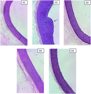
Hypercholesterolemia is one of the main causes for coronary heart disease, which occurs due to high levels of serum cholesterol. Oxidized LDL accumulation leads to atherosclerotic plaque formation, which contributes to myocardial infarction and cardiovascular diseases. Consumption of statins leads to adverse health effects such as liver and muscle toxicity; thus attention is now focused on alternative treatments using chemicals of plant origin. This study is designed to investigate the phytochemical components, hypocholesterolemic and antiatherosclerotic effects of Amaranthus viridis (A. viridis) using hypercholesterolemic rabbits. Gas Chromatography Mass Spectrometry (GC-MS/MS) analysis revealed 30 compounds, while Reverse Phase-High Performance Liquid Chromatography (RP-HPLC) detected the presence of ascorbic acid, rutin, quercetin and catechin. An animal model study was performed on twenty New Zealand white rabbits that were randomly divided into 5 groups and fed with normal diet, 2% high cholesterol diet (HCD), 2% HCD + 10 mg kg−1 simvastatin, 2% HCD + 100 mg kg−1 A. viridis extract and 2% HCD + 200 mg kg−1 A. viridis extract, respectively. The supplementation with A. viridis extract significantly reduced total cholesterol, LDL and triglyceride levels, and increased the levels of HDL and antioxidant enzymes (SOD and GPx). The elevated levels of AST, ALT and creatine kinase indicate liver and muscle injuries. Treatment with A. viridis extract also diminished the development of aortic plaques and decreased the intima
:
media ratio, as observed in simvastatin-treated rabbits. The phytocomponents of A. viridis have been reported to have therapeutic effects in treating hypercholesterolemia and atherosclerosis, and the in vivo study on A. viridis further confirms its potential as an alternative therapeutic agent.
1. Introduction
Hypercholesterolemia is a condition characterized by increased concentrations of cholesterol, particularly low-density lipoprotein (LDL) and triglycerides (TG),1 which can lead to atherosclerosis, obesity and cancer. Statins are well known synthetic drugs that are effective in lowering cholesterol. However, long term usage of statins causes side effects such as liver and muscle damage, and myopathy. Serious side effects such as rhabdomyolysis and acute renal failure may occur if myopathy is not diagnosed and the statins are continued.2 Thus, use of natural products developed from medicinal plants was encouraged for treating cardiovascular diseases.3
Medicinal plants have been widely claimed on its nutritional values and pharmacological activities such as antioxidant, anti-inflammatory, anti-thrombotic, anti-artherogenic and cardioprotective effects.4 Phytochemicals are active compounds that naturally exist in plants and known as strong effectors of biological processes. They are capable of lowering the risk of diseases via complementary and overlapping mechanisms. Meanwhile, flavonoids provide significant protections against the progression of chronic illness such as tumor,5 diabetes6 and cardiovascular diseases.7 Flavonoids were demonstrated to decrease LDL oxidation,8 inhibit lipid peroxidation and suppress the progression of atherosclerotic plaque.9
Amaranthus viridis (A. viridis) commonly known as green amaranth and “bayam pasir” locally belongs to the family of Amaranthaceae. A. viridis is widely found in tropical countries10 and has been traditionally used to treat respiratory problems, asthma, eczema and psoriasis.11 In a recent study, A. viridis showed high inhibition (about 70%) on HMG-CoA reductase, a precursor enzyme of cholesterol biosynthesis in liver.12,13 The prospective of medicinal plants for the prevention and treatment of hypercholesterolemia is still broadly unexplored. Alternative therapy for the development of potent and safe hypocholesterolemic agent is highly needed.
The present study was aimed to investigate the phytochemical components, hypocholesterolemic and antiatherosclerotic effects of Amaranthus viridis (A. viridis) using hypercholesterolemia-induced rabbits. The phytochemicals present in A. viridis leaf extract were detected using Gas Chromatography Mass Spectrometry (GC-MS/MS) and Reverse Phase-High Performance Liquid Chromatography (RP-HPLC). The identified phytocomponents have been reported to possess therapeutics effects against hypercholesterolemia and its related cardiovascular diseases. Thus, in vivo study on rabbits was performed to further investigate the potential of A. viridis as an alternative strategy for the treatment of hypercholesterolemia and atherosclerosis.
2. Materials and methods
2.1. Preparation of A. viridis methanol extract
A. viridis leaf was purchased from a local market in Selangor, Malaysia. A voucher specimen was deposited in the Institute of Bioscience, Universiti Putra Malaysia (voucher no. SK 2085/12). A. viridis leaf was washed and air dried for overnight at room temperature. The leaf was grounded using a blender (MX 8967, Panasonic) and subjected to methanol 50% (v/v) distillation for 48 hours. The mixture was filtered and isolated using a separatory funnel. The methanolic extract of A. viridis leaf was concentrated using rotary evaporator (Heidolph) under reduced pressure at 40 °C and freeze dried at −40 °C.12
2.2. Phytochemical screening
The phytocomponents of A. viridis leaf was determined qualitatively for flavonoids, phenolics, saponins, tannins, alkaloids, diterpenes and steroids. The phytochemical assays were conducted using freeze-dried A. viridis leaf extract.
2.2.1. Test for flavonoids. Ethyl acetate (10 mL) was mixed with A. viridis extract (0.5 mg) and heated over a steam bath for 3 minutes. The mixture was filtered and the filtrate (4 mL) was shaken with ammonia solution (10%; 1 mL). A yellow coloration indicates the presence of flavonoids.12
2.2.2. Test for phenolic content. Folin-ciocalteu reagent (10 fold diluted; 0.75 mL) was mixed with A. viridis extract (200 μL; 0.5 mg mL−1). After 5 minutes incubation, sodium carbonate solution (6%; 0.75 mL) was added and the mixture was further incubated for 90 minutes at room temperature. The formation of brown color indicates the presence of phenolic compounds.14
2.2.3. Test for saponins. Distilled water (5 mL) was added to A. viridis extract (0.5 g) and shaken vigorously. The formation of froth determines the presence of saponins.15
2.2.4. Test for tannins. A. viridis extract (0.5 g) was boiled in distilled water (10 mL) and filtered. A few drops of ferric chloride solution (1%) were mixed with the filtrate. The formation of blue black color shows the presence of hydrolysable tannins while brownish green precipitates regarded the presence of condensed tannins.16
2.2.5. Test for alkaloids. A. viridis extract (0.5 g) was partitioned with chloroform and followed by ammoniacal chloroform. Sulphuric acid (10%) was added to the mixture and tested with Mayer's reagent. A white precipitate formation determined the presence of alkaloids.15
2.2.6. Test for steroids/triterpenes. Chloroform (1 mL) was mixed with A. viridis extract (0.5 mg) followed by addition of a few drops of concentrated sulphuric acid and acetic anhydride. Appearance of blue or green color indicates the presence of steroids while appearance of red or brown color determines the presence of triterpenes.15
2.3. Gas chromatography mass spectrometry (GC-MS/MS) analysis
A. viridis leaf extract (1 μL) was analyzed using gas chromatography (TSQ Quantum XLS, Thermo Scientific) which is equipped with a TG-5 MS capillary column (30 m length × 0.25 mm ID × 0.25 μm thickness) and a flame ionization detector (FID). Carrier gas (helium) was used at a constant flow rate of 0.8 mL per minute. The oven temperature was fixed for 5 minutes at 40 °C and increased 2 °C per minute gradually up to 280 °C. The injector and detector temperatures were maintained at 200 °C and 250 °C, respectively. The mass spectrometer was handled in scan mode from m/z 40 to 450 Da and the mass spectra were taken at 70 eV with a 0.7 seconds scan interval. Individual compounds were identified by comparing the obtained mass spectra with the internal references in mass spectra library, NIST (National Institute of Standard and Technology).17,18
2.4. RP-HPLC analysis of standards
Stock solution of ascorbic acid and eight flavonoids standards (rutin, luteolin, catechin, quercetin, apigenin, naringin, myricetin and hesperidin) (Sigma, Missouri, US) were prepared in methanol at various concentrations (0.2, 0.4, 0.6, 0.8 and 1.0 mg mL−1). The standards were filtered through membrane filter (0.45 mm; Millipore) before subjected into HPLC separately. The linear calibration curve was plotted at A280 as peak area against standard concentration (mg mL−1).19
2.4.1. Gradient RP-HPLC. The ascorbic acid and flavonoids compounds in the sample were analyzed using a RP-HPLC method as mentioned by Wang and Helliwell20 with slight modifications. The HPLC analyses were operated with a 9486 tunable absorbance UV detector, Water 600 pump controller and equipped with an Eclipes XDR-C18 reverse phase column (25 cm × 4.6 mm ID × 5 μm; Supelco, USA) at room temperature. The samples were eluted with gradient elution of mobile phase solvent A [deionized water–trifluoroacetic acid (TFA); pH 2.5] and solvent B (HPLC-grade methanol). The gradient elution was started with 100% solvent A at 0 minute; followed by 70% solvent A: 30% solvent B for the 10 minutes; 50% solvent A: 50% solvent B for 30 minutes and finally with 100% solvent A for 40 minutes. The flow rate was fixed at 1.0 mL per minute and the injection volume was 20 μL with a post-time of 15 minutes before the next injection. Detection of flavonoids was read at absorbance 280 nm. Identification of flavonoids in the sample was determined by matching the retention time against the standard.
2.5. Animals and experimental design
The animal experiments were approved by the Institutional Animal Care and Use Committee (IACUC) of Universiti Putra Malaysia (UPM/IACUC/AUP-R011/2013). Twenty male New Zealand white rabbits (1.5–1.8 kg) were purchased from a local supplier. The rabbits were housed individually in stainless steel cages and were fed standard rabbit pellets for a week for acclimatization. The rabbits were kept in almost constant room temperature at 23–25 °C with 12 h light–dark cycle throughout the study.
The animal model study was conducted for 12 weeks. The rabbits were randomly divided into the following groups (n = 4 in each group). Group 1: control rabbits fed with standard diet (12 weeks); group 2: rabbits fed with 2% high cholesterol diet (HCD) (12 weeks); group 3: rabbits fed with 2% HCD (8 weeks) and treated with simvastatin (10 mg kg−1) (4 weeks); group 4: rabbits fed with 2% HCD (8 weeks) and treated with A. viridis leaf extract (100 mg kg−1) (4 weeks) and group 5: rabbits fed with 2% HCD (8 weeks) and treated with A. viridis leaf (200 mg kg−1) (4 weeks).
The high cholesterol diet was prepared by dissolving the cholesterol (2%; USP grade, anhydrous; Sigma Chemical Co, Missouri, USA) in chloroform (99%) and sprayed on the standard pellets. Butylated hydroxyanisole (0.02%) was dissolved in chloroform to reduce the cholesterol oxidation. The diets were exposed in well-ventilated fume hoods for overnight at room temperature to evaporate the chloroform. The diets were then vacuum-packed and stored in −20 °C freezer. All the rabbits were fed about 150 g pellets per day, with or without cholesterol supplementation and water was accessible ad libitum. Food and water intake were recorded on daily basis, while the rabbits were weighed every 2 weeks. Blood samples collection were performed at 0, 4, 8 and 12th week via ear marginal vein using butterfly needle (23-gauge) and syringes (3 mL) into EDTA and heparinised tubes. At the end of the study, the rabbits were euthanized through intravenous injection with overdose of sodium pentobarbital.
2.5.1. Measurement of serum lipids. The serum levels of total cholesterol (TC), LDL, high-density lipoprotein HDL and TG levels were evaluated using Roche kit (Penzberg, Germany) and measured spectrophotometrically by Hitachi chemistry analyzer (Tokyo, Japan).
2.5.2. Liver and muscle test. The serum levels of alanine transaminase (ALT), aspartate transaminase (AST) and creatine kinase (CK) were determined using enzymatic kit (Randox Laboratories, Crumlin, UK) by Hitachi chemistry analyzer (Tokyo, Japan).
2.5.3. Serum antioxidant activities. Superoxide dismutase (SOD) activity was evaluated using RANSOD kit (Randox Laboratories, Crumlin, UK) by Vitalab Selectra Analyzer (Merck, Darmstadt, Germany). The collected erythrocytes were washed with 0.9% NaCl solution (3 mL) for four times by centrifugation at 1000 × g for 10 minutes. Cold distilled water was added up to 2 mL to the packed erythrocytes, vortexed for 10 seconds and incubated at 4 °C for 15 minutes. The lysate was diluted with phosphate buffer (pH 7; 0.01 mol L−1) and mixed well. The absorbance of the mixture was measured at 505 nm. The glutathione peroxidase (GPx) activity was determined by diluting serum (0.05 mL) with RANSEL kit diluting agent (2 mL; Randox Laboratories, Crumlin, UK) and the mixture was read at A340 using Vitalab Selectra Analyzer (Merck, Darmstadt, Germany).
2.5.4. Histopathological analysis. The rabbits' aortic arches were carefully removed, cleaned and fixed in neutral buffer formalin (10%). The tissues were embedded in paraffin, cut in 5 μm thick sections and stained with hematoxylin and eosin. The thickness of intima, media and intima
:
media ratio of rabbits (n = 4) were analyzed under the light microscope equipped with an image analyzer system (Olympus, Germany).
2.5.5. Statistical analysis. The data was expressed as mean ± standard deviation (SD). All the groups were analyzed using SPSS program version 19.0. Multiple comparisons among the groups were determined using one way analysis of variance (ANOVA) followed by Dunnett's Post hoc test. The difference between groups was considered statistically significant when p < 0.05.
3. Results
3.1. Phytochemical screening
The phytochemical analysis of A. viridis extract revealed the presence of medically beneficial components such as phenolic compounds, flavonoids, condensed tannins and saponins while other components like hydrolysable tannins, alkaloids, steroids and triterpenes were not detected (Table 1).
Table 1 Qualitative analysis of phytochemical components of A. viridis leaf extracta
| Phytochemical components |
Results |
| +: detected, −: not detected. |
| Phenolic |
+ |
| Flavonoids |
+ |
| Hydrolysable tannins |
− |
| Condensed tannins |
+ |
| Saponins |
+ |
| Alkoloids |
− |
| Steroids |
− |
| Triterpenes |
− |
3.2. GC-MS/MS analysis
GC-MS/MS chromatogram of A. viridis leaf extract was shown in Fig. 1. A. viridis leaf extract revealed the presence of 30 phytocomponents. The molecular formula, molecular weight (MW) and peak area (%) of the phytocomponents were summarized in Table 2.
 |
| | Fig. 1 GCMS/MS chromatogram of A. viridis leaf extract. | |
Table 2 GC-MS/MS analysis of phytocomponents identified in the A. viridis leaf
| No. |
RT |
Compound name |
Molecular formula |
MW |
Peak area (%) |
| 1 |
3.42 |
Allantoic acid |
C4H8N4O4 |
176 |
0.466 |
| 2 |
17.55 |
1H-2,8a-Methanocyclopenta[a]cyclopropa[e]cyclodecen-11-one, 5,6-bis(benzoyloxy)-1a,2,5,5a,6,9,10,10a-octahydro-5a-hydroxy-4-(hydroxymethyl)-1,1,7,9-tetramethyl-,[1aR-(1aà,2à,5á,5aá, 6á,8aà,9à,10aà)]- |
C34H36O7 |
556 |
0.828 |
| 3 |
19.10 |
Benzofuran, 2,3-dihydro- |
C8H8O |
120 |
0.817 |
| 4 |
21.20 |
Tridecanoic acid, 3-hydroxy-, ethyl ester |
C15H30O3 |
258 |
0.443 |
| 5 |
21.72 |
2-Methoxy-4-vinylphenol |
C9H10O2 |
150 |
1.429 |
| 6 |
25.80 |
1-Dodecanol |
C12H26O |
186 |
1.352 |
| 7 |
26.51 |
2-Thiophenecarboxylic acid, 5-nonyl- |
C14H22O2S |
254 |
0.478 |
| 8 |
26.78 |
Phenol, 2,6-bis(1,1-dimethylethyl)- |
C14H22O |
206 |
2.808 |
| 9 |
27.09 |
Bicyclo[3.2.2]nonane-1,5-dicarboxylic acid, 5-ethyl ester |
C13H20O4 |
240 |
0.490 |
| 10 |
27.90 |
Octadecanoic acid, 4-hydroxy-, methylester |
C19H38O3 |
314 |
0.521 |
| 11 |
30.14 |
N,N′-Bis(carbobenzyloxy)-lysine methyl(ester) |
C23H28N2O6 |
428 |
0.517 |
| 12 |
30.87 |
3-Chloropropionic acid, heptadecyl ester |
C20H39ClO2 |
346 |
2.081 |
| 13 |
31.54 |
1H-Cyclopenta(b)quinoline, 2,3,5,6,7,8-hexahydro-9-amino- |
C12H16N2 |
188 |
2.322 |
| 14 |
32.32 |
Tetradecanoic acid |
C14H28O2 |
228 |
0.676 |
| 15 |
33.10 |
Cyclobuta[1,2:3,4]dicyclooctene-1,7(2H,6bH)-dione, dodecahydro-, (6aà,6bà,12aà,12bá)- |
C16H24O2 |
248 |
0.810 |
| 16 |
33.70 |
Heptanoic acid, docosyl ester |
C29H58O2 |
438 |
0.675 |
| 17 |
35.68 |
3-Octadecenoic acid, methyl ester |
C19H36O2 |
296 |
0.800 |
| 18 |
36.10 |
Benzenepropanoic acid, 3,5-bis(1,1-dimethylethyl)-4-hydroxy-, methyl ester |
C18H28O3 |
292 |
0.936 |
| 19 |
36.41 |
l-(+)-Ascorbic acid 2,6-dihexadecanoate |
C38H68O8 |
652 |
10.655 |
| 20 |
37.39 |
Propanoic acid, 3-mercapto-, dodecylester |
C15H30O2S |
274 |
0.606 |
| 21 |
39.25 |
Phytol |
C20H40O |
296 |
4.722 |
| 22 |
39.74 |
9,12,15-Octadecatrienoic acid, (Z,Z,Z)- |
C18H30O2 |
278 |
12.191 |
| 23 |
40.10 |
Octadecanoic acid |
C18H36O2 |
284 |
1.529 |
| 24 |
45.99 |
9,12,15-Octadecatrienoic acid, 2,3-bis[(trimethylsilyl)oxy]propyl ester, (Z,Z,Z)- |
C27H52O4Si2 |
496 |
1.074 |
| 25 |
46.52 |
1,3-Dioxane, 5-(hexadecyloxy)-2-pentadecyl-, trans- |
C35H70O3 |
538 |
2.591 |
| 26 |
48.00 |
Oleic acid, eicosyl ester |
C38H74O2 |
562 |
0.921 |
| 27 |
51.77 |
4H-Cyclopropa[5′,6′]benz[1′,2′:7,8]azule no[5,6-b]oxiren-4-one, 8-(acetyloxy)-1,1a,1b,1c,2a,3,3a,6a,6b,7,8,8a-dodecahydro-3a,6b,8a-trihydroxy-2a-(hydroxymethyl)-1,1,5,7-tetramethyl-, (1aà,1bá,1cá,2aá,3aá,6aà,6bà,7à,8á,8aà)- |
C26H34O11 |
522 |
1.019 |
| 28 |
53.02 |
7,8-Epoxylanostan-11-ol, 3-acetoxy- |
C32H54O4 |
502 |
3.418 |
| 29 |
57.54 |
1-Monolinoleoylglycerol trimethylsilyl ether |
C27H54O4Si2 |
498 |
4.520 |
| 30 |
59.69 |
Propanoic acid, 3,3′-thiobis-, didodecyl ester |
C30H58O4S |
514 |
36.988 |
3.3. RP-HPLC analysis
The chromatographic separations of ascorbic acid and eight flavonoid standards (rutin, luteolin, catechin, quercetin, apigenin, naringin, myricetin and hesperidin) by gradient elution at concentration of 0.2 mg mL−1 was shown in Fig. 2. The HPLC chromatogram of A. viridis extract is presented in Fig. 3. The compounds detected in A. viridis leaf were ascorbic acid, rutin, catechin and quercetin. The concentration of ascorbic acid and each flavonoid were determined from the standard calibration curve and presented as the mean of three replicates (Table 3). Ascorbic acid showed the highest concentration (0.812 mg mL−1), followed by rutin (0.399 mg mL−1), quercetin (0.256 mg mL−1) and catechin (0.173 mg mL−1).
 |
| | Fig. 2 HPLC chromatogram for ascorbic acid and flavonoids standards at 280 nm: (1) ascorbic acid, (2) rutin, (3) luteolin, (4) catechin, (5) quercetin, (6) apigenin, (7) naringin, (8) myricetin and (9) hesperidin. | |
 |
| | Fig. 3 HPLC chromatogram of A. viridis leaf extract at 280 nm: (1) ascorbic acid, (2) rutin, (3) catechin and (4) quercetin. | |
Table 3 Compounds detected in A. viridis leaf extracta
| No. |
Compound |
Retention time (RT) |
Area |
Concentration (mg mL−1) |
| All data are expressed as mean ± SD. |
| 1 |
Ascorbic acid |
4.12 ± 0.031 |
618 460 ± 529.14 |
0.812 ± 0.0041 |
| 2 |
Rutin |
5.07 ± 0.024 |
318 310 ± 1285.23 |
0.399 ± 0.0023 |
| 3 |
Catechin |
8.51 ± 0.037 |
121 357 ± 727.27 |
0.173 ± 0.0038 |
| 4 |
Quercetin |
13.40 ± 0.011 |
200 567 ± 992.03 |
0.256 ± 0.0026 |
3.4. Effect of A. viridis extract on body weight
Significant increase (p < 0.05) in body weight was noted in rabbits fed with HCD for 12 weeks compared to the normal diet group (Table 4). Treatment with simvastatin and A. viridis (100 and 200 mg kg−1) for 4 weeks showed reduction in the body weight compared to the untreated hypercholesterolemic rabbits.
Table 4 Body weight of rabbits between different groupsa
| Group |
Body weight (kg) |
| Initial |
Final |
Change |
| G1: normal control, G2: hypercholesterolemic control, G3: simvastatin (10 mg kg−1) treated, G4: A. viridis extract (100 mg kg−1) treated, G5: A. viridis extract (200 mg kg−1) treated. All data are presented as the mean ± SD (n = 4 for each group). Significantly different from others (p < 0.05). |
| G1 |
1.55 ± 0.13 |
2.10 ± 0.07 |
0.55 ± 0.17 |
| G2 |
1.68 ± 0.19 |
2.63 ± 0.15 |
0.95 ± 0.12b |
| G3 |
1.73 ± 0.14 |
2.45 ± 0.19 |
0.72 ± 0.14 |
| G4 |
1.61 ± 0.11 |
2.38 ± 0.09 |
0.77 ± 0.07 |
| G5 |
1.65 ± 0.09 |
2.39 ± 0.11 |
0.74 ± 0.21 |
3.5. Effect of A. viridis extract on serum lipids
Rabbits fed with 2% cholesterol diet showed significant increase (p < 0.05) in TC, LDL and TG levels compared to the normal diet group after 8 weeks (Table 5). Significant reductions (p < 0.05) were noted in the levels of serum TC, LDL and TG at week 12, after 4 weeks of treatment with simvastatin and A. viridis extract. Administration of simvastatin (10 mg kg−1; positive control) significantly lowered (p < 0.05) TC (55.6%), LDL (52.7%) and TG (39.5%) levels. Administration of A. viridis extract (100 and 200 mg kg−1) significantly reduced (p < 0.05) levels of TC by 44.4% and 49.9%, respectively, LDL by 43.6% and 50.3%, respectively and TG by 35.4% and 38.3%, respectively. The TC, LDL and TG lowering effects of A. viridis extract (200 mg kg−1) were statistically similar with simvastatin. A. viridis extract at dose of 200 mg kg−1 showed better hypocholesterolemic effect than that of 100 mg kg−1. Meanwhile, at week 12, HDL level in untreated hypercholesterolemic rabbits was significantly lower (p < 0.05) compared to the normal control and treatment groups. Simvastatin and A. viridis (100 and 200 mg kg−1) treated groups showed significant increase (p < 0.05) in the levels of HDL, 24.3%, 33.4% and 56.2%, respectively. A. viridis extracts raised HDL levels more efficiently than simvastatin.
Table 5 Serum total cholesterol, LDL, triglycerides and HDL levels of rabbits from various groupsa
| |
G1 |
G2 |
G3 |
G4 |
G5 |
| G1: normal control, G2: hypercholesterolemic control, G3: simvastatin (10 mg kg−1) treated, G4: A. viridis extract (100 mg kg−1) treated, G5: A. viridis extract (200 mg kg−1) treated. All data are presented as the mean ± SD (n = 4 for each group). One way ANOVA was performed followed by Dunnett's Post hoc test for multiple comparisons. Within a week, values with different superscript letters are significantly different from each other (p < 0.05). |
| Total cholesterol (mg dl−1) |
| Baseline |
37.25 ± 3.10a |
39.55 ± 3.38a |
41.65 ± 2.35a |
40.04 ± 2.38a |
41.32 ± 4.33a |
| Week 4 |
41.12 ± 3.54a |
456.08 ± 4.54b |
463.45 ± 3.42b |
476.23 ± 7.37b |
466.68 ± 3.65b |
| Week 8 |
45.51 ± 4.46a |
1089.23 ± 13.22b,c |
1112.70 ± 18.23c |
1002.53 ± 13.51b |
1038.85 ± 9.35b |
| Week 12 |
51.27 ± 2.80a |
1399.26 ± 11.29d |
494.16 ± 8.67b |
557.35 ± 6.34c |
520.14 ± 8.32b |
![[thin space (1/6-em)]](https://www.rsc.org/images/entities/char_2009.gif) |
| LDL level (mg dl−1) |
| Baseline |
27.71 ± 0.64a |
29.03 ± 0.87a |
28.51 ± 0.53a |
29.33 ± 2.35a |
26.83 ± 1.62a |
| Week 4 |
32.16 ± 0.45a |
411.13 ± 3.62b |
435.21 ± 2.69b |
419.27 ± 3.55b |
421.21 ± 2.37b |
| Week 8 |
39.42 ± 0.69a |
1007.06 ± 11.35b |
1185.62 ± 11.34b,c |
1024.78 ± 8.70b |
1109.43 ± 3.06b,c |
| Week 12 |
44.63 ± 1.12a |
1321.67 ± 13.78d |
560.73 ± 14.99b |
578.42 ± 7.41c |
551.37 ± 10.05b |
![[thin space (1/6-em)]](https://www.rsc.org/images/entities/char_2009.gif) |
| Triglyceride level (mg dl−1) |
| Baseline |
121.34 ± 3.15a |
109.21 ± 3.32a |
119.11 ± 4.27a |
117.00 ± 4.36a |
105.61 ± 4.25a |
| Week 4 |
135.21 ± 5.24a |
335.61 ± 5.45b |
325.66 ± 3.99b |
315.69 ± 2.31b |
333.34 ± 3.55b |
| Week 8 |
152.66 ± 6.30a |
621.46 ± 7.77b |
659.28 ± 10.05b |
649.04 ± 12.43b |
626.16 ± 13.01b,c |
| Week 12 |
161.34 ± 4.56a |
889.22 ± 8.54d |
398.56 ± 9.23b |
419.16 ± 7.93c |
386.46 ± 6.96b |
![[thin space (1/6-em)]](https://www.rsc.org/images/entities/char_2009.gif) |
| HDL level (mg dl−1) |
| Baseline |
44.61 ± 0.81b |
42.67 ± 4.28a,b |
39.26 ± 0.44a |
43.21 ± 1.19b |
38.43 ± 3.71a |
| Week 4 |
45.14 ± 1.15c |
39.11 ± 3.17a,b |
37.73 ± 0.51a |
41.01 ± 1.00c |
36.26 ± 4.07a |
| Week 8 |
47.77 ± 1.00b |
35.67 ± 2.91a |
35.82 ± 2.41a |
39.41 ± 2.22a,b |
35.06 ± 2.95a |
| Week 12 |
48.36 ± 0.65c |
31.74 ± 4.01a |
44.54 ± 2.32b |
52.56 ± 2.73d |
54.78 ± 3.06d |
3.6. Assessment of liver and muscle injuries
Significant increase (p < 0.05) in the levels of ALT, AST and CK were observed in the hypercholesterolemic control rabbits (Table 6). The treatment with A. viridis extracts (100 and 200 mg kg−1) significantly reduced (p < 0.05) ALT (29.2% and 33.4%, respectively), AST (31% and 43%, respectively) and CK (26.5% and 27.2%, respectively) levels. Meanwhile, the treatment with simvastatin (10 mg kg−1) revealed significant elevation (p < 0.05) in ALT (68.5%), AST (66.1%) and CK (34.7%) levels.
Table 6 Serum ALT, AST and CK levels of rabbits from various groupsa
| |
G1 |
G2 |
G3 |
G4 |
G5 |
| G1: normal control, G2: hypercholesterolemic control, G3: simvastatin (10 mg kg−1) treated, G4: A. viridis extract (100 mg kg−1) treated, G5: A. viridis extract (200 mg kg−1) treated. All data are presented as the mean ± SD (n = 4 for each group). One way ANOVA was performed followed by Dunnett's Post hoc test for multiple comparisons. Within a week, values with different superscript letters are significantly different from each other (p < 0.05). |
| ALT (U L−1) |
| Baseline |
22.01 ± 2.35a |
21.35 ± 0.77a |
21.13 ± 1.61a |
21.43 ± 1.27a |
22.45 ± 1.55a |
| Week 4 |
23.09 ± 1.36a |
45.32 ± 2.17b |
47.82 ± 1.70c |
42.52 ± 1.16b |
42.37 ± 1.42b |
| Week 8 |
24.72 ± 1.39a |
65.99 ± 2.22c |
60.08 ± 3.57b |
59.95 ± 3.06b |
68.35 ± 2.66c |
| Week 12 |
26.31 ± 1.52a |
95.43 ± 4.15c |
101.25 ± 3.63d |
42.45 ± 1.64b |
45.53 ± 3.58b |
![[thin space (1/6-em)]](https://www.rsc.org/images/entities/char_2009.gif) |
| AST (U L−1) |
| Baseline |
25.05 ± 0.93a |
26.75 ± 2.06a |
25.68 ± 2.06a |
27.32 ± 2.45a,b |
27.04 ± 3.50a,b |
| Week 4 |
32.45 ± 2.15a |
44.19 ± 3.23b |
49.44 ± 2.17c |
44.23 ± 3.17b |
42.41 ± 2.43b |
| Week 8 |
37.19 ± 1.59a |
73.98 ± 1.46b |
81.34 ± 3.51c |
68.31 ± 3.34b |
76.23 ± 3.54b,c |
| Week 12 |
40.02 ± 1.57a |
101.22 ± 3.10c |
135.11 ± 4.51d |
47.12 ± 2.99b |
43.45 ± 4.40b |
![[thin space (1/6-em)]](https://www.rsc.org/images/entities/char_2009.gif) |
| CK (U L−1) |
| Baseline |
536.28 ± 25.15a |
555.92 ± 25.27a |
538.25 ± 51.60a |
526.45 ± 35.39a |
562.13 ± 31.06a |
| Week 4 |
561.24 ± 22.67a |
875.32 ± 35.33b |
796.23 ± 83.99b |
809.20 ± 80.26b |
824.57 ± 26.30b |
| Week 8 |
583.42 ± 27.30a |
1345.00 ± 53.42b |
1480.23 ± 96.31b |
1475.58 ± 74.21b |
1359.35 ± 94.35b |
| Week 12 |
595.14 ± 16.79a |
1843.22 ± 77.37c |
1994.24 ± 83.26c |
1084.20 ± 110.67b |
990.02 ± 83.51b |
3.7. Assessment of serum antioxidant levels
As presented in Table 7, the hypercholesterolemic control group caused significant decrease (p < 0.05) in SOD and GPx levels throughout the experiment. A. viridis (100 and 200 mg kg−1) treated groups showed significant increase (p < 0.05) in SOD (2.6% and 5.8%, respectively) and GPx (21.4% and 37.5%, respectively) levels. Meanwhile, treatment with simvastatin demonstrated significant reduction (p < 0.05) in SOD and GPx, 4.8% and 7.6%, respectively.
Table 7 Serum antioxidant enzymes levels of rabbits from various groupsa
| |
G1 |
G2 |
G3 |
G4 |
G5 |
| G1: normal control, G2: hypercholesterolemic control, G3: simvastatin (10 mg kg−1) treated, G4: A. viridis extract (100 mg kg−1) treated, G5: A. viridis extract (200 mg kg−1) treated. All data are presented as the mean ± SD (n = 4 for each group). One way ANOVA was performed followed by Dunnett's Post hoc test for multiple comparisons. Within a week, values with different superscript letters are significantly different from each other (p < 0.05). |
| SOD (U mL−1) |
| Baseline |
5.56 ± 0.14a |
5.59 ± 0.32a |
5.73 ± 0.34a,b |
5.85 ± 0.18b |
5.67 ± 0.23a |
| Week 4 |
5.69 ± 0.34c |
5.45 ± 0.39a |
5.59 ± 0.11b |
5.69 ± 0.20b |
5.54 ± 0.11a |
| Week 8 |
5.73 ± 0.27c |
5.21 ± 0.10a |
5.36 ± 0.25b |
5.37 ± 0.13b |
5.39 ± 0.09a |
| Week 12 |
5.79 ± 0.35c |
4.89 ± 0.19a |
5.10 ± 0.25a |
5.51 ± 0.32b |
5.70 ± 0.25c |
![[thin space (1/6-em)]](https://www.rsc.org/images/entities/char_2009.gif) |
| GPx (U L−1) |
| Baseline |
1351.03 ± 101.31b |
1409.25 ± 125.69b |
1223.10 ± 107.98a |
1192.95 ± 192.95a |
1336.67 ± 148.03b |
| Week 4 |
1397.95 ± 123.31c |
1051.58 ± 133.73a |
1049.15 ± 121.07a |
972.17 ± 151.28a |
1142.57 ± 104.34b |
| Week 8 |
1431.05 ± 144.13c |
952.84 ± 162.43b |
975.24 ± 112.13b |
870.15 ± 134.20a |
851.30 ± 115.55a |
| Week 12 |
1532.45 ± 179.36c |
899.23 ± 146.94a |
901.24 ± 168.11a |
1056.13 ± 131.38b |
1170.36 ± 122.35b |
3.8. Evaluation of atherosclerotic lesion
The rabbits' aortic intimal surfaces from 5 groups were stained with hematoxylin and eosinas shown in Fig. 4. Normal control group showed healthy aorta with intact endothelial lining and uniform thickness. Meanwhile, hypercholesterolemic control group caused notable alteration and thickening in the intimal surface of aorta with the presence of a large atheromatous plaque. No plaques were detected in the aortic wall of simvastatin and A. viridis-treated groups. The thickness of intima and media, and intima/media ratio of the different groups were summarized in Table 8. The highest value of intima and media thickness, and intima/media ratio was noted in hypercholesterolemic control group while significant reductions (p < 0.05) were observed in the thickness of intima and media as well as intima/media ratio in simvastatin and A. viridis (100 and 200 mg kg−1) treated groups. The treatment with simvastatin and A. viridis (200 mg kg−1) showed no significant difference in the intima/media ratio.
 |
| | Fig. 4 Representative photographs of rabbits' aorta from 5 groups stained with hematoxylin and eosin. The aortic arch of a control hypercholesterolemic rabbit (G2) showing a large intimal plaque (arrow). G1: normal control, G2: hypercholesterolemic control, G3: simvastatin (10 mg kg−1) treated, G4: A. viridis extract (100 mg kg−1) treated, G5: A. viridis extract (200 mg kg−1) treated. (Magnification 50×). | |
Table 8 Thickness of intima, media and intima/media ratio of rabbits at week 12a
| Groups |
Intima thickness (μm) |
Media thickness (μm) |
Intima/media |
| G1: normal control, G2: hypercholesterolemic control, G3: simvastatin (10 mg kg−1) treated, G4: A. viridis extract (100 mg kg−1) treated, G5: A. viridis extract (200 mg kg−1) treated. All data are presented as the mean ± SD (n = 4 for each group). One way ANOVA was performed followed by Dunnett's Post hoc test for multiple comparisons. Within a column, values with different superscript letters are significantly different from each other (p < 0.05). |
| G1 |
1173.101 ± 126.29a |
3111.675 ± 183.13a |
0.377 ± 0.021a |
| G2 |
5051.567 ± 143.56d |
6123.112 ± 124.43d |
0.825 ± 0.056d |
| G3 |
1591.159 ± 115.47b |
3765.245 ± 154.25b |
0.404 ± 0.011b |
| G4 |
1840.976 ± 134.50c |
4211.556 ± 146.32c |
0.556 ± 0.038c |
| G5 |
1701.625 ± 129.05b |
3889.732 ± 102.34b |
0.459 ± 0.015b |
4. Discussion
Phenolic compounds have been reported to possess antioxidant effect,21 increase the capacity of antioxidant in plasma22 and suppress LDL oxidation.23 Flavonoids reported to exhibit anti-inflammatory and cardioprotective effect by improving the endothelial function.24,25 Meanwhile, tannins were claimed to possess antiplatelet26 and antihypercholesterolemic activities by inhibiting the cholesterol absorption.27 Condensed tannins are desirable in therapeutic treatment as they do not interfere in the iron absorption compared to hydrolysable tannins that suppress the absorption of iron which may cause anemia.28 Several reports on saponins showed that they inhibit absorption of cholesterol in the intestine and lower LDL as well as plasma cholesterol levels without altering the concentration of HDL in experimental animal models.29 In addition, saponins were also reported to lower the risk of atherosclerosis.30
GC-MS/MS offers better sensitivity and selectivity compared to GC-MS through the removal of matrix ion interferences by Selected Reaction Monitoring (SRM). SRM provides highly specific identification even in low levels of compounds with high matrix background.31 The potential components of A. viridis leaf extract that possess beneficial effects in treating hypercholesterolemia and atherosclerosis were 9,12,15-octadecatrienoic acid (peak area 12.191%); l-(+)-ascorbic acid 2,6-dihexadecanoate (peak area 10.655%); phytol (peak area 4.722%); phenol, 2,6-bis(1,1-dimethylethyl) (peak area 2.808%); 2-methoxy-4-vinylphenol (peak area 1.429%) and oleic acid, eicosyl ester (peak area 0.921%) as presented in Table 9.
Table 9 Potential effects of A. viridis leaf compounds in treating hypercholesterolemia and atherosclerosis
| No |
Retention time |
Compound name |
Compound nature |
Biological activity |
| 1 |
21.72 |
2-Methoxy-4-vinylphenol |
Phenolic compound |
Antioxidant32 – anti-inflammatory, suppresses the activation of NF-κB and MAPK33 |
| 2 |
26.78 |
Phenol, 2,6-bis(1,1-dimethylethyl)- |
Aromatic and phenolic compound |
Anti-inflammatory34 – anti-atherosclerotic, lowers plasma cholesterol level and inhibits LDL cholesterol peroxidation35 |
| 3 |
36.41 |
l-(+)-Ascorbic acid 2,6-dihexadecanoate |
Reductone |
Antioxidant and reduces the triglycerides level36 – protects LDL against peroxidation and inhibits the progression of atherosclerosis37 |
| 4 |
39.25 |
Phytol |
Diterpene |
Lowers and maintains the level of LDL cholesterol and triglycerides, possess anti-cholesterol effects and reduces the risk of atherosclerosis38 |
| 5 |
39.74 |
9,12,15-Octadecatrienoic acid |
Polyunsaturated omega-3 fatty acid |
Cholesterol reducing agent and lowers the risk of cardiovascular diseases39 – anti-inflammatory effects, lowers triglycerides level, retards the atherosclerotic plaque progression40 |
| 6 |
48.00 |
Oleic acid, eicosyl ester |
Monounsaturated omega-9 fatty acid |
Inhibits the activity of HMG COA reductase41,42 |
RP-HPLC is a chromatographic method widely used in separation and quantification of phenolic compounds. The compounds separation were carried out with a reversed-phase column. Besides ascorbic acid, three flavonoids (rutin, catechin and quercetin) were detected in RP-HPLC analysis which can be linked with the treatment of hypercholesterolemia and atherosclerosis based on previous reports. Rutin decreases hepatic triglyceride and cholesterol levels, and attenuates antioxidant enzymes (SOD and GPx) activities in obese rats.43 Voskresensky and Bobyrev44 demonstrated that rutin delays the progression of hypercholesterolemia and suppresses the atherosclerotic plaque formation in rabbit's aorta. Meanwhile, catechin was proven to protect against myocardial injury, enhance endothelial function and reduce the production of monocyte chemotactic protein-1 (MCP-1) in coronary vascular endothelial cells, which can reduce the risk of atherosclerosis.45,46 The anti-atherosclerotic effect of quercetin was revealed by Bhaskar et al.,47 notable suppression was observed in the atherosclerotic plaque of hypercholesterolemic rabbits supplemented with quercetin. Thus, this shows that several compounds of A. viridis leaf extract detected by GC-MS/MS and RP-HPLC possess beneficial effects against hypercholesterolemia and atherosclerosis. This suggests the potential of A. viridis extract as an alternative therapeutic agent in treating hypercholesterolemia and atherosclerosis.
The present study is the first report that investigated the oral administration of A. viridis extract in hypercholesterolemia-induced rabbits. Rabbit is known to be a better model than rat or mouse to study on hypercholesterolemia and its related diseases since rabbit's metabolism and lipoprotein profile are much similar to humans.48 A synthetic hypocholesterolemic drug, simvastatin, was used as a positive control as its mechanism in inhibiting HMG-CoA reductase is well-known.49
Feeding with HCD caused elevated levels of TC, LDL and TG, which increase peroxidation of lipid and influence the progress of atherosclerosis, as also demonstrated in several studies.50 Meanwhile, a significant decrease observed in HDL level of hypercholesterolemia-induced rabbits, was also reported by Baskaran et al.51 Administration of simvastatin and A. viridis extract showed reduction in body weight as well as TC, LDL and TG levels, while significant increase was noted in the level of HDL. LDL cholesterol is a primary target in cholesterol-lowering therapy. Deposition of oxidized LDL plays a role in infiltrating and damaging arterial walls, thus leading to the development of atherosclerotic plaque formation. High cholesterol diet has been reported to lower fatty acid oxidation, resulted in the elevation of TG level which is also considered as risk factor for cardiovascular diseases.52 In present study, A. viridis extract (100 and 200 mg kg−1) exhibited beneficial effects by reducing the serum levels of TC, LDL and TG in the treated rabbits. A. viridis extract (200 mg kg−1) attenuates the level of cholesterol including LDL and TG as effective as simvastatin.
HDL plays an important role in transporting excess cholesterol from cells back to the liver through reverse cholesterol transport system. HDL also involves in protecting the biological membranes against oxidative damage.53 Clinical and epidemiological studies have proven that low HDL level leads to atherogenic development,54 thus therapeutic approaches to raise the level of HDL is widely encouraged.55 Remarkable elevation in HDL level as shown in A. viridis-treated rabbits is a preferable criteria for an ideal hypercholesterolemic and atherosclerotic agent.
The liver is the main organ that responsible in the regulation of cholesterol homeostasis. The activities of liver marker enzymes (ALT and AST) were assessed in order to detect the liver injury while CK was evaluated to diagnose the muscle damage. These enzymes were proven to leak into the blood stream when injury occurs in the cell membranes.56 Significant elevations were observed in the enzymes' levels of hypercholesterolemic control and simvastatin-treated groups compared to the normal rabbits. This indicates that high level of cholesterol and the administration of simvastatin caused liver and muscle injuries.57,58 In contrast, treatment with A. viridis extract for 4 weeks lowered the elevations of ALT, AST and CK suggests its hepatic and muscle-protective effect.
Antioxidant enzymes (SOD and GPx) play crucial roles in enhancing the dismutation of oxygen free radicals and sustaining the physiological concentrations of oxygen and hydrogen peroxide.59 A significant reduction in SOD and GPx levels was noted in simvastatin-treated and hypercholesterolemic control rabbits. Trocha et al.60 also reported reduced GPx activity in simvastatin-treated group, which may be due to the diminished antioxidant capacity in the serum of animal model. High cholesterol feeding increases the oxygen radicals and changes the serum antioxidant status in vivo that leads to lipid peroxidation.61 The present study showed that HCD for 8 weeks causes reduction in SOD and GPx activities. Several reports have also demonstrated that hypercholesterolemia attenuates the antioxidant capacity of SOD62,63 and GPx,64 which can be associated with the increased risk of cardiovascular diseases.65,66 The result obtained in this study showed that A. viridis leaf extract is capable of improving the activities of SOD and GPx in hypercholesterolemia-induced rabbits, the effect may be due to the presence of polyphenolic compounds. Moreover, plant polyphenols are able to regulate the anti-oxidative status by enhancing the activities of antioxidant enzymes.60 Thus, this suggests that A. viridis extract is capable of ameliorating the antioxidant status and could be beneficial in inhibiting lipid peroxidation and managing oxidative damages.
Oxidized LDL particles are commonly accumulated in subendothelial surface layer. Accumulation of oxidized LDL in the macrophages stimulates proliferation of monocytes, endothelial cells and smooth muscle cells. When the macrophages' scavenging receptor for oxidized LDL is upregulated, it causes formation of foam cell which are the primary component of fatty streaks. This further leads to formation of atheromatous plaque and intimal layer thickening.67,68 The histological examination of rabbits' aortas is in agreement with the serum biochemical data. The severity of atherosclerotic plaque correlates with the level of hypercholesterolemia as observed in the aorta of hypercholesterolemic control rabbits. Supplementation with simvastatin and A. viridis showed a remarkable reduction in intimal thickening and aortic plaque. In addition, no significant difference in intima/media ratio was observed between A. viridis (200 mg kg−1) and simvastatin-treated groups. This indicates that A. viridis extract (200 mg kg−1) is as efficient as simvastatin in managing atherosclerosis.
The phytocomponents identified through GS-MS/MS and RP-HPLC support the findings from in vivo study. The mechanism by which A. viridis extract suppresses the atherosclerotic plaque are not known but could be due to its phytocomponents that possess antioxidant, hypocholesterolemic and antiatherosclerotic effects like diminishing oxidative stress, lowering serum cholesterol, inhibiting LDL peroxidation, reducing inflammation and preventing macrophage accumulation. Therefore, it can be inferred that A. viridis leaf extract possesses beneficial therapeutic effects against hypercholesterolemia and atherosclerosis.
5. Conclusion
The potential phytochemicals of A. viridis leaf extract that can be associated with the treatment of hypercholesterolemia and atherosclerosis are 9,12,15-octadecatrienoic acid; l-(+)-ascorbic acid 2,6-dihexadecanoate; phytol; phenol, 2,6-bis(1,1-dimethylethyl)-; 2-methoxy-4-vinylphenol; oleic acid, eicosyl ester; rutin; quercetin and catechin. In present study, A. viridis extract has been proven to lower the levels of TC, LDL and TG, and enhance the HDL and antioxidant enzymes (SOD and GPx) levels. A. viridis extract showed non toxic and protective effects on liver and muscle by reducing the levels of AST, ALT and creatine kinase. The atherosclerotic plaque formation in hypercholesterolemic rabbits was successfully inhibited by A. viridis extract. Therefore, the in vivo study further proved the potential of A. viridis leaf extract as an effective alternative strategy for hypercholesterolemia and atherosclerosis. Further study can be carried out on the mechanisms of A. viridis extract in suppressing the aortic plaque formation. Investigations on the bioactive compounds of A. viridis leaf extract could be useful in the development of prophylactic agent against hypercholesterolemia and atherosclerosis.
Acknowledgements
This research is supported by Putra Grants 9438200 from Universiti Putra Malaysia. Shamala Salvamani is supported by Ministry of Higher Education of Malaysia.
References
- T. Yokozawa, E. J. Cho, S. Sasaki, A. Satoh, T. Okamoto and Y. Sei, Biol. Pharm. Bull., 2006, 29, 760–765 CAS.
- D. J. Maron, S. Fazio and M. F. Linton, Circulation, 2000, 101, 207–213 CrossRef CAS PubMed.
- M. F. Ismail, M. Z. Gad and M. A. Hamdy, Pharmacol. Res., 1999, 39, 157–166 CrossRef CAS PubMed.
- S. Salvamani, B. Gunasekaran, N. A. Shaharuddin, S. A. Ahmad and M. Y. Shukor, BioMed Res. Int., 2014, 480258 Search PubMed.
- Z. Feng, W. Hao, X. Lin, D. Fan and J. Zhou, OncoTargets Ther., 2014, 7, 947–956 Search PubMed.
- S. Kumar, R. Malhotra and D. Kumar, Indian J. Pharm. Sci., 2010, 72, 533–537 CrossRef CAS PubMed.
- P. C. Hollman, M. G. Hertog and M. B. Katan, Biochem. Soc. Trans., 1996, 24, 785–789 CrossRef CAS PubMed.
- M. J. Tikkanen, K. Wähälä, S. Ojala, V. Vihma and H. Adlercreutz, Proc. Natl. Acad. Sci. U. S. A., 1998, 95, 3106–3110 CrossRef CAS.
- G. T. McAnlis, J. McEneny, J. Pearce and I. S. Young, Eur. J. Clin. Nutr., 1999, 53, 92–96 CAS.
- K. Girija and K. Lakshman, Asian Pac. J. Trop. Biomed., 2011, 1, S62–S65 CrossRef.
- B. A. Kumar, K. Lakshman, K. N. Jayaveera, C. V. Murgan, P. A. Kumar, R. V. Kumar, H. Meghda and S. M. Sridhar, Asian Pac. J. Trop. Med., 2010, 3, 527–530 CrossRef.
- G. Baskaran, S. Salvamani, S. A. Ahmad, N. A. Shaharuddin, P. D. Pattiram and M. Y. Shukor, Drug Des., Dev. Ther., 2015, 9, 509–517 CrossRef PubMed.
- S. Salvamani, B. Gunasekaran, M. Y. Shukor, N. A. Shaharuddin, M. K. Sabullah and S. A. Ahmad, J. Evidence-Based Complementary Altern. Med., 2016, 8090841 Search PubMed.
- Y. Velioglu, G. Mazza, L. Gao and B. Oomah, J. Agric. Food Chem., 1998, 46, 4113–4117 CrossRef CAS.
- J. B. Harborne, Phytochemical Methods, Springer, London, UK, 1984 Search PubMed.
- W. C. Evans and D. Evans, Trease and Evans Pharmacognosy, Elsevier, St Louis, MO, 2002 Search PubMed.
- S.-J. Lee, K. Umano, T. Shibamoto and K.-G. Lee, Food Chem., 2005, 91, 131–137 CrossRef CAS.
- O. Lasekan, J. Sci. Food Agric., 2013, 93, 1055–1061 CrossRef CAS PubMed.
- R. Alves, S. Casal and M. B. P. Oliveira, Food Sci. Technol. Int., 2009, 15, 57–63 CrossRef CAS.
- H. Wang and K. Helliwell, Food Res. Int., 2001, 34, 223–227 CrossRef CAS.
- N. Salah, N. J. Miller, G. Paganga, L. Tijburg, G. P. Bolwell and C. Rice-Evans, Arch. Biochem. Biophys., 1995, 322, 339–346 CrossRef CAS PubMed.
- G. G. Duthie, M. W. Pedersen, P. T. Gardner, P. C. Morrice, A. M. Jenkinson, D. B. McPhail and G. M. Steele, Eur. J. Clin. Nutr., 1998, 52, 733–736 CAS.
- N. L. Kerry and M. Abbey, Atherosclerosis, 1997, 135, 93–102 CrossRef CAS PubMed.
- D. Rein, T. G. Paglieroni, T. Wun, D. A. Pearson, H. H. Schmitz, R. Gosselin and C. L. Keen, Am. J. Clin. Nutr., 2000, 72, 30–35 CAS.
- Z. Wang, Y. Huang, J. Zou, K. Cao, Y. Xu and J. M. Wu, Int. J. Mol. Med., 2002, 9, 77–80 CAS.
- J. A. Vita, Am. J. Clin. Nutr., 2005, 81, 292S–297S CAS.
- K. Tebib, P. Besançon and J.-M. Rouanet, J. Nutr., 1994, 124, 2451–2457 CAS.
- M. Brune, L. Rossander and L. Hallberg, Eur. J. Clin. Nutr., 1989, 43, 547–557 CAS.
- H. J. Harwood, C. E. Chandler, L. D. Pellarin, F. W. Bangerter, R. W. Wilkins, C. A. Long, P. G. Cosgrove, M. R. Malinow, C. A. Marzetta and J. L. Pettini, J. Lipid Res., 1993, 34, 377–395 CAS.
- H. G. Rodrigues, Y. S. Diniz, L. A. Faine, C. M. Galhardi, R. C. Burneiko, J. A. Almeida, B. O. Ribas and E. Novelli, Int. J. Food Sci. Nutr., 2005, 56, 79–85 CrossRef CAS PubMed.
- A. Garrido Frenich, M. J. Gonzalez-Rodriguez, F. J. Arrebola and J. L. Martínez Vidal, Anal. Chem., 2005, 77, 4640–4648 CrossRef PubMed.
- S. Fukai, S. Tanimoto, A. Maeda, H. Fukuda, Y. Okada and M. Nomura, J. Oleo Sci., 2009, 58, 213–219 CrossRef CAS PubMed.
- J. B. Jeong, S. C. Hong, H. J. Jeong and J. S. Koo, Arch. Pharmacal Res., 2011, 1(34), 2109–2116 CrossRef PubMed.
- L. Costantino, C. Parenti, M. Di Bella, P. Zanoli and M. Baraldi, Pharmacol. Res., 1993, 27, 349–358 CrossRef CAS PubMed.
- S. J. Busch, K. S. Chen, R. A. Parker, P. S. Wright and M. T. Yates, US pat. US 6, 114, 572, 2000.
- J. A. Vinson and J. Jang, J. Med. Food, 2001, 4, 187–192 CrossRef CAS PubMed.
- N. Sakuma, M. Yoshikawa, T. Hibino, T. Sato, Y. Kamiya, N. Ohte, N. Tamai, M. Kunimatsu, G. Kimura and M. Inoue, J. Nutr. Sci. Vitaminol., 2001, 47, 28–31 CrossRef CAS PubMed.
- P. Olofsson, H. Malin and H. Rikard, US. Pat. Application No. 13/726,725, 2012.
- B. Haber, H. Stephan and R. Matthias, US. Pat. Application No. 10/539,174, 2003.
- D. Good, C. J. Lavie and H. O. Ventura, Ochsner J., 2009, 9, 32–36 Search PubMed.
- W. Siefken, H. Höppner and I. Harris, Exp. Dermatol., 2000, 9, 138–145 CAS.
- F. Natali, L. Siculella, S. Salvati and G. V. Gnoni, J. Lipid Res., 2007, 48, 1966–1975 CrossRef CAS PubMed.
- C.-L. Hsu, C.-H. Wu, S.-L. Huang and G.-C. Yen, J. Agric. Food Chem., 2009, 57, 425–431 CrossRef CAS PubMed.
- O. N. Voskresensky and V. N. Bobyrev, Farmakologiyai Toksikologiya, 1979, 42, 378–382 Search PubMed.
- H. Y. Ahn, Y. Xu and S. T. Davidge, Life Sci., 2008, 82, 964–968 CrossRef CAS PubMed.
- M. A. Potenza, F. L. Marasciulo, M. Tarquinio, E. Tiravanti, G. Colantuono, A. Federici, J. A. Kim, M. J. Quon and M. Montagnani, Am. J. Physiol.: Endocrinol. Metab., 2007, 292, E1378–E1387 CrossRef CAS PubMed.
- S. Bhaskar, K. S. Kumar, K. Krishnan and H. Antony, Nutrition, 2013, 29, 219–229 CrossRef CAS PubMed.
- A. B. Waqar, T. Koike, Y. Yu, T. Inoue, T. Aoki, E. Liu and J. Fan, Atherosclerosis, 2010, 213, 148–155 CrossRef CAS PubMed.
- Y. Kureishi, Z. Luo, I. Shiojima, A. Bialik, D. Fulton, D. J. Lefer, W. C. Sessa and K. Walsh, Nat. Med., 2000, 6, 1004–1010 CrossRef CAS PubMed.
- Y. Zhang, L. Li, J. You, J. Cao, X. Fu and Y. Zhang, Drug Des., Dev. Ther., 2013, 7, 233–242 CrossRef CAS PubMed.
- G. Baskaran, S. Salvamani, A. Azlan, S. A. Ahmad, S. K. Yeap and M. Y. Shukor, J. Evidence-Based Complementary Altern. Med., 2015, 751714 Search PubMed.
- O. A. Adaramoye, V. O. Nwaneri, K. C. Anyanwo, E. O. Farombi and G. O. Emerole, Clin. Exp. Pharmacol. Physiol., 2005, 32, 40–46 CrossRef CAS PubMed.
- J.-R. Nofer, B. Kehrel, M. Fobker, B. Levkau, G. Assmann and A. V. Eckardstein, Atherosclerosis, 2002, 161, 1–162 CrossRef CAS PubMed.
- G. Assmann and A. M. Gotto Jr, Circulation, 2004, 109, 111–118 CrossRef PubMed.
- O. A. Adaramoye, O. Akintayo, J. Achem and M. A. Fafunso, Vasc. Health Risk Manage., 2008, 4, 235–241 CrossRef.
- F. Martinello, S. M. Soares, J. J. Franco, A. C. Santos, A. Sugohara, S. B. Garcia, C. Curti and S. A. Uyemura, Food Chem. Toxicol., 2006, 44, 810–818 CrossRef CAS PubMed.
- S. Bolkent, R. Yanardag, S. Bolkent and M. M. Doger, Biol. Trace Elem. Res., 2004, 101, 219–229 CrossRef CAS PubMed.
- R. S. Scott, C. J. Lintott and M. J. Wilson, N. Z. Med. J., 1991, 104, 493–495 CAS.
- M. I. Kazeem, M. A. Akanji, M. T. Yakubu and A. O. T. Ashafa, J. Evidence-Based Complementary Altern. Med., 2013, 935486 Search PubMed.
- M. Trocha, A. Merwid-Ląd, E. Chlebda, M. Pieśniewska, T. Sozański and A. Szeląg, Pharmacol. Rep., 2010, 62, 757–762 CrossRef CAS PubMed.
- K. Prasad and J. Kalra, Am. Heart J., 1993, 125, 958–973 CrossRef CAS PubMed.
- M. A. Mikail, I. A. Ahmed, M. Ibrahim, N. Hazali, M. S. B. A. Rasad, R. A. Ghani, R. Hashim, R. A. Wahab, S. J. Arief, M. L. M. Isa and S. Draman, Eur. J. Clin. Nutr., 2015, 1–10 Search PubMed.
- I. Fki, M. Bouaziz, Z. Sahnoun and S. Sayadi, Bioorg. Med. Chem., 2005, 13, 5362–5370 CrossRef CAS PubMed.
- J. Balkan, S. Doğru-Abbasoğlu, G. Aykaç-Toker and M. Uysal, Nutr. Res., 2004, 24, 229–234 CrossRef CAS.
- A. Crawford, R. G. Fassett, D. P. Geraghty, D. A. Kunde, M. J. Ball, I. K. Robertson and J. S. Coombes, Gene, 2012, 501, 89–103 CrossRef CAS PubMed.
- B. Buijsse, D. H. Lee, L. Steffen, R. R. Erickson, R. V. Luepker, D. R. Jacobs Jr and J. L. Holtzman, PLoS One, 2012, 7, e38901 CAS.
- L. Yao, J. E. Heubi, D. D. Buckley, H. Fierra, K. D. Setchell, N. A. Granholm, P. Tso, D. Y. Hui and L. A. Woollett, J. Lipid Res., 2002, 43, 654–660 CAS.
- V. M. Homer, A. D. Marais, F. Charlton, A. D. Laurie, N. Hurndell, R. Scott, F. Mangili, D. R. Sullivan, P. J. Barter, K. A. Rye and P. M. George, Atherosclerosis, 2008, 196, 659–666 CrossRef CAS PubMed.
|
| This journal is © The Royal Society of Chemistry 2016 |
Click here to see how this site uses Cookies. View our privacy policy here.