
Sequential delivery of BMP-7 and IGF-I to enhance the osteoinductive property of deproteinized bovine bone
Ning Liu and
Zuolin Wang*
Shanghai Engineering Research Center of Tooth Restoration and Regeneration, Department of Oral Implant, School of Stomatology, Tongji University, Shanghai, 200072, China. E-mail: zuolin@tongji.edu.cn; Fax: +86-21-66524025; Tel: +86-21-66313725
First published on 9th May 2016
Abstract
As an alternative to autogenous bone grafting, deproteinized bovine bone (DBB) is a widely used scaffold for bone repair. However, it lacks intrinsic osteoinductivity. The aim of this study was to improve the osteoinductivity of DBB for clinical bone repair through sequential delivering bone morphogenetic protein-7 (BMP-7) and insulin-like growth factor-I (IGF-I). By culturing adipose-derived mesenchymal stem cells (ASCs) with different concentrations of BMP-7 and IGF-I, the optimal dose was determined to enhance cell proliferation and osteogenic differentiation. Osteogenic related genes (Runx2, OPN and ALP) were investigated to evaluate the effects of sequential application of growth factors on osteogenic differentiation of ASCs. Then the optimal delivery system with sequential release of BMP-7 and IGF-I was developed on DBB by IGF-I incorporated calcium phosphate (CaP) coating and BMP-7 adsorption (IGF-I inc. & BMP-7 ads.). ASCs were cultivated on the scaffolds to investigate cell adhesion and osteogenic differentiation in vitro. IGF-I inc. & BMP-7 ads. DBB increased the alkaline phosphatase (ALP) activity of ASCs. In vivo, ectopic bone formation was examined using rat models. Osteoid tissue was observed to be greatest in the IGF-I inc. & BMP-7 ads. group compared with the other groups six weeks after implantation. In vitro and in vivo results of this study suggested that sequential release of BMP-7 and IGF-I could enhance the osteoinductive property of DBB.
1. Introduction
Autogenous bone grafting is considered to be the gold standard for the treatment of bone defects. However, it often requires an additional bone harvest surgery, which is always accompanied with multiple complications, such as bleeding, pain, infection, edema, and morbidity of the donor site.1,2 This has led to the exploration and development of various bone graft substitute materials. Deproteinized bovine bone (DBB) is one of the most widely used scaffolds for bone repair and augmentation.3–5 It comprises protein-deprived biphasic calcium phosphate with the same chemical composition as human bone. However, the material intrinsically lacks the property of osteoinductivity that induces the osteogenic differentiation of pluripotent mesenchymal stem cells and the capability for formation of bone at an ectopic site in itself.6To overcome this limitation, trials have been conducted to combine the scaffold with growth factor to enhance its bone regeneration performance.7–9 Natural bone development and repair are orchestrated by a cascade of numerous biologically active proteins. Cytokines and growth factors, such as bone morphogenetic proteins (BMPs), insulin-like growth factor-I (IGF-I), fibroblast growth factor (FGF) and vascular endothelial growth factor (VEGF) have been observed in the course of bone healing.10 Some biomolecules are expressed early in the wound-healing process, some are synthesized later, and some are produced in between.
BMPs, which belong to the transforming growth factor-β (TGF-β) superfamily, were well established of their ability to induce bone formation.11,12 Among them, BMP-2 and BMP-7 were the most effective ones and has been approved by FDA for clinical applications.13,14 BMP-7, also known as osteogenic protein-1 (OP-1), was found able to regulate the expression of a variety of molecular events contributing in bone formation.15–17 Multiple studies demonstrated that BMP-7 induced osteogenic differentiation of mesenchymal stem cells (MSC) and bone regeneration.18,19 BMP-7 could upregulate BMP-2 mRNA expression and was primarily responsible for osteogenic differentiation of primary MSC in vitro, by inducing the expression of osteogenic associated genes and matrix mineralization independently of endogenous BMP-2 expression.20 In addition, Fahd et al. reported that BMP-7 was a powerful inducer of osteogenic differentiation and mineralization of adipose-derived mesenchymal stem cells (ASCs), over and above growth factors present in osteogenic medium.21 Similarly, IGF-I is an important growth factor for the acquisition of bone mass during adolescence as well as maintenance of bone remodeling during adult life.22 IGF-I influences cell proliferation and chemotaxis, promotes bone matrix formation, and has the ability to reinforce BMP-7.23–25 It was reported that IGF-I enhanced alkaline phosphatase (ALP) activity in BMP-7 treated fetal rat calvaria cells.26 Moreover, the use of BMP-7 and IGF-I had better ability to promote osteogenic differentiation of human periodontal ligament cells (hPDLCs) in vitro and bone-like structures formed in vivo.27 Therefore, it has gained interest for us to use BMP-7 and IGF-I in combination or sequential to improve the functional performance of DBB.
In order to combine DBB with these growth factors, surface coating is considered to be a proper method. Among various coating approaches, the biomimetic calcium phosphate (CaP) coating acts in a more efficient and similar way to the natural system. Biomimetic coating is a technique that an osteoconductive amorphous calcium phosphate layer is formed on the surface of a substratum immersed in the supersaturated simulated body fluid (SBF) solution. The substratum can be rendered osteoinductive property through the co-precipitation of osteogenic agents added to the SBF solution and consequently they were incorporated into the crystalline latticework of the coating, then the incorporated agents could be delivered in a temporally regulated manner,28–31 thus this approach was introduced in this study.
The aim of this study was to improve the osteoinductivity of DBB for clinical bone repair. Sequential delivery of growth factors might enhance osteogenic differentiation of cells compared with single factor. The null hypothesis was that a sequential release of BMP-7 and IGF-I would not improve the osteoinductivity of DBB.
2. Experimental
2.1. Cell culture and identification
The 2 week-old male C57BL/6 mice from Laboratory Animal Center of Tongji University were used in this study, following International Guiding Principles for Animal Research (1985). The experimental protocol was approved by the Laboratory Animal Center of Tongji University. For isolation of ASCs, inguinal fat pads were dissected from mice, successively cleaned with sterile phosphate buffered saline (PBS). Specimens were then full excised and incubated with 0.075% type I collagenase (Sigma-Aldrich, St. Louis, MO, USA) for 60 min at 37 °C with vigorous agitation. After neutralization of collagenase, cells released from adipose specimens were filtered and collected by centrifugation at 1000 rpm for 5 min. Resulting pellets were resuspended and seeded in tissue culture-treated flasks in basic culture medium (Minimum Essential Medium alpha plus 10% fetal bovine serum and 1% penicillin-streptomycin). ASCs were maintained in a humidified atmosphere of 5% CO2 at 37 °C and passaged two times prior to all assays.ASCs were characterized by fluorescence activated cell sorting (FACS) analysis. For FACS, ASCs were collected and resuspended at 106 cells per ml and then washed with PBS containing 1% BSA. Cells were stained using antibodies CD29-FITC, CD90.2-PERCP5.5, CD105-APC and CD45-PE, isotype-matched normal IgG (BioLegend®, San Diego, CA, USA) were used as controls. After incubation at 4 °C for 30 min protected from light, cells were washed and resuspended, then detected by flow cytometry. Quantitative fluorescence analysis was performed using a FACS Calibur cytometer (Becton Dickinson, Franklin Lakes, N.J., USA) and CellQuest software program. To test the multi-potency of the cells, ASCs were cultured with either osteogenic or adipogenic media (Cyagen Biosciences Inc., Guangzhou, China) for 21 days. Standard Alizarin red and oil red O staining were used to identify osteoblast and adipocyte-like cells respectively.
2.2. Cell proliferation assay
To evaluate the proliferation of ASCs in response to different concentrations of BMP-7 and IGF-I (R&D Systems, Minneapolis, Minnesota, USA), the cell counting kit-8 (CCK-8) was used according to the manufacturer's instruction (Dojindo Laboratories, Kumamoto, Japan). In brief, ASCs were harvested and seeded in 96-well plates at an initial density of 3 × 10³ cells per well in 100 μl basic culture medium for 24 h at 37 °C, then cells were treated with different concentrations of BMP-7 at 100, 200, 400 ng ml−1 or IGF-I at 25, 50, 100 ng ml−1 for 4 days and 7 days. Cells in basic culture medium served as control. Culture medium was changed every 2 days. At the time point, 10 μl of CCK-8 solution was added into each well, and incubated for 2 h at 37 °C. All experiments were carried out in triplicate. The absorbance at 450 nm was read with a microplate reader (Tecan infinite M200, Tecan Inc., Switzerland).2.3. Alkaline phosphatase (ALP) activity
To determine the early osteogenic differentiation of ASCs stimulated by BMP-7 and IGF-I at different concentrations, the activity of intracellular ALP and protein content were measured at 7 days after cell culture. For osteogenic differentiation, ASCs (2 × 10⁵/well) were seeded in 6-well plates and cultured in basic culture medium in the presence of different concentrations of BMP-7 (100, 200, 400 ng ml−1) or IGF-I (25, 50, 100 ng ml−1) supplemented with 10 mM β-glycerolphosphate and 50 μg ml−1 ascorbic acid (Sigma-Aldrich, St. Louis, MO, USA). At designated time point, cells were washed twice with PBS and then lysed with 0.5 ml of 1% Triton X-100 at 37 °C for 1 h. ALP activity was evaluated with a commercial phosphatase substrate kit (Jiancheng Technology, Nanjing, China). The cell number was measured by determining total protein content, which was analyzed at 562 nm using a commercial BCA Protein Quantitation Assay Kit (KeyGEN BioTECH Inc., Nanjing, China). ALP activity was expressed as nanomoles of pnitrophenol liberated per microgram of total cellular protein.2.4. Real-time polymerase chain reaction (RT-PCR) analysis
To evaluate the sequential delivery of growth factors on cell responses, we established a group of growth factor-cell response models (Table 1). Osteogenic relative gene expressions for Runt-related transcription factor 2 (Runx2), osteopontin (OPN) and alkaline phosphatase (ALP) were assessed by real-time polymerase chain reaction (RT-PCR). In this experiment, ASCs were seeded in 6-well plates at a density of 2 × 10⁵ cells per well and cultured in 5% CO2 at 37 °C in a humidified atmosphere. On days 1 and 4, 200 ng ml−1 BMP-7, 100 ng ml−1 IGF-I or their combination was separately added to the culture medium (Table 1). On day 7, cells were lysed with Trizol (Roche, Basel, Switzerland) and thoroughly vortexed. Total RNA was isolated, the reverse transcription was performed with a Transcriptor First Strand cDNA Synthesis Kit (Roche, Basel, Switzerland) to produce the required cDNA. RT-PCR was performed for the quantification of gene expression using the primers listed in Table 2 and a FastStart Universal SYBR Green Master (Roche, Basel, Switzerland) in a Real-Time PCR System (LightCycler®96, Roche, Switzerland). Target genes were normalized against glyceraldehydes-3-phosphatedehydrogenase (GAPDH) expression.| A | B | C | D | E | F | |
|---|---|---|---|---|---|---|
| Day 1 | N/A | BMP-7 | IGF-I | BMP-7 | IGF-I | BMP-7 + IGF-I |
| Day 4 | N/A | BMP-7 | IGF-I | IGF-I | BMP-7 | BMP-7 + IGF-I |
| Gene | Direction | Sequence (5′–3′) |
|---|---|---|
| Runx2 | Forward | CCAACCCACGAATGCACTATC |
| Reverse | TAGTGAGTGGTGGCGGACATAC | |
| Osteopontin | Forward | AGCAAGAAACTCTTCCAAGCAA |
| Reverse | GTGAGATTCGTCAGATTCATCCG | |
| ALP | Forward | GCCCTCTCCAAGACATATA |
| Reverse | CCATGATCACGTCGATATCC | |
| GAPDH | Forward | GACGGCCGCATCTTCTTGTGC |
| Reverse | TGCAAATGGCAGCCCTGGTGA |
2.5. Biomimetic calcium phosphate coating and surface morphology
Geistlich Bio-Oss® granules (Geistlich Pharma AG, Wolhusen, Switzerland), a porous deproteinized bovine bone (DBB), ranging from 0.25 to 1 mm in size were used. First, a dense layer of amorphous calcium phosphate was formed which served as a seeding substratum for the deposition of a crystalline layer. 0.1 g DBB granules were immersed in 10 ml of five-fold-concentrated simulated body fluid (684 mM NaCl; 12.5 mM CaCl2·2H2O; 21 mM NaHCO3; 5 mM Na2HPO4·2H2O; 7.5 mM MgCl2·2H2O) for 24 h at 37 °C. Afterwards, the granules were immersed in 5 ml of a supersaturated solution of calcium phosphate (40 mM HCl; 2 mM Na2HPO4·2H2O; 4 mM CaCl2·2H2O; and 50 mM TRIS base) for 48 h at 37 °C to form the crystalline layer. The entire procedure was conducted under sterile conditions. DBB granules with or without CaP coating were dehydrated in increasing concentrations of ethanol (70–100%), sputtered with a gold film and imaged with a field scanning electron microscope (FESEM: JSM-6700F, JEOL, Japan).2.6. Cell adhesion and morphology on scaffolds
ASCs (5 × 10⁴ cells per well) were cultured on either uncoated or CaP-coated DBB in 96-well plates containing basic culture medium for 4 h. After 4 h culture period, the scaffolds were washed with PBS to remove non-adhered cells and then trypsinized. The number of attached cells was measured with a hemocytometer and the assay was performed in triplicate. The surface characteristic and cell morphology on DBB with or without CaP coating were observed using scanning electron microscope (SEM) after 4 h or 24 h culture. The specimens were washed with PBS and fixed with 2.5% glutaraldehyde solution for 30 min at room temperature. After being thoroughly washed with PBS, the samples were dehydrated in ascending concentrations of ethanol (30, 50, 70, 80, 90, 95, 100%) for 15 min. Then the samples were lyophilized and sputter-coated with gold. Cell morphology was examined under a scanning electron microscope (SEM: S-3000N, Hitachi, Japan).2.7. Alkaline phosphatase (ALP) activity of ASCs cultured on scaffolds
To further evaluate cell osteogenic differentiation on scaffolds, we studied ALP activity as an indicator to four groups (Table 3): (i) native DBB granules (DBB); (ii) DBB granules bearing a IGF-I incorporated calcium phosphate coating (IGF-I inc.); (iii) DBB granules bearing a calcium phosphate coating upon which BMP-7 was superficially adsorbed (BMP-7 ads.); (iv) DBB granules bearing a IGF-I incorporated calcium phosphate coating upon which BMP-7 was superficially adsorbed (IGF-I inc. & BMP-7 ads.). Briefly, IGF-I incorporated calcium phosphate coating was achieved by introducing IGF-I in the supersaturated solution of calcium phosphate at a concentration of 1 μg ml−1. BMP-7 was incorporated by dropping 200 ng ml−1 BMP-7 solution onto the CaP-coated DBB, making sure that the scaffolds were homogeneously wetted and no unabsorbed liquid left. The samples were freeze dried after each process and the entire procedure was conducted under sterile conditions. ASCs were seeded on scaffolds in 12-well plates at a density of 5 × 10⁵ cells per well and cultured for 7 days. After removing the culture medium, cells were washed three times with PBS and then lysed with 1 ml of 1% Triton X-100 following three freeze–thaw cycles, which consisted of freezing at −80 °C for 30 min immediately followed by thawing at 37 °C for 15 min. The lysates obtained were centrifuged at 3000 rpm for 3 min, the supernatants were used to determine ALP activity according to the manufacturer's instructions (Jiancheng Technology, Nanjing, China). The cell number was measured by determining total protein content using a commercial BCA Protein Quantitation Assay Kit (KeyGEN BioTECH Inc., Nanjing, China).| A | B | C | D | |
|---|---|---|---|---|
| CaP coating | — | IGF-I | + | IGF-I |
| Adsorption | — | — | BMP-7 | BMP-7 |
2.8. Animal model and implantation procedures
As an experimental animal model, Six Sprague-Dawley rats (males, 6 weeks old) from Laboratory Animal Center of Tongji University were used. All animal procedures were conducted in accordance with the International Guiding Principles for Animal Research (1985). The experimental protocol was approved by the Laboratory Animal Center of Tongji University. The rats were anesthetized by intraperitoneal administration of 1% pentobarbital. The surgical site was shaved and prepared with a 70% ethanol solution, then a portion of inguinal fat pad was dissected from each rat and ASCs were cultured as aforementioned. After sutured the incision, penicillium was injected. Two weeks later, four subcutaneous dorsal pockets were prepared and four groups of scaffolds (Table 3, n = 6, 0.1 g granules per sample) containing autologous ASCs were implanted in each rat under anesthesia. Six weeks after implantation, the animals were sacrificed with an overdose of pentobarbital and all implants were retrieved.2.9. Histological evaluation
The harvested specimens were fixed in 4% paraformaldehyde for 24 h and then decalcified in 10% ethylene diamine tetraacetic acid (EDTA) for two weeks. After dehydrated in a graded series of alcohol, specimens were embedded in paraffin and sectioned at a thickness of 5 μm. The sections were treated with hematoxylin and eosin (H&E) and Masson's Trichrome stain (KeyGEN BioTECH Inc., Nanjing, China).2.10. Statistical analysis
Data were expressed as the mean ± standard deviation (SD). Statistical analyses were performed by one-way ANOVA followed by a Student-Newman-Keuls test. Differences were considered statistically significant for P < 0.05.3. Results
3.1. Cell culture and identification
ASCs (passage 0) were isolated successfully from adipose tissue by collagenase digestion, they appeared as spindle-shaped cells both as scattered individuals and in small colonies (Fig. 1A). To identify the multi-potency of the isolated cells, the ASCs were cultured in osteogenic and adipogenic media, and the differentiation into these lineages was confirmed by positive Alizarin red staining and oil red O staining respectively (Fig. 1B and C). For mesenchymal stem cell markers (CD29, CD90, and CD105), more than 90% of the cells expressed CD29 and CD105, 75.9% of the cells expressed CD90.2. Most cells were negative for CD45 (4.82%), the hematopoietic stem cell marker (Fig. 1D). | ||
| Fig. 1 Isolation and characterization of ASCs. (A) Morphology of ASCs at passage 0. (B) Alizarin red staining showed osteogenic differentiation of ASCs. (C) Oil red O staining showed adipogenic differentiation of ASCs. (D) Flow cytometry histograms of the expression of the indicated cell surface markers related to ASCs. | ||
3.2. Cell proliferation assay
The cell proliferation in different concentrations of IGF-I and BMP-7 was assessed on day 4 and 7 with the CCK-8 assay (Fig. 2). The cell proliferation was stimulated by the treatment of cells with varying concentrations (25, 50, and 100 ng ml−1) of IGF-I (Fig. 2I), and there was no significant differences between them on day 4. On day 7, the maximum stimulation of the cell proliferation was obtained in 50 and 100 ng ml−1 IGF-I treated groups and they were significant higher than the group treated with 25 ng ml−1 IGF-I (P < 0.05). The control group (0 ng ml−1) was always significantly lower than the IGF-I treated groups on day 4 and 7 (P < 0.05). As measured by assay for the effects of BMP-7 treatments (Fig. 2II), there was no significant differences between cell proliferations cultured in different doses (100, 200, and 400 ng ml−1) of BMP-7 on day 4 and 7, but the cell proliferation in wells treated by BMP-7 were significantly higher than that of the control group (P < 0.05).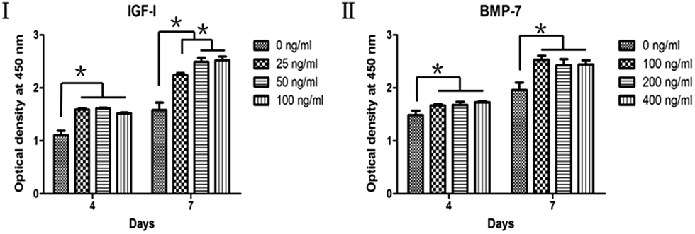 | ||
| Fig. 2 Effects of IGF-I (I) and BMP-7 (II) on proliferation of ASCs with various concentrations. * denotes significant difference (P < 0.05). | ||
3.3. Alkaline phosphatase (ALP) activity
Osteogenic differentiation of ASCs treated with graded concentrations of IGF-I and BMP-7 was assessed by measuring ALP activity (Fig. 3). The effects of IGF-I and BMP-7 on ALP activity were enhanced in a dose-dependent manner. The control group (0 ng ml−1) was significantly lower than any of the IGF-I and BMP-7 treated groups (P < 0.05), and 3-fold of ALP activity beyond the control group was observed at the concentration of 400 ng ml−1 BMP-7. However, no significant difference of ALP activity was found between BMP-7 at 200 ng ml−1 and 400 ng ml−1. | ||
| Fig. 3 Effects of IGF-I and BMP-7 on alkaline phosphatase (ALP) activity of ASCs with various concentrations. * denotes significant difference (P < 0.05). # denotes significant difference compared with DBB group (P < 0.05). | ||
3.4. Real-time polymerase chain reaction (RT-PCR) analysis
Fig. 4 showed the expression of osteogenic genes (Runx2, OPN and ALP) after 7 days incubation of ASCs in the medium. The Runx2 expression was significantly increased with treatment that began with BMP-7 on day 1 followed by BMP-7 or IGF-I on day 4 (P < 0.05). The OPN expression in well treated with BMP-7 on day 1 followed by IGF-I on day 4 was significantly higher than that of the BMP-7 group (P < 0.05). The expression of ALP was significantly improved by the treatment of BMP-7 followed by IGF-I compared to the other groups (P < 0.05). | ||
| Fig. 4 Effects of sequential application of soluble growth factors on osteogenic gene expressions of ASCs in monolayer culture. Osteogenic gene markers including Runx2, OPN and ALP were elevated. * denotes significant difference (P < 0.05). | ||
3.5. Scaffold characterization by SEM
In the scanning electron microscope, DBB granules were present in irregular shape (Fig. 5A) and the calcium phosphate mineral layer was deposited on DBB granules (Fig. 5C). The high magnified SEM images revealed that the surface of DBB was parallel arrays of ridges and troughs (Fig. 5B), and the biomimetic calcium phosphate coating on DBB displayed an interlacing network of crystalline surface (Fig. 5D).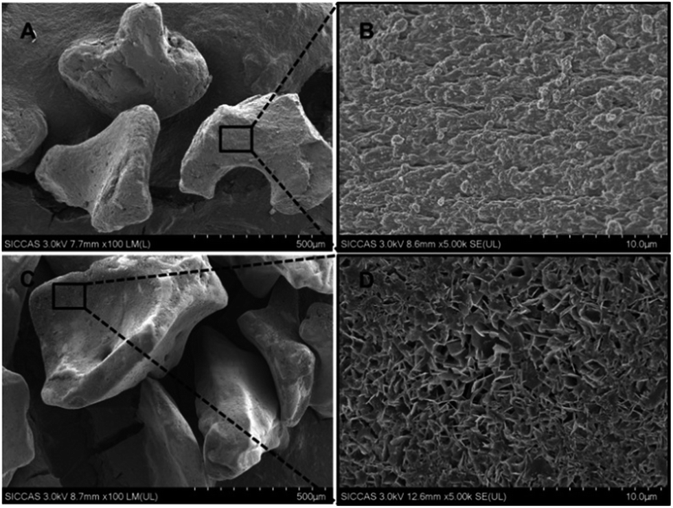 | ||
| Fig. 5 SEM micrographs on the surface of scaffolds: (A, B) native DBB granules; (C, D) DBB granules bearing a calcium phosphate coating. The black square corresponds to a higher magnification of the area. | ||
3.6. Cell adhesion and morphology on scaffolds
After 4 h of culture, (2.57 ± 0.42) × 104 cells of the seeded ASCs (5 × 10⁴ cells) adhered to the CaP-coated scaffold, whereas (1.02 ± 0.8) × 104 cells adhered to the uncoated scaffold (Fig. 6I), the adherent cell number between the two groups was significantly different (P < 0.05). Under the SEM (Fig. 6II), ASCs were sparse and present a round morphology with few pseudopodia on the uncoated DBB, whereas the ASCs were elongated or flatted on the CaP-coated DBB 4 h after seeding. Cells showed an elongated shape and were spread over the scaffolds of both groups after 24 h, but cells covered almost entirely on the CaP-coated DBB. These observations suggested that cells adhered well on both of the scaffolds and biomimetic CaP coating promoted faster adhesion of ASCs. This may relate to the surface structure of the calcium phosphate coating, which provide anchors for the portion of cells. | ||
| Fig. 6 Cell adhesion on uncoated and CaP-coated DBB granules. (I) Quantification of the number of ASCs adhesion on scaffolds, * denotes significant difference between groups (P < 0.05). (II) Morphology and distribution of ASCs cultured on uncoated and CaP-coated DBB granules for 4 h and 24 h. Constructs were observed by SEM. | ||
3.7. Alkaline phosphatase (ALP) activity of ASCs cultured on scaffolds
The ALP activities of ASCs cultured on four groups of scaffolds were investigated (Table 3). The ALP activity of ASCs cultured on IGF-I inc. & BMP-7 ads. DBB was significantly higher than that on the BMP-7 ads. and IGF-I inc. DBB (P < 0.05). However, the ALP activities of ASCs cultured on BMP-7 ads. and IGF-I inc. scaffolds were significantly higher than that on native DBB (P < 0.05) (Fig. 7).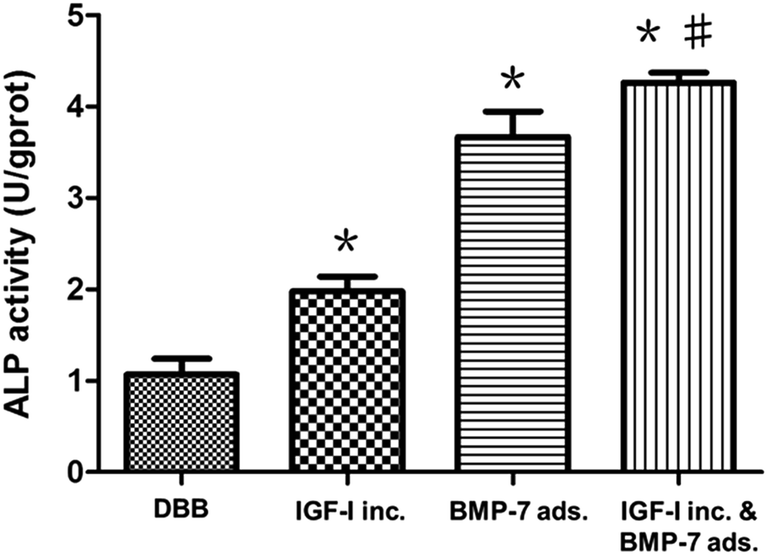 | ||
| Fig. 7 Effect of sequential delivery systems on alkaline phosphatase (ALP) activity of ASCs cultured on scaffolds. DBB: native DBB granules; IGF-I inc.: DBB granules bearing a IGF-I incorporated calcium phosphate coating; BMP-7 ads.: DBB granules bearing a calcium phosphate coating upon which BMP-7 was superficially adsorbed; IGF-I inc. & BMP-7 ads.: DBB granules bearing a IGF-I incorporated calcium phosphate coating upon which BMP-7 was superficially adsorbed. * denotes significant difference compared with the native DBB group (P < 0.05). # denotes significant difference of IGF-I inc. & BMP-7 ads. group compared with the other groups (P < 0.05). | ||
3.8. Histological analysis
Histological staining with H&E and Masson's Trichrome was performed to compare tissue morphology and composition of the ectopic sections at six weeks (Fig. 8). In HE stained images (Fig. 8I), there was no new bone tissue but fibrous tissue observed in the native DBB group. The image of the ectopic sections of IGF-I inc. group showed a little osteoid tissue around granules. In the BMP-7 ads. group, a large osteoid tissue was observed in middle of the image. In the IGF-I inc. & BMP-7 ads. group, there was more osteoid tissue formed on the surface of the scaffold compared with the other groups in the images. Similar results were found on Masson's Trichrome stain (Fig. 8II). No osteoid tissue (pink) but fibrous tissue (green) was present in the native DBB group, while more osteoid tissue was surrounded the granules in the IGF-I inc. & BMP-7 ads. group. | ||
| Fig. 8 H&E (I) and Masson's Trichrome stain (II) micrographs of cross-sections through DBB granules with ASCs in the four groups, six weeks after subcutaneous implantation in rats. (A) native DBB granules; (B) DBB granules bearing a IGF-I incorporated calcium phosphate coating; (C) DBB granules bearing a calcium phosphate coating upon which BMP-7 was superficially adsorbed; (D) DBB granules bearing a IGF-I incorporated calcium phosphate coating upon which BMP-7 was superficially adsorbed. DBB granules (red arrowheads) and osteoid tissue (black arrows). | ||
4. Discussion
It is well-recognized that an ideal scaffold should possess both osteoconductivity and osteoinductivity for favorable attachment, proliferation and differentiation of the osteoblasts or progenitor cells. DBB, a commercially available bone graft substitute, has many fascinating properties and has been used widely for the treatment of bone defects.32,33 Nevertheless, it was reported that DBB delays the early bone formation34 and lacks intrinsic osteoinductivity.35 In this study, we aimed to improve the osteoinductivity of deproteinized bovine bone through sequential delivery of BMP-7 and IGF-I. The results of this study demonstrated that DBB with a sequential release of BMP-7 and IGF-I has superior properties on osteogenic differentiation of ASCs and ectopic bone formation, the null hypothesis was rejected.Adipose tissue is a rich source of multipotent progenitor cells, from which ASCs are easily accessible and available in large quantities. ASCs can transform into osteoblasts after osteogenic induction and regenerate bone tissue after implantation in vivo.36,37 Multiple pre-clinical and clinical studies so far have been published demonstrating the safety and efficacy of ASCs administration to treat different diseases.38–40 Moreover, ASCs were applied to enhance jaw bone height with scaffold in patients with pronounced deficit of the maxillary and mandibular bone tissue.41 Recently, there is an increasing recognition of the importance of seeding ASCs in a three-dimensional environment to generate tissues with increased functionality and engraftment capacity.42,43 Given these factors we used ASCs to evaluate the biological capabilities of the release system formed in this study.
In order to employ an optimal concentration of BMP-7 and IGF-I, the proliferation and osteogenic differentiation of ASCs in response to different concentrations of BMP-7 and IGF-I was evaluated. In the present study, BMP-7 and IGF-I both stimulated cell proliferation and osteogenic differentiation of ASCs. Considering the cell response stimulated by BMP-7, there was no significant difference between the effects of BMP-7 at concentrations of 200 ng ml−1 and 400 ng ml−1, thus the optimal concentration of BMP-7 might be 200 ng ml−1. Regarding to IGF-I, the proliferation rate of ASCs was at the peak at concentrations of 50 ng ml−1 and 100 ng ml−1, no significant difference was found between them. However, ALP activity was significantly greater at 100 ng ml−1 than 50 ng ml−1 (P < 0.05), therefore concentration of IGF-I at 100 ng ml−1 was employed in the following experiments. Previous study showed that BMP-7 stimulated cell proliferation in the early stage of cell culture, but down-regulated it before the onset of events associated with osteogenic differentiation.44 In this study, BMP-7 stimulated cell proliferation in the early culture stage, the effects began to attenuate at high concentrations in the late culture stage. In addition, IGF-I can also affect cell proliferation and osteogenic differentiation of ASCs in our study. Walsh et al. reported that IGF-I did not affect the proliferation or early osteogenic differentiation of bone marrow stromal cells (BMSCs).45 In another study, Gao et al. reported that IGF-I-immobilized PLGA/HA microcarriers significantly increased ALP activity and expressions of osteogenetic related genes of ASCs.46 That may due to the differences of cell types, cell maturity or culture conditions.
To investigate the effects of sequential application of BMP-7 and IGF-I on osteogenic differentiation of ASCs, the RT-PCR analysis of several genes typically associated with mineralization during osteogenesis was conducted. The RT-PCR results showed that treatment which began with BMP-7 on day 1 followed IGF-I on day 4 induced higher Runx2, OPN and ALP expressions. Thus we selected this group as the experimental group and employed the biomimetic composite coating and adsorption approaches to load BMP-7 and IGF-I on DBB to establish a delivery system. The ALP activity of ASCs on DBB with an initial release of BMP-7 followed by IGF-I was significantly higher than that of the others (P < 0.05), which was consistent with the bone-related gene expressions induced by the soluble growth factors. Finally, we used an ectopic model to verify the effect of the sequential release system with ASCs on bone formation in vivo.
Raiche et al. used two layers of glutaraldehyde cross-linked gelatin coatings for controlled release of BMP-2 and IGF-I.47 Similarly, Kim developed a chitosan gel/gelatin microsphere dual delivery system for sequential release of BMP-2 and IGF-I.24 In their study, the sequential release of BMP-2 and IGF-I significantly stimulated the ALP activity of bone marrow stromal cells and they emphasized the importance of the sequential release of growth factors to optimize their efficacy.24,47 However, gelatin is an amphoteric protein which easily dissolves in water at body temperature and releases amino acids, most of the incorporated osteogenic growth factors diffuse away rapidly.24 In contrast to this, the biomimetic calcium phosphate coating exhibited a controlled and slow release profile.28 Previous studies showed that biomimetic calcium phosphate coating with growth factor had a positive effect on bone formation in vivo.48–50 The results of this study suggested that sequential release of BMP-7 and IGF-I could enhance osteogenic differentiation of ASCs in vitro and ectopic bone formation in vivo, which agreed with previous researches.
With respect to fracture repair, BMPs production peaks around day 3 post-fracture and is followed by a slow decline to the baseline levels by day 10, whereas IGFs synthesis increases between days 5 and 10.51,52 So there is an increasing need for sophisticated delivery systems capable of delivering growth factors in a sequential delivery manner at different release rates to induce cooperative and synergistic processes that mimic natural wound healing. In this study, we developed a delivery system on deproteinized bovine bone to release BMP-7 and IGF-I in a temporally controlled manner. The initial release of BMP-7 might serve as a strategy to kick off regime and steer stem cells into osteogenic lineage differentiation, then the sustained releases of IGF-I has the ability to enhance matrix mineralization. In addition, the dissolving of calcium phosphate could locally generate high calcium and phosphate concentrations, which is needed in the process of mineralization.
A subcutaneous site is routinely used to evaluate the osteoinductivity of biomaterials.53–55 However, the mechanical stress that is imposed on the granules and subsequent movement could compromise osteogenic activity and promote a foreign-body response. Further in vivo studies are needed to verify the efficacy of this strategy for promoting bone regeneration in bone defect models, which mimic the clinical applications.
5. Conclusions
In this study, we employed the biomimetic calcium phosphate coating and adsorption technique to deliver BMP-7 and IGF-I in a temporally manner. The results demonstrated that an initial release of BMP-7 followed by a sustained release of IGF-I significantly enhanced osteogenic differentiation of ASCs in vitro and bone formation activity in vivo. Therefore, it is a promising approach to improve the osteoinductive property of DBB.Acknowledgements
This study was financially supported by the grants from the National Natural Science Foundation of China (No. 81271110), Natural Science and Technology Support Program (No. 2014BAI04B07) and the Fundamental Research Funds for the Central Universities (No. 20152957) of China.References
- A. S. Herford and J. S. Dean, Oral Maxillofac. Surg. Clin., 2011, 23, 433–442 CrossRef PubMed.
- J. A. Goulet, L. E. Senunas, G. L. DeSilva and M. L. Greenfield, Clin. Orthop. Relat. Res., 1997, 76–81 CAS.
- A. Mordenfeld, C. Lindgren and M. Hallman, Clin. Implant Dent. Relat. Res., 2015 DOI:10.1111/cid.12374.
- J. W. Moon, D. S. Sohn, J. U. Heo and J. S. Kim, Implant Dentistry, 2015, 24, 19–24 CrossRef PubMed.
- M. Merli, M. Moscatelli, G. Mariotti, U. Pagliaro, E. Raffaelli and M. Nieri, European Journal of Oral Implantology, 2015, 8, 271–281 Search PubMed.
- J. B. Huh, J. J. Yang, K. H. Choi, J. H. Bae, J. Y. Lee, S. E. Kim and S. W. Shin, Int. J. Mol. Sci., 2015, 16, 16034–16052 CrossRef CAS PubMed.
- M. R. Oliveira, S. A. DeC, S. Ferreira, C. C. Avelino, I. J. Garcia and R. C. Mariano, Int. J. Oral Maxillofac. Surg., 2015, 44, 649–655 CrossRef CAS PubMed.
- C. Schmitt, R. Lutz, H. Doering, M. Lell, J. Ratky and K. A. Schlegel, Clin. Oral Implants Res., 2013, 24, 450–460 CrossRef PubMed.
- D. Cardaropoli, International Journal of Periodontics and Restorative Dentistry, 2009, 29, 289–295 Search PubMed.
- M. Varkey, S. A. Gittens and H. Uludag, Expert Opin. Drug Delivery, 2004, 1, 19–36 CrossRef CAS PubMed.
- A. C. Mitchell, P. S. Briquez, J. A. Hubbell and J. R. Cochran, Acta Biomater., 2016, 30, 1–12 CrossRef CAS PubMed.
- G. Litwack, Vitam. Horm., 2015, 99, xiii–xiv Search PubMed.
- P. Yilgor, K. Tuzlakoglu, R. L. Reis, N. Hasirci and V. Hasirci, Biomaterials, 2009, 30, 3551–3559 CrossRef CAS PubMed.
- D. W. Lee, Y. P. Yun, K. Park and S. E. Kim, Bone, 2012, 50, 974–982 CrossRef CAS PubMed.
- I. H. Ali and D. P. Brazil, Br. J. Pharmacol., 2014, 171, 3620–3632 CrossRef CAS PubMed.
- M. C. Papanna, N. Al-Hadithy, B. V. Somanchi, M. D. Sewell, P. M. Robinson, S. A. Khan and R. A. Wilkes, Injury, 2012, 43, 1135–1140 CrossRef CAS PubMed.
- J. Leach and R. G. Bittar, J. Clin. Neurosci., 2009, 16, 1417–1420 CrossRef CAS PubMed.
- V. P. Mantripragada and A. C. Jayasuriya, Mater. Sci. Eng., C, 2016, 63, 596–608 CrossRef CAS PubMed.
- G. Burastero, S. Scarfi, C. Ferraris, C. Fresia, N. Sessarego, F. Fruscione, F. Monetti, F. Scarfo, P. Schupbach, M. Podesta, G. Grappiolo and E. Zocchi, Bone, 2010, 47, 117–126 CrossRef CAS PubMed.
- K. Lavery, S. Hawley, P. Swain, R. Rooney, D. Falb and M. H. Alaoui-Ismaili, Bone, 2009, 45, 27–41 CrossRef CAS PubMed.
- F. Al-Salleeh, M. W. Beatty, R. A. Reinhardt, T. M. Petro and L. Crouch, Arch. Oral Biol., 2008, 53, 928–936 CrossRef CAS PubMed.
- A. Filus and Z. Zdrojewicz, Pediatr. Endocrinol., Diabetes Metab., 2015, 20, 161–169 CrossRef PubMed.
- M. Nakasaki, K. Yoshioka, Y. Miyamoto, T. Sasaki, H. Yoshikawa and K. Itoh, Bone, 2008, 43, 869–879 CrossRef CAS PubMed.
- S. Kim, Y. Kang, C. A. Krueger, M. Sen, J. B. Holcomb, D. Chen, J. C. Wenke and Y. Yang, Acta Biomater., 2012, 8, 1768–1777 CrossRef CAS PubMed.
- F. M. Chen, M. Zhang and Z. F. Wu, Biomaterials, 2010, 31, 6279–6308 CrossRef CAS PubMed.
- L. C. Yeh and J. C. Lee, Biochim. Biophys. Acta, 2006, 1763, 57–63 CrossRef CAS PubMed.
- L. Yang, Y. Zhang, R. Dong, L. Peng, X. Liu, Y. Wang and X. Cheng, J. Periodontal Res., 2010, 45, 532–540 CAS.
- X. Lin, K. de Groot, D. Wang, Q. Hu, D. Wismeijer and Y. Liu, Open Biomed. Eng. J., 2015, 9, 56–64 CrossRef CAS PubMed.
- T. Kokubo and H. Takadama, Biomaterials, 2006, 27, 2907–2915 CrossRef CAS PubMed.
- X. Yu and M. Wei, J. Biomed. Mater. Res., Part B, 2011, 97, 345–354 CrossRef PubMed.
- Y. Liu, G. Wu and K. de Groot, J. R. Soc., Interface, 2010, 7(Suppl. 5), S631–S647 CrossRef CAS PubMed.
- M. Ayna, Y. Acil and A. Gulses, International Journal of Periodontics & Restorative Dentistry, 2015, 35, 541–547 Search PubMed.
- M. Hallman and L. Zetterqvist, Clinical Implant Dentistry and Related Research, 2004, 6, 82–89 CrossRef PubMed.
- M. Araujo, E. Linder and J. Lindhe, Clin. Oral Implants Res., 2009, 20, 1–6 CrossRef CAS PubMed.
- Z. Schwartz, T. Weesner, S. van Dijk, D. L. Cochran, J. T. Mellonig, C. H. Lohmann, D. L. Carnes, M. Goldstein, D. D. Dean and B. D. Boyan, J. Periodontol., 2000, 71, 1258–1269 CrossRef CAS PubMed.
- D. Dufrane, P. L. Docquier, C. Delloye, H. A. Poirel, W. Andre and N. Aouassar, Medicine, 2015, 94, e2220 CrossRef CAS PubMed.
- W. Zhang, X. Zhang, S. Wang, L. Xu, M. Zhang, G. Wang, Y. Jin, X. Zhang and X. Jiang, J. Dent. Res., 2013, 92, 1136–1141 CrossRef CAS PubMed.
- I. Kim, S. I. Bang, S. K. Lee, S. Y. Park, M. Kim and H. Ha, Stem Cells Transl. Med., 2014, 3, 1312–1321 CrossRef CAS PubMed.
- M. Tobita, S. Tajima and H. Mizuno, Stem Cell Res. Ther., 2015, 6, 215 CrossRef PubMed.
- E. M. Perez-Merino, J. M. Uson-Casaus, C. Zaragoza-Bayle, J. Duque-Carrasco, L. Marinas-Pardo, M. Hermida-Prieto, R. Barrera-Chacon and M. Gualtieri, Vet. J., 2015, 206, 385–390 CrossRef CAS PubMed.
- A. A. Kulakov, D. V. Goldshtein, A. S. Grigoryan, A. A. Rzhaninova, I. S. Alekseeva, I. V. Arutyunyan and A. V. Volkov, Bull. Exp. Biol. Med., 2008, 146, 522–525 CrossRef CAS PubMed.
- D. Sadeghi, H. Nazarian and H. Nojehdehian, Med. Hypotheses, 2014, 82, 54–56 CrossRef CAS PubMed.
- G. K. Sandor, J. Numminen, J. Wolff, T. Thesleff, A. Miettinen, V. J. Tuovinen, B. Mannerstrom, M. Patrikoski, R. Seppanen, S. Miettinen, M. Rautiainen and J. Ohman, Stem Cells Transl. Med., 2014, 3, 530–540 CrossRef CAS PubMed.
- F. Zhang, L. F. Ren, H. S. Lin, M. N. Yin, Y. Q. Tong and G. S. Shi, Arch. Oral Biol., 2012, 57, 460–468 CrossRef CAS PubMed.
- S. Walsh, C. M. Jefferiss, K. Stewart and J. N. Beresford, Bone, 2003, 33, 80–89 CrossRef CAS PubMed.
- T. Gao, N. Zhang, Z. Wang, Y. Wang, Y. Liu, Y. Ito and P. Zhang, Macromol. Biosci., 2015, 15, 1070–1080 CrossRef CAS PubMed.
- A. T. Raiche and D. A. Puleo, J. Biomed. Mater. Res., Part A, 2004, 69, 342–350 CrossRef CAS PubMed.
- M. Ramazanoglu, R. Lutz, P. Rusche, L. Trabzon, G. T. Kose, C. Prechtl and K. A. Schlegel, Journal of Cranio-Maxillo-Facial Surgery, 2013, 41, 826–835 CrossRef PubMed.
- T. Liu, G. Wu, Y. Zheng, D. Wismeijer, V. Everts and Y. Liu, Clin. Oral Implants Res., 2014, 25, 1412–1421 CrossRef PubMed.
- M. Dadsetan, T. Guda, M. B. Runge, D. Mijares, R. Z. LeGeros, J. P. LeGeros, D. T. Silliman, L. Lu, J. C. Wenke, B. P. Brown and M. J. Yaszemski, Acta Biomater., 2015, 18, 9–20 CrossRef CAS PubMed.
- W. T. Bourque, M. Gross and B. K. Hall, Int. J. Dev. Biol., 1993, 37, 573–579 CAS.
- Y. Yu, J. L. Yang, P. J. Chapman-Sheath and W. R. Walsh, J. Biomed. Mater. Res., 2002, 60, 392–397 CrossRef CAS PubMed.
- J. Leotot, L. Coquelin, G. Bodivit, P. Bierling, P. Hernigou, H. Rouard and N. Chevallier, Acta Biomater., 2013, 9, 6630–6640 CrossRef CAS PubMed.
- J. Zhang, H. Zhou, K. Yang, Y. Yuan and C. Liu, Biomaterials, 2013, 34, 9381–9392 CrossRef CAS PubMed.
- J. L. Bain, B. K. Culpepper, M. S. Reddy and S. L. Bellis, Int. J. Oral Maxillofac. Implants, 2014, 29, 1437–1445 Search PubMed.
| This journal is © The Royal Society of Chemistry 2016 |