Fabrication of a selective 4-amino phenol sensor based on H-ZSM-5 zeolites deposited silver electrodes

Mohammed M. Rahman*a, Bahaa M. Abu-Zieda, Mohammad M. Hasanb, Abdullah M. Asiria and Mohammad A. Hasnat*b
aCenter of Excellence for Advanced Material Research (CEAMR) and Chemistry Department, Faculty of Science, King Abdulaziz University, P. O. Box 80203, Jeddah 21589, Saudi Arabia. E-mail: mmrahman@kau.edu.sa; Fax: +966-59-642-1830; Tel: +966-59-642-1830
bDepartment of Chemistry, Shahjalal University of Science and Technology, Sylhet 3114, Bangladesh. E-mail: mah-che@sust.edu; Fax: +88-0821-715257; Tel: +88-0821-715257

Received 15th February 2016 , Accepted 3rd May 2016

First published on 4th May 2016


Abstract

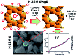
H-ZSM-5 zeolite is an inorganic material with large surface area and well-defined internal structure with porous uniform cages, cavities or channels. In this study, H-ZSM-5 was synthesised by calcination of the NH4-form at 500 °C for 3 h in air flow. This protonated H-ZSM-5 has been characterized in detail, which includes its optical, structural, morphological, and elemental properties by various conventional methods. For probable chemical sensor development, H-ZSM-5 was deposited on a silver electrode (AgE, surface area, 0.0216 cm2) to fabricate a sensor with a fast response towards selective 4-amino phenol (4-AMP) in the liquid phase. The sensor exhibited good sensitivity and long-term stability and enhanced electrochemical responses. The calibration plot was linear (r2 = 0.9979) over the 0.1 nM to 1.0 mM 4-AMP concentration ranges. The sensitivity was ∼2.085 μA cm−2 nM−1 and the detection limit was 0.02 nM (at a signal-to-noise ratio (SNR) of 3). By employing CV and EIS techniques, it was unveiled that the sensor is not well-operative in the absence of air. This shows a promising future for sensitive sensor development using mesoporous H-ZSM-5 by IV methods for applications in the detection of hazardous and carcinogenic phenolic compounds in environmental and health care fields.


Introduction

Zeolite-Socony-Mobil (abb., ZSM-5) is a synthetic material consisting of Si, Al, and O atoms arranged in an exceptional framework (MFI type) together with water molecules located within the interior pores.1 ZSM-5 zeolite has unique physico-chemical properties. For instance, it has a pore diameter of 0.54–0.56 nm and adjustable Si/Al ratios varying in the range from 10 to several hundred.2 The framework structure of ZSM-5 consists of several pentasil units, each of which consists of eight five-membered rings, connected with each other by oxygen bridges. The various pentasil chains are interconnected to form intersecting straight and sinusoidal channels.3 In addition to its unique framework structure, ZSM-5 zeolite has outstanding thermal and hydrothermal stability, high acidity, molecular shape selectivity and resistance to deactivation.4 These features nominated ZSM-5 zeolite to be applied as active catalysts in various industrial reactions in refining and petroleum processing. For instance, ZSM-5 has been widely exploited for conversion of methanol to gasoline and olefins,5 paraffin isomerization,6 hydroxylation of benzene to phenol7 and ethylbenzene production.8

The crystalline microporous alumosilicates zeolites are composed of [TO4] tetrahedral (T = Si, Al), e.g. Na-form zeolites are Na+-ion conductors9,10 and H-form zeolites are protonic conductors owing to the mobility of protons which counteract the negative charge of the [AlO4]-tetrahedra by means of bridging Si–OH–Al groups. By means of impedance measurements11–13 and quantum chemical calculations14,15 of H-ZSM-5, it has already been established that protons can move through the zeolite framework by a thermally activated hopping process. The probability of proton hopping depends on the spatial distance between an occupied and the next unoccupied lattice-site and therefore on the SiO2/Al2O3 ratio (module) in the zeolite lattice. In nitrogen and oxygen containing atmospheres, the presence of solvate molecules, such as H2O or NH3 in concentrations of 100 ppm and above, leads to an increase of the proton mobility up to 420 °C.16 This involves either the vehicle or the Grotthus mechanism. While the vehicle mechanism is based on diffusive motion of solvated protons, namely, H3O+ or NH4+, in the case of the Grotthus mechanism, the local dynamic of the supporting solvate molecules play an important role. If a proton binds to a solvate molecule, it can be transferred to another solvate molecule or to a free lattice site via a rotational motion of the solvent molecule. This mechanism requires a sufficient number of solvate molecules between the [AlO4]-tetrahedra. According to temperature programmed desorption (TPD) measurements on H-ZSM-5, a complete desorption is expected at temperatures above 450 °C.17 From this result, it is preliminarily concluded that the supporting effect of the solvate molecules vanishes at temperatures above the desorption temperature. Since it was demonstrated that H-ZSM-5 is stable also in an oxygen-free, hydrogen containing atmosphere,18 H-ZSM-5 zeolite was chosen as a promising candidate for impedance measurements at elevated temperatures in order to detect traces of toxic chemical in pure environments. In this article, new limits are defined for the chemical sensitivity of an H-ZSM-5 type zeolite, which gives rise to the prospect of new chemical sensor application areas for this versatile implementable material. Owing to the optical and morphological properties outlined above, zeolites are well-established in a number of industrial applications, such as ion exchangers for water purification,19–21 as drying agents or absorbents for organic vapors,22 as molecular sieves and separation membranes,23 or as catalyst beds in the petrochemical industry or in the synthesis of fine chemicals.24,25 The use of natural zeolites for environmental remediation (organic compounds and heavy-metal ions) has also been established. Moreover, natural zeolites have been found to be helpful in the control of malodors emanating from confined livestock-rearing areas, kennels, pet shops, zoos and pet-litter trays. Their high surface area, and therefore their high adsorption capacity, as well as their ion-exchange properties make them very useful in these fields.26

Recently, the developments of reliable and sensitive methods were used for the detection of phenolic compounds for environmental safety, protection, food quality control, and health.27 Several phenolic compounds have been classified as “priority pollutants” by both the United States Environmental Protection Agency (USEPA) and the European Commission. Phenolic compounds are comparatively general in waste streams of diverse large-scale processing and manufacturing, where they serve as precursor materials in various industries such as coal mining, crude oil refining, paper bleaching, and production of dyes, resins, plastics, explosives, detergents, pharmaceuticals, pesticides, and herbicides.28,29 In contrast, a few phenols of plant origin have been shown to display a broad range of attractive physiological characteristics as antioxidants, anti-inflammatories, and cardiovascular prophylactics, and their use as additives in some alcoholic beverages and food products has been promoted.30,31 Recent determination methods such as spectrophotometry, fluorimetry, gas or liquid chromatography, mass spectrometry and capillary electrophoresis are usually accurate and consistent but possess limitations such as being expensive, time-consuming and requiring pre-concentration and extraction steps that increase the risk of sample loss and generation of other hazardous byproducts.32 Electrochemical sensing of phenolic compounds represents a promising approach that can be utilized to complement already existing methods owing to the collective characteristics of high sensitivity and selectivity, low cost, simple instrumentation and potential for miniaturization.33,34 Herein, the H-ZSM-5 zeolite has significant properties such as large surface area (surface-to-volume ratio), non-toxicity, chemical stability, and high electrical conductivity, which offer high quality electron communication features that enhance the direct electron communication towards the target toxic analytes. As 4-AMP is highly carcinogenic and pose a serious threat to health and the environment, fabricating a simple and reliable chemical sensor is urgently required. Although numerous papers have been published on the application of H-ZSM-5 zeolite as catalysts and adsorbents, there is a lack of information about its environmental sensing applications in general and for 4-AMP sensing in particular. Accordingly, in this paper the 4-AMP sensing properties of H-ZSM-5 films have been investigated and explained in term of fabrication and their typical electrochemical characteristics. The simple fabrication technique is used for the preparation of H-ZSM-5 zeolite on flat AgE with conducting coating binders, which is measured by the simple and reliable IV technique. The mechanism has been discerned by means of Cyclic Voltammetry (CV) and Electrochemical Impedance Spectroscopy (EIS). To the best of our knowledge, this is the first report for the sensitive detection of 4-AMP with H-ZSM-5 zeolite using the simple IV technique in short response time.

Experimental

Materials and methods

Analytical grade 4-AMP, ethyl acetate, acetone, methanol, 3-methoxy phenol, disodium phosphate (Na2HPO4), dichloromethane, butyl carbitol acetate, tetrahydrofuran, chloroform, 1,2-dichlorobenzene, and monosodium phosphate (NaH2PO4) were used and purchased from Sigma-Aldrich Company, USA. Stock solution of 9.0 mM 4-AMP was prepared from the purchased chemical. Initially, NH4-ZSM-5 zeolites (Si/Al ratio of 11.4), prepared via the template-free synthesis method, were obtained from ALSI Penta Zeolithe GmbH (Germany). Later, the H-form (H-ZSM-5) was obtained by calcining NH4-form at 500 °C for 3 h in air flow. The morphology of the H-ZSM-5 zeolite was checked on an FESEM instrument (FESEM; JSM-7600F, Japan). Elemental analysis was examined for H-ZSM-5 by XEDS from JEOL, Japan. The powder X-ray diffraction (XRD) prototypes were assessed for the H-ZSM-5 zeolite with an X-ray diffractometer (X'pert Explorer, PANalytical diffractometer) equipped with Cu-Kα1 radiation (λ = 1.5406 nm) using a generator voltage of 40.0 kV and a generator current of 35.0 mA. FT-IR spectra were obtained for the H-ZSM-5 zeolite using a spectrophotometer (Spectrum-100 FT-IR) in the mid-IR range, which was purchased from Thermo-Nicolet iS50 FT-IR spectrometer using an Attenuated Total Reflectance (ATR) sampling accessory. The X-ray photoelectron spectroscopy (XPS) measurements were executed on a Thermo Scientific K-Alpha KA1066 spectrometer for H-ZSM-5 zeolites. A monochromatic AlKα X-ray radiation source was used as the excitation source, where beam-spot size was kept at 300.0 μm. The spectra were obtained in the fixed analyzer transmission mode, where pass energy was maintained at 200.0 eV. The scanning of the spectra was performed at pressures less than 10−8 Torr. Atomic absorption spectroscopy (AAS) was used to evaluate the silver ions released in the bulk solution after consecutive IV measurements. The IV technique was executed by using an Electrometer (Keithley, 6517A, Electrometer, USA) for measuring the current responses in two electrode systems for target 4-AMP chemical sensor based on H-ZSM-5 zeolite in buffer phase at room temperature conditions, where flat-AgE and Pd-wire were used as the working and counter electrodes, respectively.

Preparation and fabrication of flat-AgE with H-ZSM-5 zeolite

0.1 M phosphate buffer solution (PBS) at pH 7.0 is prepared by mixing equi-molar quantities of 0.2 M Na2HPO4 and 0.2 M NaH2PO4 solutions in 100.0 mL de-ionized water at room temperature. The silver electrode (AgE) was fabricated with H-ZSM-5 zeolite using butyl carbitol acetate (BCA) and ethyl acetate (EA) as a conducting binder. Then, it was kept in the oven at 50.0 °C for 1 hour until the film was completely dry, stable, and smooth. A cell was assembled with H-ZSM-5/AgE and Pd-wire as working and counter electrodes, respectively. As received 4-AMP (99.9%) was diluted to make various concentrations (0.1 nM to 0.1 M) in DI water and used as a target analyte. The ratio of current versus concentration (slope of calibration curve) was used to calculate the 4-AMP sensitivity. The detection limit was evaluated from the ratio of 3N/S (ratio of noise × 3 vs. slope) from the linear dynamic range of the calibration curve. An electrometer was used as a constant voltage source for IV measurement in a simple two electrode system. Amount of 0.1 M PBS was kept constant in the beaker as 10.0 mL throughout the chemical investigation. IV response was measured with a H-ZSM-5/AgE film. The electrochemical experiments (CV and EIS) were carried out under thermostatic and N2-atmosphere conditions using an Autolab PGSTAT128N instrument (Metrohm Autolab B.V.). All potentials in these measurements have been reported with respect to Ag|AgCl (sat. KCl) reference electrodes.

Evaluation of Ag+ released by various analytical techniques

Flat-AgE was used to evaluate the sensor performances in common phosphate buffer in the presence of target analyte at neutral pH. In general, AgE is used for the electrochemical investigation of biosensors, sensors, catalysis, and other purposes. AgE is a highly stable electrode, particularly in the buffer, and no Ag+ ions leach from the electrode surface during electrochemical performances. In this approach, H-ZSM-5 materials were immobilized on the flat AgE-surface, which was covered with conducting coating binders such as carbitol acetate (BCA) and ethyl acetate (EA). This procedure eventually resulted in the leaching of Ag-particles from the interior region of the electrode matrix. Even on examining SEM images of the used AgE surface, it was noticed that no erosion or corrosion of the AgE surface occurred in this system. For environmental safety, several direct additional experiments were performed and are presented below to confirm that Ag-ions were not released by the fabricated electrode (H-ZSM-5/binders/AgE) during the several consecutive IV measurements. First, AgE was fabricated with H-ZSM-5 using binders and used for the measurement of IV plots in PBS buffer after injecting various concentrations of target analytes. After measuring the IVs, the resultant solution was tested for the presence of Ag+ ions. The tests may be summarized as follows:
Ice-cold borohydrate test. No Ag-NPs were formed (confirmed by UV/vis spectrum) during the electrochemical study. No peak was appeared at ∼415 nm for Ag-NPs formation. Therefore, it was confirmed that no Ag+ existed in the solution phase.
Silver iodide test. NaI solution was added into the test solution electrolytes obtained from I–​V measurement, no yellow color/ppt was formed. Therefore, it was confirmed that no Ag+ was existed in the leaching solution.
Tollen test (Ag+ + NH4OH + aldehyde). No Ag-mirror formation was observed on the wall of test-tube even after warming the electrolyte solution. Therefore, it was confirmed that no Ag+ existed in the solution.
AAS test. Finally, an Ag+ test was performed using atomic absorption spectrophotometry. No absorption band was observed around ∼328 nm, which is the characteristic wave for the atomized Ag-species.

Finally, it was confirmed that Ag+ ions were not released from the interior of the electrode matrix.

Results and discussion

Morphological and optical characterization of H-ZSM-5 zeolite

High resolution FESEM images of calcined H-ZSM-5 zeolite are exhibited in Fig. 1a and b. The FESEM images display H-ZSM-5 nano-structural zeolite materials. The average diameter of H-ZSM-5 is calculated in the range of 0.65–1.2 μm, which is close to 0.82 ± 0.2 μm. It is exhibited noticeably from the FESEM images that the simple blending methodology of prepared products are H-ZSM-5 zeolites, which revealed in aggregated shape, high-density, and obtained protonated zeolites. It is also proposed that approximately all the structures are composed in aggregated protonated H-ZSM-5 zeolites.35 The X-ray electron dispersive spectroscopy (XEDS) investigation of H-ZSM-5 zeolite indicates the presence of Al, Si, and O in the aggregated zeolite materials.
image file: c6ra04124h-f1.tif
Fig. 1 Study of morphological and elemental analysis of zeolites. (a and b) Low to high magnified FESEM images of H-ZSM-5 zeolite, (c) selected area of FESEM image of H-ZSM-5 zeolite, and (d) elemental analysis (XEDS) of H-ZSM-5 zeolite. (Inset) elemental compositions (wt% and at%) of H-ZSM-5 zeolite.

It is clearly displayed that the as prepared H-ZSM-5 zeolite contained only aluminium, silicon, and oxygen, which is presented in Fig. 1c and d. The composition of O, Al, and Si was 58.63%, 3.12%, and 38.25%, respectively. Comparative elemental analysis data are also included in Fig. 1d (inset). No other peak related with any impurity has been detected in the XEDS, which confirms that the H-ZSM-5 zeolite products are composed only of Al, Si, and O.36

The X-ray diffraction patterns obtained for the as-received and the calcined zeolites are shown in Fig. 2a. Inspection of this figure reveals that the H-ZSM-5 zeolite samples exhibit similar patterns of original NH4-ZSM-5 zeolite (not shown). All the obtained reflections can be assigned to the pentasil type framework structure (JCPDS card file no. 43-0321). Moreover, the obtained pattern confirms that the characteristics of the H-ZSM-5 zeolite remained intact after the thermal treatment of NH4-ZSM-5 at 500 °C for the conversion of H-ZSM-5. The the fundamental vibration characteristic framework structure of H-ZSM-5 zeolite appear at the IR region of 1400–400 cm−1. The FTIR spectrum of the H-ZSM-5 zeolite sample is shown in Fig. 2b. The obtained spectrum for the H-ZSM-5 shows absorption bands at 1066 cm−1, 792 cm−1 and 450 cm−1, which can be assigned to the vibrations of (Si, Al)O4 siliceous materials and can also be observed in siliceous materials (silica, quartz and cristobalite).37 The two bands located at 1216 cm−1 and 542 cm−1 could be attributed to the asymmetric stretching of SiO4 and AlO4 tetrahedra and the double-rings tetrahedra vibration in the H-ZSM-5 zeolite framework, respectively. The band appearing at 1630 cm−1 can be assigned to the bending vibration of H2O molecules inside the H-ZSM-5 framework, which reflects the hydrophilicity of the zeolite material.


image file: c6ra04124h-f2.tif
Fig. 2 Study of structural and optical properties of zeolite. (a) X-ray diffraction patterns (XRD) and (b) Fourier-transform infra-red (FTIR) spectrum obtained for H-ZSM-5 zeolite.

X-ray photoelectron spectroscopy (XPS) is a quantitative spectroscopic method that determines the elemental-composition, empirical-formula, chemical-state and electronic-state of the elements that are present within materials. XPS spectra are attained by irradiating materials with a beam of X-rays while simultaneously determining the kinetic energy and number of electrons that get away from the top 1 to 10.0 nm of the material being analysed. Herein, XPS measurements were measured for H-ZSM-5 zeolite to investigate the chemical states of aluminium, silicon, and oxygen. The full XPS spectrum of H-ZSM-5 zeolite is presented in Fig. 3a. In Fig. 3b, the spin–orbit peak of the Al 2p binding energy for the samples appeared at around 80.7 eV, which is in good agreement with the reference data for aluminium.38 The Si 2p spectrum shows a distinguished peak at 103.6 eV in Fig. 3c, which indicates the presence of silicon in the H-ZSM-5 zeolites.39 The O 1s spectrum shows a distinguished peak at 532.9 eV in Fig. 3d. The peak at 532.9 eV is assigned to oxygen, which indicates the presence of oxygen (i.e. O2) in the H-ZSM-5.40 Therefore, it is concluded that the H-ZSM-5 zeolites have three different elements.


image file: c6ra04124h-f3.tif
Fig. 3 Study of X-ray photoelectron spectroscopy (XPS) for binding energy analysis of H-ZSM-5 zeolite. (a) Full spectrum, (b) Al 2p level, (c) Si 2p level, and (d) O 1s level of acquired with MgKα1 radiations. X-ray beam-spot size is 300.0 μm; pass-energy is 200.0 eV; pressures less than 10−8 Torr.

Detection of 4-AMP sensor using H-ZSM-5 zeolite by AgE

The probable application of H-ZSM-5 zeolite assembled onto flat-AgE as chemical sensors (particularly 4-AMP analyte in a buffer system) has been investigated for measuring and detecting a target chemical. Use of H-ZSM-5/AgE as chemical sensors is in the initial stage of development and no other reports are available. The H-ZSM-5/AgE sensors have advantages such as stability in air, non-toxicity, chemical inertness, electro-chemical activity, simplicity to assemble, ease in fabrication, and chemo-safe characteristics. As in the case of 4-AMP sensors, the current response in the IV method of H-ZSM-5/AgE changes considerably when aqueous phenolic analytes are adsorbed. The fabricated-surface of the H-ZSM-5 sensor was prepared with conducting binders (EC and BCA) on the flat AgE surface, which is presented in Scheme 1a. The IV signals of the 4-AMP chemical sensor were anticipated to have H-ZSM-5/AgE on thin-film as a function of current versus potential. The resultant electrical responses of target 4-AMP are investigated by a simple and reliable IV technique using H-ZSM-5/AgE. The holding time of the electrometer was set for 1.0 s. A significant amplification in the current response with applied potential was confirmed.
image file: c6ra04124h-s1.tif
Scheme 1 Schematic view of (a) H-ZSM-5 coated AgE with conducting coating binders, (b) detection IV method (theoretical), (c) observed IV response by H-ZSM-5/AgE, and (d) proposed adsorption mechanisms of 4-AMP detection in the presence of H-ZSM-5 zeolite onto flat-AgE. Surface area of AgE: 0.0216 cm2; method: IV. Delay time: 1 s.

The concentration of 4-AMP was varied from 0.1 nM to 0.1 M by adding de-ionized water in different proportions. Herein, Fig. 4a represents the IV responses for uncoated-AgE (gray-dotted) and H-ZSM-5-coated-AgE (red-dotted) electrodes.


image file: c6ra04124h-f4.tif
Fig. 4 Study of control experiment. IV responses of (a) AgE and H-ZSM-5/AgE; (b) H-ZSM-5/AgE (without 4-AMP) and H-ZSM-5/AgE (with 4-AMP). Injected analyte: 25.0 μL in 10.0 mL buffer.

In the PBS system, the H-ZSM-5/AgE electrode shows that the reaction rate is reduced slightly owing to the presence of H-ZSM-5 on the bare-AgE surface. A considerable enhancement of current value with applied potential is demonstrated with fabricated H-ZSM-5/AgE in the presence of target 4-AMP analyte, which is presented in Fig. 4b. The blue-dotted and black-dotted curves indicate the responses of the fabricated film after injecting 25.0 μL 4-AMP in 10.0 mL PBS solution, measured at different time intervals.

The IV responses of variable 4-AMP concentration (0.1 nM to 0.1 M) on thin H-ZSM-5/AgE were investigated (time delaying, 1.0 s) and are presented in Fig. 5a. Analytical parameters (such as sensitivity, detection limit, linearity, and linear dynamic range) were calculated from the calibration curve (current vs. concentration), which are presented in Fig. 5b. A wide range of 4-AMP concentration was selected to study the possible detection limit (from the calibration curve), which was examined in the range of 0.1 nM to 0.1 M (linearly, r2: 0.9978). The sensitivity was calculated as close to ∼2.085 μA cm−2 nM−1, where the detection limit was 0.02 nM (ratio, 3N/S). The H-ZSM-5/AgE exhibited mesoporous behaviour, where the electrical resistance decreases in the presence of target 4-AMP in the PBS phase. The film resistance decreased gradually (increasing the resultant current) upon increasing the 4-AMP concentration in the bulk system.


image file: c6ra04124h-f5.tif
Fig. 5 Study of chemical response in the presence of different concentrations. IV responses of (a) concentration variations (0.1 nM to 0.1 M) of 4-AMP and (b) calibration plot of H-ZSM-5 fabricated flat-AgE electrode (at +0.5 V). Injected analyte: 25.0 μL in 10.0 mL buffer.

In a two-electrode system, the IV characteristic of the H-ZSM-5 was activated as a function of 4-AMP concentration at room temperature. Similar phenomena for toxic chemical detection have also been reported in earlier articles.41–45 For a low concentration of 4-AMP in a liquid medium, there was a smaller surface coverage of 4-AMP molecules on the H-ZSM-5/AgE film and hence the surface reaction proceeded steadily. By increasing the 4-AMP concentration, the surface reaction rate increased significantly (the response gradually increased as well) owing to the large amount of surface area making contact with 4-AMP molecules. A further increase of 4-AMP concentration on the H-ZSM-5/AgE surface (low-dimensional crystalline size and low-lattice disorder) led to a more rapid increase of current responses due to a larger surface covered by 4-AMP molecules.

Interference (for selectivity) was studied for the 4-AMP sensor in the presence other chemicals such as acetone, methanol, 3-methoxy phenol, dichloromethane, tetrahydrofuran, chloroform, 1,2-dichlorobenzene, and blank (only buffer) using the H-ZSM-5 embedded on flat AgE, which is presented in Fig. 6a. The concentrations of analytes were kept constant at a level of 0.1 μM in the PBS system. From the current response of each individual analytes, the percentile of responses at +0.5 V of 4-AMP (64.2%), acetone (4.8%), methanol (2.3%), 3-methoxy phenol (1.6%), dichloromethane (1.9%), tetrahydrofuran (4.9%), chloroform (1.8%), 1,2-dichlorobenzene (2.3%), and blank (0%) with H-ZSM-5/AgE sensors was calculated. Herein, it is clearly demonstrated that the H-ZSM-5/AgE electrode sensor is the most selective toward 4-AMP (64.2%) compared with other chemicals, which is presented in Fig. 6b.


image file: c6ra04124h-f6.tif
Fig. 6 Selectivity study with various analytes by H-ZSM-5/AgE electrodes. (a) IV responses and (b) current responses in percentile of analytes at +0.5 V (4-AMP); analyte concentration was taken at 0.1 μM. Potential range: 0 to +1.5 V; delay time: 1.0 s.

To check the reproducibly and storage stabilities, the IV response for the H-ZSM-5/AgE sensor was examined (see Fig. 7a). After each experiment (each run), the fabricated H-ZSM-5/AgE substrate was washed thoroughly with the phosphate buffer solution and it was observed that the current response did not significantly decrease. The current loss in each experiment was calculated and is presented in Fig. 7b. The 4-AMP chemical sensor based on H-ZSM-5/AgE displayed good reproducibility and stability for over a week and no major changes in sensor responses were found. After a week, the chemical sensor response of the sensor decreased slowly, which may be due to the weak-interaction between the fabricated H-ZSM-5 zeolites active surfaces and the 4-AMP molecules.


image file: c6ra04124h-f7.tif
Fig. 7 Reproducibility study with H-ZSM-5/AgE. (a) IV responses of all reproducible signals (run-1 to run-6) and (b) current responses of analyte at +0.5 V (0.1 μM 4-AMP); analyte concentration was taken at 0.1 μM. Potential range: 0 to +1.5 V; delay time: 1.0 s.

The significant result was achieved by H-ZSM-5/AgE, which can be employed as proficient electron mediators for the development of efficient chemical sensors. Actually, the response time was around 10.0 s for the fabricated H-ZSM-5/AgE to reach the saturated steady-state level. The higher sensitivity of the fabricated H-ZSM-5/AgE could be attributed to the excellent absorption (porous surfaces in H-ZSM-5/binders/AgE) and adsorption ability, high catalytic-decomposition activity, and good biocompatibility of the H-ZSM-5 zeolites. The estimated sensitivity of the fabricated sensor is relatively higher and the detection limit is comparatively lower than previously reported chemical sensors based on other nano-composite or nano-material-modified electrodes measured by the IV technique.48–52

Due to its high specific surface area, the H-ZSM-5 zeolite provides a favourable nano-environment for 4-AMP high quantity detection. The high sensitivity of H-ZSM-5/AgE provides excellent electron communication features, which enhanced the direct electron transfer between the active sites of H-ZSM-5 zeolite and coated-AgE. The H-ZSM-5/AgE system demonstrated a simple and reliable approach for the detection of toxic chemicals. It is also revealed that there is significant applicability to a large group of chemicals for a wide-range of ecological and biomedical applications in environmental and health-care fields, respectively. Table 1 shows some selected applications of zeolites for sensing inorganic and organic compounds with various analytical methods.

Table 1 Selected application of zeolites for sensing inorganic and organic compounds by various analytical approaches
Sensing layer Analyte Transduction Performances Ref.
Zeolite A (or Y)-polymer Cs+ Potentiometry Detection limit: 8 × 10⁵ M Cs+, fast response (15 s) 53
Zeolite Y/zeolite A/mordenite pressed discs Na+, Cs+ Potentiometry Nernstian response/at low concentration ratios 54
Zeolite-entrapped Ru(CN)64− Glucose Amperometry Flow-injection analysis, DL: 0.1 μM 55
Zeolite polymer cast/gold film Surfactant Potentiometry Surfactant titration 56
Zeolite H-ZSM5 thick film NH3 Change in electrical impedance of zeolite Tested on diesel engine test bench 420.0 °C 57
Zeolite layer cast/gold film SO2 QCM Non-specific binding 58
H-ZSM-5 zeolites/AgE 4-AMP IV method Sens. 2.085 μA cm−2 nM−1; DL: 0.02 nM LDR: 0.1 nM to 1.0 mM; r2 = 0.9979 res. time: 10 s selective detection This work


In order to explain the interactions of H-ZSM-5/AgE towards 4-AMP molecules, cyclic voltammograms of this analyte were recorded in the presence of oxygen and nitrogen atmospheres, as shown in Fig. 8. It is seen from the CV profiles that with reference to the blank solution (PBS), 4-AMP molecules exhibited redox peaks where the anodic and cathodic peaks developed at +0.10 and +0.05 V, respectively. Note that (i) the anodic peak is relatively more intense compared to its counterpart cathodic peak both in the aerated and N2 saturation conditions and (ii) performance under the inert N2 atmosphere resulted in a decrease in current intensities of both peaks. Thus, in order to attain a clearer information about the redox behaviour of 4-AMP molecules, concentration dependent CVs were recorded in an air saturated environment.


image file: c6ra04124h-f8.tif
Fig. 8 Cyclic voltammograms of 4-AMP (1.0 mM) in the presence of air and N2 atmosphere at a scan rate of 25 mV s−1 using an H-ZSM-5/AgE electrode and at a pH of 6.5.

Fig. 9 shows that irrespective of concentration (0.25 mM to 1.25 mM), ΔEp remains less than 59/n mV although the Ipa/Ipc ratio is always greater than unity. This observation suggests that the electrode process was quasi-reversible. From this point, it was assumed that some of the species, which generated during the positive going scan, experienced some chemical reactions that were either electrochemically inactive during the reverse scan and or diffused from the electrode surface to the bulk solution. This means that in addition to redox reactions, probably an EC mechanism was also involved on the H-ZSM-5/AgE surface. According to several published reports,59,60 the 4-AMP molecules are easily oxidized to p-quinoneimine, which may be gradually hydrolysed to benzophenon as shown in Scheme 2. This chemical reaction might be responsible for quasi-reversibility. In order to have quantitative justification of electrochemical reversibility, the standard rate constant (ks) was next calculated by exploiting Nicolson's equation:61

 
Ψ = ksDnνF/(RT)]−1/2 (1)
where the symbols have their usual meanings. By inserting the tabular data of Ψ,61 which depends on ΔEp × n, (here, ‘n’ has been considered as 2 as per Scheme 2), D (4.55 × 10⁻⁶ cm2 s−1)62 and other parameters into eqn (1), ks was calculated at various scan rates (Fig. 10). It was found that ks increased from 1.93 × 10⁻³ cm s−1 to 7.8 × 10⁻³ cm s−1 when the scan rate was varied from 5 mV s−1 to 500 mV s−1. It is to note that the standard rate constant is simply a measure of the kinetic competency of a redox couple. A process with a large ks (≥0.1 cm s−1) will achieve equilibrium faster than a system with a smaller ks. Thus, in the present case, quasi-reversibility is diagnostically proven where a chemical step removed some electrogenerated p-quinoneimine in relevance with Scheme 2. However, as can be seen in Fig. 8, under an inert (N2) atmosphere, electrode performance declines compared to that in an aerated atmosphere. Thus, to explain this incongruity, further investigations were required. Impedance spectroscopy is a strong tool to explain various interfacial properties of the electrochemical systems.


image file: c6ra04124h-f9.tif
Fig. 9 Concentration dependent CVs of 4-AMP under aerated condition. Other conditions are same as mentioned in Fig. 7.

image file: c6ra04124h-s2.tif
Scheme 2 Reaction trail of electrochemical oxidation of 4-AMP molecules.

image file: c6ra04124h-f10.tif
Fig. 10 Dependency of the scan rate (5 mV s−1 to 500 mV s−1) on the CV profiles of 4-AMP (1.0 mM) at room temperature using an H-ZSM-5/AgE electrode under aerated condition.

Fig. 11a shows the complex plane plots of the impedance, which was recorded with respect to 1.0 mM 4-AMP (in PBS, pH ∼ 6.5) under aerated and N2-saturated conditions. It can be seen that sigmoidal curves appeared with respect to the real or x-axis, where Rs was observed at 2.07 kΩ and 0.71 kΩ under aerated and inert atmosphere conditions, respectively. The capacitive impedance contributes to the negative imaginary part of the impedance. At the low frequency edge (ω → 0), the capacitive impedance was sufficiently large that the EIS spectra did not exhibit any complete semicircle. Therefore, the dc resistance of the system was sufficiently large and there was no significant dc current to flow through the system. As the frequency increased, the capacitive impedance decreased. At the edge of very high frequency (ω → ∞), the capacitor became short-circuited and only solution resistance Rs remained. However, a Bode plot represents the associated frequency of the impedance (Fig. 11b). Note that the plot of impedance versus frequency exhibited a break point, which corresponded to the characteristic frequency ω = 1/RsCdl. Using this relationship, Cdl was estimated as 2.34 μF and 0.73 μF in presence of air and N2, respectively. This observation thus tells us that the capacitive nature of H-ZSM-5/AgE declined under the inert conditions. This means that dissolved O2 molecules interacted with H-ZSM-5/AgE, which increased the charging capacity of the electrode. According to some published articles, it is reported that oxygen (dissolved) is chemisorbed on the H-ZSM-5/AgE surfaces when the zeolite-film is immersed in a buffer system. During the chemi-sorption, dissolved oxygen forms ionic species such as O2 and O, which gain electrons from the conduction band.46,47 The reaction trails are as follows [eqn (2) to (4)].

 
O2(diss) → O2(ads) (2)
 
O2(ads) + e → O2(ads) (3)
 
O2(ads) + e → 2O(ads) (4)


image file: c6ra04124h-f11.tif
Fig. 11 EIS spectra of 4-AMP containing system at the working potential of 0.075 V under aerated and N2 atmospheres. (a) Complex plane plots and (b) Bode magnitudes.

Thus, the reaction between 4-AMP and ionic oxygen species may take place by following eqn (5), which is schematically presented in Scheme 3a and b.

 
4-AMP (ads-ox/H-ZSM-5) + nO(ads) → 4-AMP (des-red/H-ZSM-5) + 2e (5)


image file: c6ra04124h-s3.tif
Scheme 3 Mechanism of 4-AMP detection with H-ZSM-5/AgE zeolites. (a) Oxygen adsorbed onto the H-ZSM-5/AgE after immersing in a buffer system; (b) absorbed oxygen released electrons by reducing the target analytes onto the H-ZSM-5/AgE surfaces, (c) selected FE-SEM image, and (d) practically IV response of target chemical-sensors in presence of target 4-AMP analytes.

Herein, the reaction (eqn (5)) depends on the concentration of 4-AMP in the medium. On H-ZSM-5/AgE surfaces, the analyte is oxidized and then releases electrons into the conduction band, therefore decreasing the resistance (increasing the conduction current) of the H-ZSM-5/AgE film upon presence of 4-AMP. It is consequently clear that reactions (2) to (5) are not possible to catalyse 4-AMP oxidation in the inert atmosphere, which is justified by the CV and EIS measurements.

In the presence of dissolved oxygen, the simple and possible reaction mechanism is generalized in Scheme 3 in presence of 4-AMP on H-ZSM-5 sensor surfaces by the IV method. In the presence of H-ZSM-5 zeolites, the electrons are released in the presence of 4-AMP by adsorbing reduced oxygen, which improved and enhanced the current responses against the potential during the IV measurement at room temperature.

Conclusions

A chemical sensor based on protonated thin film H-ZSM-5 zeolite type material was fabricated on a flat-silver electrode. The analytical performances of a 4-AMP chemical sensor using H-ZSM-5/AgE were investigated by the reliable IV method in terms of sensitivity, detection limit in short response time as well as reproducibility. The mechanism of the electrode process was investigated by CV and EIS measurements. This extensive research was performed in terms of preparation and characterization of H-ZSM-5 zeolites and applied for the 4-AMP sensor using the IV method. It has been shown that the oxygen interference catalyses the electrochemical oxidation of 4-AMP phenol. Thus, the presence of air was a required condition in this research, which was advantageous for using H-ZSM-5 zeolites as electrochemical based sensors. Hence, this approach was introduced as a new route for efficient chemical sensor development in environmental and healthcare fields. Herein, we highlighted the analytical potential as a chemical sensor of this H-ZSM-5 zeolite materials and suggest avenues for further research.

Acknowledgements

Center of Excellence for Advanced Materials Research (CEAMR), King Abdulaziz University, Jeddah, Saudi Arabia is highly acknowledged. The World Academy of Sciences (TWAS) is acknowledged greatly for a partial financial support (Ref: 14-050 RG/CHE/AS_G; UNESCO FR 34028605) to M. A. Hasnat.

References

  1. R. J. Argauer and G. R. Landolt. Crystalline zeolite ZSM-5 and method of preparing the same, US Pat., 3 702 886, November 14 1972.
  2. T. Xue, Y. M. Wang and M. Y. He, Microporous Mesoporous Mater., 2012, 156, 29 CrossRef CAS.
  3. L. B. McCusker and C. Baerlocher, Stud. Surf. Sci. Catal., 2005, 157, 41 CrossRef CAS.
  4. D. B. Shukla, V. P. Pandya and F. Fetting, Mater. Chem. Phys., 1993, 33, 50 CrossRef CAS.
  5. P. Losch, M. Boltz, B. Louis, S. Chavan and U. Olsbye, C. R. Chim., 2015, 18, 330 CrossRef CAS.
  6. I. E. Maxwell and W. Stork, Stud. Surf. Sci. Catal., 2001, 137, 747 CrossRef CAS.
  7. S. Gopalakrishnan, A. Zampieri and W. Schwieger, J. Catal., 2008, 260, 193 CrossRef CAS.
  8. P. O. Graf and L. Lefferts, Chem. Eng. J., 2009, 64, 2773 CAS.
  9. U. Simon and U. Flesch, J. Porous Mater., 1999, 6, 33 CrossRef CAS.
  10. S. Neumeiera, T. Echterhofb, R. Bolling, H. Pfeiferb and U. Simona, Sens. Actuators, B, 2008, 134, 171 CrossRef.
  11. M. E. Franke and U. Simon, Solid State Ionics, 1999, 118, 311 CrossRef CAS.
  12. M. E. Franke and U. Simon, Phys. Status Solidi B, 2000, 218, 287 CrossRef CAS.
  13. U. Simon and M. E. Franke, Microporous Mesoporous Mater., 2000, 41, 1 CrossRef CAS.
  14. M. E. Franke, M. Sierka, U. Simon and J. Sauer, Phys. Chem. Chem. Phys., 2002, 4, 5207 RSC.
  15. M. E. Franke, M. Sierka, J. Sauer and U. Simon, Mater. Res. Soc. Symp. Proc., 2001, 658, GG7.4.1 CAS.
  16. M. E. Franke and U. Simon, ChemPhysChem, 2004, 5, 465 CrossRef CAS PubMed.
  17. L. Rodrıguez-Gonzalez, F. Hermes, M. Bertmer, E. Rodrıguez-Castellon, A. Jimenez-Lopez and U. Simon, Appl. Catal., A, 2007, 328, 174 CrossRef.
  18. M. E. Franke, U. Simon, F. Roessner and U. Roland, Appl. Catal., A, 2000, 202, 179 CrossRef CAS.
  19. K. Sahner, G. Hagen, D. Schönauer, S. Reiß and R. Moos, Solid State Ionics, 2008, 179, 2416 CrossRef CAS.
  20. S. Babel and T. Kurniawan, J. Hazard. Mater., 2003, 97, 219 CrossRef CAS PubMed.
  21. M. M. Scherer, S. Richter, R. L. Valentine and P. J. J. Alvarez, Crit. Rev. Environ. Sci. Technol., 2000, 30, 363 CrossRef CAS.
  22. F. Khan and A. Ghoshal, J. Loss Prev. Process Ind., 2000, 13, 527 CrossRef.
  23. M. Arruebo, J. Coronas, M. Menéndez and J. Santamaría, Sep. Purif. Technol., 2001, 25, 275 CrossRef CAS.
  24. J. Weitkamp, Solid State Ionics, 2000, 131, 175 CrossRef CAS.
  25. J. V. Waal and H. V. Bekkum, J. Porous Mater., 1998, 5, 289 CrossRef.
  26. M. G. Valdes, A. I. Pérez-Cordoves and M. E. Dıaz-Garcıa, TrAC, Trends Anal. Chem., 2006, 25, 24 CrossRef CAS.
  27. D. A. Oriero, I. O. Gyan, B. W. Bolshaw, I. F. Cheng and D. E. Aston, Microchem. J., 2015, 118, 166 CrossRef CAS.
  28. F. Karim and A. N. M. Fakhruddin, Rev. Environ. Sci. Bio/Technol., 2012, 11, 261 CrossRef CAS.
  29. Rajesh, W. Takashima and K. Kaneto, Sens. Actuators, B, 2004, 102, 271 CrossRef CAS.
  30. N. Balasundram, K. Sundram and S. Samman, Food Chem., 2006, 99, 191 CrossRef CAS.
  31. J. Yang, J. Lee and S. Choi, J. Sens., 2009, 916515, 1 Search PubMed.
  32. A. Arecchi, M. Scampicchio, S. Drusch and S. Mannino, Anal. Chim. Acta, 2010, 659, 133 CrossRef CAS PubMed.
  33. N. J. Ronkainen, H. B. Halsall and W. R. Heineman, Chem. Soc. Rev., 2010, 39, 1747 RSC.
  34. D. W. Kimmel, G. LeBlanc, M. E. Meschievitz and D. E. Cliffel, Anal. Chem., 2012, 84, 685 CrossRef CAS PubMed.
  35. M. J. Rice, A. K. Chakraborty and A. T. Bell, J. Phys. Chem. A, 1998, 102, 7498 CrossRef CAS.
  36. J. L. Tallon and R. G. Buckley, J. Phys. Chem., 1987, 91, 1469 CrossRef CAS.
  37. S. Narayanan, J. Judith Vijaya, S. Sivasanker, L. John Kennedy and S. K. Jesudoss, Powder Technol., 2015, 274, 338 CrossRef CAS.
  38. T. Hauffman, O. Blajiev, J. Snauwaert, C. V. Haesendonck, A. Hubin and H. Terryn, Langmuir., 2008, 24, 13450 CrossRef CAS PubMed.
  39. B. Ulgut and S. Suzer, J. Phys. Chem. B, 2003, 107, 2939 CrossRef CAS.
  40. M. M. Rahman, S. B. Khan, M. Faisal, M. A. Rub, A. O. Al-Youbi and A. M. Asiri, Talanta, 2012, 99, 924 CrossRef CAS PubMed.
  41. M. M. Rahman, S. B. Khan and A. M. Asiri, PLoS One, 2014, 9, e85036 Search PubMed.
  42. X. L. Cheng, H. Zhao, L. H. Huo, S. Gao and J. G. Zhao, Sens. Actuators, B., 2004, 102, 248 CrossRef CAS.
  43. J. K. Srivastava, P. Pandey, V. N. Mishra and R. Dwivedi, J. Nat. Gas Chem., 2011, 20, 179 CrossRef CAS.
  44. M. M. Rahman, S. B. Khan and A. M. Asiri, Microchim. Acta, 2015, 182, 487 CrossRef CAS.
  45. M. M. Rahman, S. B. Khan and A. M. Asiri, Electrochim. Acta, 2013, 112, 422 CrossRef CAS.
  46. M. M. Rahman, A. Jamal, S. B. Khan and M. Faisal, J. Phys. Chem. C, 2011, 115, 9503 CAS.
  47. M. M. Rahman, A. Jamal, S. B. Khan and M. Faisal, ACS Appl. Mater. Interfaces, 2011, 3, 1346 CAS.
  48. M. M. Rahman, S. B. Khan, A. M. Asiri, K. A. Alamry, A. A. P. Khan, A. Khan, M. A. Rub and N. Azum, Microchim. Acta, 2013, 180, 675 CrossRef CAS PubMed.
  49. M. M. Rahman, A. Jamal, S. B. Khan, M. Faisal and A. M. Asiri, Microchim. Acta, 2012, 178, 99 CrossRef CAS.
  50. M. M. Rahman, S. B. Khan, G. Gruner, M. S. Al-Ghamdi, M. A. Daous and A. M. Asiri, Electrochim. Acta, 2013, 103, 143 CrossRef CAS.
  51. E. Jordan, R. G. Bell, D. Wilmer and H. Koller, J. Am. Chem. Soc., 2006, 128, 558 CrossRef CAS PubMed.
  52. M. M. Rahman, A. Jamal, S. B. Khan and M. Faisal, Biosens. Bioelectron., 2011, 28, 127 CrossRef CAS PubMed.
  53. M. Arvand, M. Mousavi, M. Zanjanchi and M. Shamsipur, Sens. Actuators, B., 2003, 96, 560 CrossRef.
  54. A. J. King, G. C. Lillie, V. W. Y. Cheung, S. M. Holmes and R. A. W. Dryfe, Analyst, 2004, 129, 157 RSC.
  55. C. Chen and C. Wang, J. Electroanal. Chem., 1999, 466, 82 CrossRef CAS.
  56. S. Matysik, F. Matysik and W. Einicke, Sens. Actuators, B., 2002, 85, 104 CrossRef CAS.
  57. R. Moos, R. Müller, C. Plog, A. Knezevic, H. Leye, E. Irion, T. Braun, K.-J. Marquardt and K. Binder, Sens. Actuators, B., 2002, 83, 181 CrossRef CAS.
  58. M. Osada, I. Sasaki, M. Nishioka, M. Sadakata and T. Okubo, Microporous Mesoporous Mater., 1998, 23, 287–294 CrossRef CAS.
  59. H. J. Salavagione, J. Arias, J. L. Vazquez, M. C. Miras, E. Morallon and C. Barbero, Electrochim. Acta., 2005, 50, 5414 CrossRef CAS.
  60. Y. Song, Spectrochim. Acta, Part A, 2007, 67, 611 CrossRef PubMed.
  61. R. S. Nicholson, Anal. Chem., 1965, 37, 1351 CrossRef CAS.
  62. X. Zhang, S. Wang and Q. Shen, Microchim. Acta, 2005, 149, 37 CrossRef CAS.

This journal is © The Royal Society of Chemistry 2016