Haiqin Liang‡
a,
Zhiyi Yao‡*b,
Wenqi Gea,
Yadong Qiaoc,
Li Zhangc,
Zhong Cao*a and
Hai-Chen Wu*b
aCollaborative Innovation Center of Micro/nano Bio-sensing and Food Safety Inspection, Hunan Provincial Key Laboratory of Materials Protection for Electric Power and Transportation, School of Chemistry and Biological Engineering, Changsha University of Science and Technology, Changsha 410114, China. E-mail: zhongcao2004@163.com; Fax: +86 731 85258733; Tel: +86 731 85258736
bKey Laboratory for Biomedical Effects of Nanomaterials & Nanosafety, Institute of High Energy Physics, Chinese Academy of Sciences, Beijing 100049, China. E-mail: yaozy@ihep.ac.cn; haichenwu@ihep.ac.cn; Fax: +86 10 88235745; Tel: +86 10 88235745
cSchool of Materials Science and Engineering, Zhengzhou University, Zhengzhou 450052, China
First published on 11th April 2016
We have developed a fluorescent sensor based on a cationic pyrene derivative for the rapid detection of picric acid in aqueous media. It can be conveniently applied on test strips for visual detection of picric acid. The detection limit of this approach can be as low as 23.2 nM by fluorescence measurements.
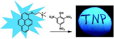
Pyrene derivatives, which are a type of organic semiconductor materials, are well known for their applications in sensing, organic photovoltaic system and supramolecular building blocks.21,22 They feature large π system, high quantum yield, good chemical stability under ambient conditions and long fluorescence lifetime.23 Especially, pyrene derivatives possess high fluorescence efficiency in both solution and film status, which is very favourable for further device applications.4,24 So far, many pyrene-based sensors have been developed for the detection of TNP by harnessing these advantages.25–31 Although good sensitivity and selectivity may be achieved in these sensors, most of them suffered from complicated syntheses and poor water-solubility. In this work, we design and synthesize a very simple pyrene derivative, N,N,N-trimethyl-2-(pyren-1-yloxy)ethanaminium bromide (PyOEA, Scheme 1) for the rapid detection of PA in 100% aqueous media. Moreover, this probe can be conveniently loaded on test papers irradiated with a UV lamp for the field detection of PA. In this strategy, pyrene moiety was functionalized with a quaternary ammonium group to improve its water solubility and the resulting positive charge could promote the interaction between PyOEA and PA which is negatively charged. Simultaneously, π–π stacking and charge transfer interactions would also cooperate with electrostatic interactions, thereby leading to efficient fluorescence quenching of the probe (Scheme 1). This approach affords a rapid detection of PA in 100% aqueous solution with high selectivity and sensitivity. Sensing performance of the probe will be discussed in detail in the following sections.
![]() | ||
Scheme 1 Chemical structure of PyOEA and schematic illustration of the proposed sensing mechanism for the detection of PA. |
Water-soluble PyOEA was prepared by a nucleophilic substitution reaction between 1-hydroxypyrene and 1,2-dibromoethane and then quaternized with trimethylamine (Fig. S1–S4†). It exhibits good solubility in aqueous media and a photoluminescent quantum yield of 37.04% (Fig. S5†). Fig. 1A shows the emission spectra of PyOEA in the absence and presence of PA in HEPES (2-[4-(2-hydroxyethyl)-1-piperazinyl]ethanesulfonic acid) buffer (10 mM, pH 7.4). It was shown that the emission maxima of PyOEA appeared at 381 nm, 402 nm and 425 nm, respectively, corresponding to the fluorescence of pyrene monomer.32 Upon the addition of increasing amounts of PA, the fluorescence intensity gradually decreased. The quenching efficiency reached 80% at the concentration of 30 μM of PA. This prominent fluorescent response substantiated that PyOEA could serve as a promising probe for the detection of PA. To obtain an optimal sensing system toward PA, we investigated the influence of several parameters in the sensing system including pH, buffer species and concentrations. Fig. S6–S8† compared the fluorescence responses of PyOEA in the presence of a series of PA concentrations under different conditions. The results suggested that pH, buffer species and concentrations had little effects on the sensing performance of this probe. HEPES buffer (10 mM, pH 7.4) was chosen for the following studies. The response time of the probe toward PA was also examined in Fig. S9.† It can be seen that the interaction between PyOEA and PA reaches equilibrium within 10 seconds. We measured the fluorescence changes of PyOEA in the presence of different concentrations of PA. As shown in Fig. 1B, there is a good linear relationship between the ratio of fluorescence intensity at 402 nm (I/I0) and the concentration of PA (R = 0.994 from 50 to 400 nM), indicating that this probe can be applied for the quantitative detection of PA within the above range. The limit of detection (LOD) of this sensor for PA in water solution was calculated to be 23.2 nM according to the IUPAC method. As a cationic probe, this result was comparable to most LODs reported in previous methods which were established based on positively charged fluorophores (Table S1†).
To examine the selectivity of PyOEA toward PA, emission spectra of PyOEA in HEPES buffer upon addition of PA, TNT, DNT, methylbenzene (MB), nitrobenzene (NB), 3-nitropropionic acid (NPA), phenol (PHE) and common ions in water samples including SO42−, CO32−, NO3−, K+, Na+, Ca2+ and Mg2+ were recorded under the same conditions. All the results are depicted in Fig. S10,† where the most obvious fluorescence quenching of PyOEA was observed in the presence of PA, while all the other analytes could barely affect the fluorescence of PyOEA. The intensity ratios of (I0/I) of the analytes were shown in Fig. 2A to evaluate the selectivity of the probe. All the values of (I0/I) are less than 1.5 except that of PA which is as high as 50. These results demonstrated the excellent specificity of this method. With a view to understanding the mechanism behind the remarkable fluorescence quenching observed for PyOEA in the presence of PA, the fluorescence lifetimes of PyOEA in the presence of various concentrations of PA were investigated (Fig. S11†). The time-resolved decay of PyOEA in 10 mM HEPES buffer (pH 7.4) showed that in the absence and presence of different concentrations of PA, the lifetime of PyOEA remained almost constant under 340 nm light excitation. These results indicate that the quenching of PyOEA fluorescence is not caused by a dynamic mechanism, but due to the formation of the complex between PyOEA and PA.33
More insight into the mechanism of the fluorescence quenching of PyOEA by PA could be gained by comparison of the emission spectra of PyOEA in the presence of PA, TNT, PHE and NPA, and the structures of the compounds (Fig. 2B). First, the negative result of TNT indicated that electrostatic interactions between the positive charge on the ammonium group of PyOEA and the negative charge on the phenol group of PA would be the primary driving force for the formation of PyOEA/PA. Second, π–π stacking and charge transfer interactions also play key roles in promoting the fluorescence quenching process, because PHE without nitro-groups and NPA without aromatic ring cannot induce the fluorescence responses. To confirm this point, we investigated the fluorescence characteristics of PyOEA in the presence of other nitrophenol species, 4-nitrophenol (NP) and 2,4-dinitrophenol (DNP). Fig. S12† clearly shows that the quenching efficiency increases in the order of NP < DNP < PA, which is in accordance with the number of nitro- groups. This result could be further quantitatively treated with the Stern–Volmer equation and the Stern–Volmer constants (KSV) for PA, DNP and NP were calculated to be 1.16 μM−1, 0.31 μM−1 and 0.087 μM−1, respectively. The value of PA is much higher than that of DNP and NP, validating the key role of the nitro-groups. All these results suggested that PA could bind PyOEA specifically though cooperative non-covalent interactions including electrostatic, π–π stacking and charge transfer interactions and then induce the fluorescence quenching.
The feasibility of this approach for the detection of PA in real-world samples was also investigated. First, we conducted anti-interference experiments by mixing the interferents with PyOEA/PA and examined the fluorescence responses (Fig. S13†). It was found that the sensing performance of PyOEA was hardly affected by the introduction of the interferents. Then, we further employed PyOEA for the detection of PA in tap water. In this test, certain amounts of PA were spiked in water samples and the emission spectra of PyOEA were recorded (Fig. 3). Interestingly, the PA induced spectral responses of the probe in tap water were largely consistent with those in HEPES buffer. Prompted by the favourable performance of this probe in solution, we attempted to fabricate test strips with PyOEA for the convenient monitoring of PA. To this end, filter papers were immersed in PyOEA solution (0.5 mM) for a few seconds and then dried to obtain the fluorescent test strip. Fig. 4A shows the responses of the paper sensor to the presence of PA under the irradiation of a hand-held UV lamp (365 nm). The blank paper strip exhibited bright blue emission upon irradiation, but writing on the paper with PA solution left clear black handwriting discernible to naked eyes due to the quenching of PyOEA fluorescence. To check the visual detection limit of the assay based on paper strips, 20 μL different concentrations of PA were spotted onto these paper sensors. As shown in Fig. 4B, when the concentration of PA increased, the spot turned darker. When [PA] ≤ 0.1 μM, changes of the strip fluorescence cannot be distinguished. Therefore, 1.0 μM was set as the low detection limit for the visual detection of PA. These findings indicated that PyOEA can be integrated in a paper strip-based fluorescent sensor for the field testing of PA with high selectivity and sensitivity.
![]() | ||
Fig. 4 Photographs of the test strips under a UV lamp (irradiation of 365 nm) for PA visual detection by handwriting (A) and dropping PA solutions with different concentrations (mM) as indicated (B). |
Footnotes |
† Electronic supplementary information (ESI) available: Detailed experimental procedures, NMR and additional spectral data. See DOI: 10.1039/c6ra04080b |
‡ These authors contributed equally. |
This journal is © The Royal Society of Chemistry 2016 |