Ji-Hun Seoacd,
Mitsuhi Hiratabd,
Sachiro Kakinokibd,
Tetsuji Yamaokabd and
Nobuhiko Yui*ad
aInstitute of Biomaterials and Bioengineering, Tokyo Medical and Dental University, Tokyo 101-0062, Japan. E-mail: yui.org@tmd.ac.jp
bDepartment of Biomedical Engineering, National Cerebral and Cardiovascular Center Research Institute, Osaka 565-8565, Japan
cDepartment of Materials Science and Engineering, Korea University, Seoul 136-701, Republic of Korea
dJST-CREST, Tokyo 102-0076, Japan
First published on 29th March 2016
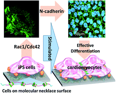
The effect of increasing molecular mobility, on hydrated polyrotaxane (PRX)-coated surfaces, on differentiation of mouse induced pluripotent stem cells (iPS cells) into cardiomyocytes was examined. PRX is composed of α-cyclodextrin (α-CD) threaded on linear poly(ethylene glycol) (PEG)-capped terminals with bulky end-groups. The degree of molecular mobility at the hydrated state (Mf) on the PRX surfaces can be varied by changing the number of threaded α-CDs. Rac1 expression was significantly upregulated for adhering iPS cells on the PRX surface with high Mf value, while it was downregulated on surfaces with low Mf value. Furthermore, the expression of N-cadherin, which is an important marker protein for cardiomyogenic differentiation of stem cells, was greatly upregulated for adhering iPS cells on the PRX surface with high Mf value, while those on surfaces with low Mf value showed low N-cadherin expression. Finally, the PRX surface with higher Mf value was found to be higher in cardiomyogenesis and beating colony formation from iPS cells, the extent of which was much higher than that on gelatin-coated surfaces. This suggests that surface hydrated molecular mobility, varied by varying a supramolecular PRX architecture on materials, plays a significant role in controlling cytoskeletal signaling pathways, eventually contributing to the direction of stem cell commitment.
We have previously demonstrated that polyrotaxane (PRX)-coated surfaces with varying extent of hydrated molecular mobility (molecular factor, Mf), estimated by quartz crystal microbalance-dissipation (QCM-D) measurement, strongly influence the fate of adhering cells. PRX is a molecular necklace-like supermolecule that contains host molecules (e.g. α-cyclodextrin [α-CD]) threaded on a linear guest molecule, e.g. poly(ethylene glycol) [PEG].8 When a determined composition of hydrophobic n-butyl groups and anti-biofouling phosphorylcholine groups were introduced at the end of the PRX segment, the PRX segment could be stably immobilized on the various cell culture dishes due to the moderate hydrophobic interaction induced by the n-butyl groups. Moreover, we have observed that changing the number of threaded α-CDs is effective for the modulation of the Mf value of PRX surfaces, and the morphologies of various cells are greatly affected by the Mf value.9 Narrower and more protruded morphology, with a disrupted actin fiber orientation, was observed for mouse fibroblasts, human umbilical vein endothelial cells, and rat-MSCs cultivated on the PRX surfaces with higher Mf value.10–12 Furthermore, increasing the Mf value on PRX surfaces was found to be favorable for the adipogenesis of rMSCs, while osteogenesis on the PRX surfaces is more pronounced when their Mf value is decreased.13
It is well recognized that the focal adhesion kinase (FAK) of cells phosphorylates α-actinin or p190RhoGEF in the course of binding with the extracellular matrix through integrins and it eventually determines their morphology.14 When FAK tends to phosphorylate α-actinin rather than p190RhoGEF, the expression level of the cytoskeletal signaling pathway, Ras homolog gene family A (RhoA), and the downstream Rho-associated protein kinase (ROCK), which is known as a molecular switch to direct multipotent stem cells for preferential differentiation into hard tissue cells (e.g. osteogenic cells) rather than soft tissue cells (e.g. adipogenic cells), is greatly downregulated.15–18 We have also confirmed that the RhoA-ROCK signaling pathway is significantly downregulated for rMSCs on the PRX surface with high Mf value, which leads rMSCs to preferentially differentiate into adipogenic cells.13 In general, cells with a downregulated RhoA-ROCK signaling pathway are known to easily upregulate another important cytoskeletal signaling pathway, Rac1/Cdc42, accompanied by the formation of a characteristic morphology such as filopodia or lamellipodia.19,20 Recently, it was reported that cells with an upregulated Rac1/Cdc42 expression tend to form a strong cell–cell junction by expressing the specific junction protein, N-cadherin, and that this provides conditions that promote stem cell differentiation into cardiomyocytes.21–23
From this perspective, we further hypothesize that stem cells adhering to the PRX surface with high Mf value may be effectively differentiated into cardiomyocytes, by activating Rac1/Cdc42 with downstream N-cadherin expression. In this study, differentiation characteristics of mouse induced pluripotent stem cells (iPS cells) into cardiomyocytes were evaluated on PRX surfaces with different Mf values.
:
1) mixed solvent using 0.820 mg of AIBN as an initiator. After 24 h, the polymer was precipitated by centrifugation and washed with ethanol, acetone, dimethyl sulfoxide, and acetone to remove residual monomers. The methylation of the PRX block copolymers were conducted as previously reported.10–13
The polymers were dispersed in 5 mL of ethanol and 5 mL of distilled water was added to prepare 0.05 wt% polymer solutions. One hundred microliters of each polymer solution was cast on a glass-bottom dish (p = 27 mm; Iwaki Glass, Tokyo, Japan), dried overnight in a clean box, and used for subsequent applications.
To prepare the gelatin-coated surface, gelatin powder (Life Technologies, Carlsbad, CA, USA) was dissolved in ultrapure water (0.1 wt%), and 200 μL of gelatin solution was placed in contact with a glass-bottomed dish at 37 °C for 30 min. After washing with fresh water, the surface was used for subsequent applications.
α-MHC: F-5′-ACGGTGACCATAAAGGAGGA-3′, R-5′-TGTCCTCGATCTTGTCGAAC-3′;
HCN4: F-5′-CGACAGCGCATCCATGACTA-3′, R-5′-GCTGGAAGACCTCGAAACGC-3′;
TnT2: F-5′-GAAACAGGATCAACGACAACCA-3′, R-5′-CGCCCGGTGACTTTGG-3′;
GATA4: F-5′-AAAACGGAAGCCCAAGAACCT-3′, R-5′-TGCTAGTGGCATTGCTGGAGT-3′;
PAX6: F-5′-TGCCCTTCCATCTTTGCTTG-3′, R-5′-TCTGCCCGTTCAACATCCTTAG-3′;
NeuroD1: F-5′-ATGACCAAATCGTACAGCGAG-3′, R-5′-GTTCATGGCTTCGAGGTCGT-3′;
EOMES: F-5′-CACCGCCACCAAACTGAGAT-3′, R-5′-CGAACACATTGTAGTGGGCAG-3′;
Fibronectin: F-5′-ACCGAAGCCGGGAAGAGCAA-3′, R-5′-GGTCCGTTCCCACTGCTGAT TTATC-3′;
Plastin3: F-5′-CCTTCCGTAACTGGATGAACTC-3′, R-5′-GGATGCTTCCCTAATTCAACAG-3′;
GAPDH: F-5′-TGCGACTTCAACAGCAACTC-3′, R-5′-CTTGCTCAGTGTCCTTGCTG-3′.
The collected active proteins (10 μL) were mixed with 10 μL Laemmli Buffer (161-0737, Bio-Rad, Richmond, CA, USA) containing 2.5% 2-mercaptoethanol and boiled at 98 °C for 5 min. The samples were loaded onto 4–20% gradient acrylamide gel (Mini-Protean TGX, Bio-Rad) and electrophoresis was conducted for 30 min at 200 V. The samples were then transferred to PVDF membrane (170-4156, Bio-Rad) using the Trans-Blot Turbo Transfer System (Bio-Rad), and incubated in blocking solution (5% skim milk in TBST, 20 mM Tris, 0.5 M NaCl, 0.05% Tween-20, pH 7.5) at room temperature for 1 h. Furthermore, the blocking solution was exchanged with the primary antibody solution (included in the assay kit, diluted to 1/1000 in TBST blocking solution) and allowed to react overnight at 4 °C. The membrane was then washed with fresh TBST for 5 min, three times. The membrane was then brought into contact with HRP-conjugated goat anti-rabbit antibody (7074, Cell Signaling Technology) solution (1/1000 diluted in 5% skim milk TBST) and allowed to react at room temperature for 90 min. After being washed with fresh TBST, the protein band was detected by ImageQuant LAS 500 (GE Healthcare, Uppsala, Sweden), using the ECL Select™ Western Blotting Detection System (GE Healthcare).
| PRX block copolymers | MPC (mol% in PMB, 1H NMR) | nBMA (mol% in PMB, 1H NMR) | Number of α-CD (/PEG chain, 1H NMR)a | Methylation (%, 1H NMR) | OMe (wt%, 1H NMR) | Viscoelasticity (Mf, ×10−6) |
|---|---|---|---|---|---|---|
a The number of α-CDs per PEG (number average molecular weight = 20 000). |
||||||
| PRX-A | 12.0 | 88.0 | 12 | >90 | 6.1 | 0.95 |
| PRX-B | 24.6 | 75.4 | 104 | 61.1 | 13.5 | 0.60 |
| Random copolymers | MPC (mol%, 1H NMR) | nBMA (mol%, 1H NMR) | HEMA (mol%, 1H NMR) | MEA (mol%, 1H NMR) | OMe (wt%, 1H NMR) | Viscoelasticity (Mf, ×10−6) |
|---|---|---|---|---|---|---|
| Random | 6.60 | 33.1 | 10.6 | 49.7 | 10.6 | 0.16 |
The optical microscopy images and the following Nanog-GFP images of iPS cells cultured in non-differentiation medium were taken after 8 days incubation. Spheroid aggregates of iPS cells with strong Nanog-GFP expression were clearly observed on the feeder cells (SNL cells) and the PRX-A surface for 8 days, while spread morphologies with weak Nanog-GFP expression were observed on the other surfaces (Fig. 2).
To confirm the degree of cell–cell junction for spheroid aggregation, we analyzed the expression level of E- and N-cadherins in adhering iPS cells by enzyme-linked immunosorbent assay (ELISA). No significant E- and N-cadherin expression was observed for iPS cells on SNL cells. In contrast, E- and N-cadherin expression was significantly upregulated on bare glass and polymer-coated surfaces. Characteristically, N-cadherin expression was significantly upregulated on the PRX-A surface, compared to the other polymer surfaces. The representative gene expression in the germ layer was analyzed by real-time PCR after 8 days in a non-differentiated state. No significant marker gene expression was observed for iPS cells cultured on the SNL feeder cells, while GATA4 (cardiac mesoderm) and fibronectin (mesenchymal marker gene) expression was strongly observed on the bare glass or polymer-coated surfaces.
The morphology of adhering iPS cells and the following GFP-Nanog fluorescence images in both non-differentiating and cardiomyogenic differentiation mediums after 3 days of cultivation were shown in Fig. 3. Herein, the gelatin-coated surface was chosen as a positive control for the cardiomyogenic differentiation of iPS cells, because gelatin has been known as an effective surface for directing iPS cells towards cardiomyogenic differentiation.24 With the exception of cells adhering to the gelatin-coated surface, cells on most of the surfaces showed a certain level of Nanog-GFP expression during the cultivation in non-differentiating medium, and this expression almost disappeared when they were cultivated in a cardiomyogenic differentiation medium.
To confirm the expression level of the cytoskeletal signaling pathway in differentiated cells, immunostaining and immunoblotting was conducted. Fig. 4A shows the images of confocal laser microscopy taken after immunostaining of Rac1 after 3 days-post differentiation. The cells on the PRX-A and gelatin-coated surfaces showed positively stained Rac1/Cdc42 proteins. The expression level of active Rac1 and other cytoskeletal signaling pathways, i.e., active RhoA-ROCK, was analyzed by immunoblotting (Fig. 4B). The expression of active-Rac1 was the highest in the cells adhering onto the PRX-A surface, while the active RhoA-ROCK pathway was significantly downregulated. The expression of active-Rac1 became moderate when PRX was changed to that of lower Mf value, but the level of RhoA-ROCK expression was slightly increased. When the surface was changed to the random copolymer, which did not contain the PRX segment, the expression of active Rac1 was almost negligible, with a slight increase in active RhoA-ROCK expression.
To confirm whether the upregulated state of Rac1 expression (initial cell–materials interaction) affects intercellular interaction (later cellular responses), E- and N-cadherin expression was quantitatively analyzed by ELISA, after 8 and 17 days post-differentiation (Fig. 5). The expression level of E-cadherin in differentiated cells was decreased when the culturing time was increased from 8 days to 17 days, except in cells on the gelatin-coated surface. The expression levels of N-cadherin after 8 days were not significantly different from those on the sample surfaces. However, a significant increase (∼2 fold) in N-cadherin expression was observed only for the cells on the PRX-A and gelatin-coated surfaces, 17 days post-differentiation. As the decrease in E-cadherin and increase in N-cadherin may represent characteristics of cardiomyogenic differentiation lineage or tumor cell formation, the gene expression of a representative marker protein of mesenchymal transition (fibronectin) and tumor cell formation (plastin3) was estimated. Significant gene expression of fibronectin was observed for the cells on all sample surfaces, whereas no significant expression of plastin3 was observed.
Further onto the ELISA analysis, we estimated the distribution of N-cadherin on differentiated cells by immunostaining, and the resulting confocal laser microscopy images are shown in Fig. 6. After 3 days post-differentiation, no significant N-cadherin expression was observed for differentiated cells on any of the sample surfaces. After 8 days, the fluorescence intensity of the secondary antibody began to increase on all sample surfaces. However, the distribution of N-cadherin was not specifically localized in cells. After 17 days, N-cadherin-mediating cell–cell junction was clearly observed, only in the cells on the PRX-A and gelatin-coated surfaces.
![]() | ||
| Fig. 6 Confocal laser microscopy images of differentiated cells after 3, 8, and 17 days, respectively. Blue: nucleus, green: N-cadherin (bar = 50 μm). | ||
The appearance of a beating colony during cardiomyogenic differentiation was monitored by optical microscopy. Fig. 7 shows the optical microscopy images of cultured cells taken at 8 days post-differentiation and the fluorescence image of the Quest-Rhod4™ assay for Ca2+ staining on the beating colonies. The characteristic cell aggregation, i.e., large areas of crowded cell aggregates (the white region in the images), was observed on the PRX-A and gelatin-coated surfaces and the beating colonies appeared only on these two surfaces. In contrast, the size of the cell aggregates was much smaller and the beating colony did not appear on the other sample surfaces. When the Quest-Rhod4™ assay was conducted for staining the Ca2+ generated as a part of the beating process, the beating area was more clearly observed on the PRX-A and gelatin-coated surfaces.
The number of beating colonies per culturing well was monitored over 17 days and the result is shown in Fig. 8. On the PRX-A surface, the number of beating colonies significantly increased over 17 days and a large area of beating colonies was formed. However, the beating colonies were almost saturated at the early stages of differentiation on the gelatin-coated surface and the beating area also significantly decreased with culturing time.
The specific gene expression of cardiomyocytes was analyzed by quantitative real-time PCR (Fig. 9). The overall expression level of specific genes in cardiomyocytes (HCN4, TNT2, α-MHC, GATA4) on the PRX-A surface gradually increased as differentiation progressed and the levels were the highest at 17 days post-differentiation. In the case of differentiated cells on the gelatin-coated surface, gene expression specific for cardiomyocytes was heterogeneously observed after 17 days. Although several marker genes, such as α-MHC and HCN4, were highly expressed during differentiation, important marker genes for cardiomyocyte maturation, i.e., GATA4 (cardiac progenitor) and TNT-2 (cardiomyocyte), were not significantly expressed. Moreover, the expression levels were saturated at 12 days and gradually decreased with the differentiation time.
PRX block copolymer surfaces containing a larger number of threading α-CDs (PRX-B) showed much lower viscoelasticity in the hydrated state than that of PRX-A in the hydrated state when the viscoelasticity was measured using QCM-D.10–13 Because surface hydrophilicity was not significantly different between PRX-A and PRX-B, this result was considered to be due to the difference in dynamic properties of different numbers of threading α-CDs in PRX surfaces.10 The present study clearly demonstrates a continuous increase in the number of beating colonies (Fig. 8) and homogeneous expression of marker genes for cardiomyocytes on the PRX-A surface (Fig. 9). This finding strongly suggests the effectiveness of increasing hydrated molecular mobility of PRX surfaces for differentiating cells into cardiomyocytes.
Stem cell lineage commitment induced by mechanotransduction has been widely studied over the last decade.25,26 The ECM formed on different types of materials generally induce different integrin-mediated intracellular signaling pathways. Transcriptional activators YAP/TAZ then translate different types of physical information into different expression levels of messenger RNA for determining stem cell fate.27,28 The relationship between the cytoskeletal signaling pathway and the formation of cell–cell junctions is considered a critical issue, as the formation of a beating colony by cardiomyocytes requires strong cell–cell junctions, mediated by N-cadherin.29 It was reported that if the intercellular interaction is enhanced by increasing the expression level of cell junction proteins, such as E- or N-cadherin, non-differentiated iPS cells tend to form spheroid aggregations with increased pluripotency.30,31 Interestingly, iPS cells on the PRX-A surface with high Mf value showed spheroid aggregation with strongly increased N-cadherin expression, in contrast to embryonic spheroid aggregation with negligible expression level of N-cadherin on SNL cells. The maintenance of stemness and embryonic spheroid formation is dependent on the expression level of cytoskeletal proteins such as cadherins and catenins. In the present study, slight differences in Nanog-GFP expression and the shapes of spheroid aggregation among the polymer surfaces are probably due to the different expression levels of the cytoskeletal signaling pathway resulting in different expression levels of cadherin.32
As cells on the PRX-A surface have been found to significantly downregulate the RhoA-ROCK signaling pathway,13 we may hypothesize that the downregulated state of RhoA-ROCK on the PRX-A surface is an important condition for the formation of a cell–cell junction by expressing N-cadherin. It has been reported that the Rac1/Cdc42 signaling pathway is easily upregulated for characteristic cell morphologies, such as lamellipodia and filopodia, when cells encounter conditions in which RhoA-ROCK is downregulated.19,20 It has also been reported that N-cadherin-mediated cell–cell junction development, which is an essential condition for the formation of cardiac tissue, was more easily observed for Rac1/Cdc42 upregulated cells.21–23 Based on these findings, it is hypothesized that the PRX-A surface may provide an optimized condition for iPS cells to express N-cadherin by activating the Rac1/Cdc42 signaling pathway, which would lead iPS cells to preferentially differentiate into cardiomyocytes. The activation of Rac1, and the subsequent increased expression of N-cadherin, was successfully observed on the PRX-A surface (Fig. 4B and 5B). Interestingly, a decrease in E-cadherin and an increase in N-cadherin expression were observed only for adhering iPS cells on the PRX-A surface. When embryonic stem cells are stimulated into mesodermal lineage, e.g., during cardiomyogenesis, several transition steps are required such as the epithelial-mesenchymal transition (EMT) step. An exchange in cadherin, i.e., a decrease in E-cadherin and an increase in N-cadherin expression, is a critical step for this transition process.33 The characteristic N-cadherin expression, with decreased E-cadherin expression on the PRX-A surface, likely indicates that iPS cells are in the position for a successful transition towards cardiomyogenic differentiation. It has also been reported that N-cadherin expression is upregulated when embryonic stem cells are differentiated into tumor-like cells.34,35 Because no significant expression of plastin3 was observed for iPS cells on any of the sample surfaces, it is suggested that the PRX-A surface is an effective surface for directing the differentiation of iPS cells into cardiomyocytes. On the gelatin-coated surface, the rapid appearance of the beating colony was characteristic; however, the expression of marker genes and cadherin exchange were not observed to a significant extent. The rapid passing through of the cardiac progenitor states on the gelatin surface possibly result in decreased gene expression compared to that of PRX-A. Although the underlying reason is not yet clearly understood, incomplete cadherin exchange (i.e. continuous strong E-cadherin expression, as shown in Fig. 5) during differentiation may suggest the possibility of incomplete transition of stem cells into cardiac mesoderm, which is an essential step in the differentiation into cardiac progenitor cells and finalized cardiomyocytes, on the gelatin-coated surface. As gelatin can induce strong surface–cell interaction at the embryonic stem cell stage, we hypothesize that this acts as an obstacle to the completion of the EMT step for differentiation into cardiomyocytes. In any event, it is confirmed that the PRX-A surface with high Mf value is very effective for directing iPS cells into cardiomyocytes, by controlling cell–material interactions and downstream formation of cell–cell junctions.
| This journal is © The Royal Society of Chemistry 2016 |