
Clay nanocomposites as engineered drug delivery systems
Maryam Jafarbeglou
 a,
Majid Abdouss
*b,
Ahmad Mousavi Shoushtari
c and
Majid Jafarbeglou
d
a,
Majid Abdouss
*b,
Ahmad Mousavi Shoushtari
c and
Majid Jafarbeglou
d
aDepartment of Nanotechnology, Amirkabir University of Technology (Tehran Polytechnic), 424 Hafez Ave., Tehran, Iran. E-mail: jafarbeglou@aut.ac.ir
bDepartment of Chemistry, Amirkabir University of Technology (Tehran Polytechnic), 424 Hafez Ave., Tehran, Iran. E-mail: phdabdouss44@aut.ac.ir
cDepartment of Textile Engineering, Amirkabir University of Technology (Tehran Polytechnic), 424 Hafez Ave., Tehran, Iran. E-mail: amousavi@aut.ac.ir
dDepartment of Pathobiology, Faculty of Veterinary Medicine, Islamic Azad University, Karaj Branch, Karaj, Iran. E-mail: m.jafarbeglou@kiau.ac.ir
First published on 5th May 2016
Abstract
Clays belong to a category of silica layered ingredients that are commonly used in the pharmaceutical industry, either as ingredients or in combination in composites and hybrids. The specific structure of clay provides effective drug delivery systems with the ability to interact with drugs and to control release. Numerous research articles have been published in this respect and valuable results reported, mostly for oral routes. However, overview and discussion of the recent and rapidly growing research along with their theoretical principles are required for understanding and exploring the great potential of this method. To further this aim, we have provided an illustrated and comprehensive study from the point of view of a nanotechnologist, interested in investigating clay aggregate effects at the nano and meso scales for the development of nano-composites and hybrids accomplished with clay. In the first stage, a review of the molecular structure and physicochemical properties of clay are discussed and then we continue with a survey of clay and nanoclay origins, preparation methods, modification and incorporation as nano-hybrids, nano-composites, and nano and microfibers. Also their effects on drug overdose toxicity and on the mechanism of drug loading and release are discussed. Finally, this review summarizes further expansions of their progressive usage in biopharmaceutical applications with the help of integration of release methods and engineering of nanostructured clays in polymeric nanocomposites.
 Maryam Jafarbeglou | Maryam Jafarbeglou is a PhD candidate of Nanotechnology in Amirkabir University of Technology (Tehran Polytechnic) and studying on polymer–clay nanocomposites. She led Nanokurt research group funded by Kurt Co. foundation grant on nanocompounds. Her last studies and interest was mostly nanomaterial applications in biomedical and biosensor fields. She has been working in industries related to petrochemicals and polymers. |
 Majid Abdouss | Majid Abdouss is a Professor of Chemistry and Eminent Endowed Chair on Nanomedicine in the Nanotechnology Institute at Amirkabir University of Technology. Majid Abdouss is among the pioneer researchers on drug delivery and nanomedicine, catalyst, environment, sustainable development and green industries. He has published several books and papers in organic chemistry, drug delivery, catalyst, solar cells, and mineral chemistry. His research publications have been cited more than 1200 times. |
 Ahmad Mousavi Shoushtari | Ahmad Mousavi Shoushtari is Associate Professor of Textile Engineering at Amirkabir University of Technology and holds the Chair in Nanotechnology Institute. He has published several books and papers about textile, polymers, nanotechnology and environment. He has studied nanocomposites, nanofibers, porous material, minerals, surface phenomena, sustainable technologies and aerogels. |
 Majid Jafarbeglou | Majid Jafarbeglou is a DVM intern at the Faculty of Veterinary Medicine in Islamic Azad University, Karaj Branch. He gained the first score of national pre-internship exam in 2015 and passed pre-clinical courses with high grades. He is the head of exotic and wild animal medicine (Zoological Medicine) in the workgroup of Scientific Community Faculty. He is also interested in pharmacology and pathology. |
1. Introduction
Controlled release systems have been developed to improve the temporal and spatial presentation of drugs in the body, to protect drugs from physiological degradation or elimination, to improve patient compliance, and to enhance quality control in the manufacturing of drug products. For designing controlled release systems, it is important to identify and understand the specific mechanisms involved in the release process. Often, more than one mechanism is involved at a given time or different mechanisms may dominate at different stages of the drug delivery process.1 Conventional release dosage forms are known to provide an immediate release of the drug, with little or no control over the release rate. In order to reach and maintain plasmatic concentrations, therapeutically effective dosage control is necessary, to avoid significant fluctuations in the plasmatic levels. The use of conventional drugs may lead to fluctuations, so that the drug level in the organism may reach levels lower than the minimum effective concentration (MEC), or exceed the maximum toxic concentration (MTC), resulting in undesirable side effects, or the lack of therapeutic benefits intended for the patient. The use of materials that modify the release of drugs helps in reducing the undesirable levels of these fluctuations, decreasing the side effects and/or enhancing the drug's therapeutic effect, increasing the patient's compliance with the treatment, as well as adding commercial value to medicines sold, by the extension of the patient protection.2–4 Clays are commonly used materials in the pharmaceutical industry, either as net ingredients or as activated ingredients.5 It was observed that when they are administered simultaneously, they may interact with drugs, reducing their absorption. Therefore, such interactions can be used to achieve technological and biopharmaceutical advantages, regarding the control of release.6 Swelling clay minerals, such as montmorillonites (MMT), are included in this category and they produce a homogeneous dispersion of hydrated nanocrystals in pure water. Clay minerals are composed of alternating negatively charged aluminosilicate layers, and exchangeable cations lie between the layers in order to compensate for the ionic balances.7 In general, most of the clay–drug complexes are intercalation complexes with cationic or relatively polar substances accompanied by the expansion of their basic spaces. Nonpolar hydrophobic drugs are also incorporated into nanosized pores in the amorphous state without formation of intercalation complexes.8,9 However, colloidal dispersions of clay particles tend to flocculate and precipitate in ion-containing solutions. Under physiological conditions, clay dispersions are unstable due to the high salt concentration and the presence of poly-electrolytes like proteins. Dispersion stability is an important factor for drug carriers because it plays a key role in absorption and bioavailability. Thus, clays are not relevant for practical pharmaceutical uses in spite of their beneficial properties and several researchers have studied this aspect.10–13 However, nonionic or cationic nanoclays can be applied as delivery carriers, depending on the charge of the intercalated molecules, fundamentally because of their unique layered structure.Bioactive molecules are stabilized into the interlayer spaces through electrostatic interaction and this phenomenon provides controlled release ability, by exchanging the bioactive molecules with other ions if enough exist in the biological environment.14 The net effect of the attractive and repulsive forces (DLVO) between two clay particles is attractive in dilute solution. The two particles will tend to move toward each other and become attached-flocculate. With control of some parameters, like electrolyte concentration and pH that affect the attractive force, the clay layers tend to move away.15
Among the several approaches proposed to achieve controlled release formulations, the ion exchange process has received considerable attention from researchers and it may take place by mixing solid substrates (namely ion exchangers) with ionic drugs in solution. In biological fluids, “counter-ions” can displace the drug from the substrate and deliver it into the body. The exchanger may then be eliminated or biodegraded (Fig. 1). Clay minerals are naturally inorganic cationic exchangers and so they may undergo ion exchange with basic drugs in solution.16
 | ||
| Fig. 1 Idealization of clay–drug complexation and in vivo drug release mechanisms (clay mineral surface charge (−); compensating cations (a+); cationic drug (X+); drug associated anions (Y−); in vivo counter ions (A+); anions associated with the counter ions (B−)) (redrawn from ref. 16). | ||
Isolated clay particles suspended in a liquid, where the particles have identical size, have defined charges. The electro-kinetic potential is highest at the surface of the particles and decreases as the distance from the surface increases in the suspending liquid. The zeta potential, or electrostatic potential, is generated by the accumulation of ions at the surface of the clay colloidal particle, which is surrounded by an electrical double layer (the Stern layer and the diffuse layer) (Fig. 2).
 | ||
| Fig. 2 Schematic presentation of clay particle in a fluid, surrounding double layer (Stern layer and diffuse layer), surface potential and zeta potential with respect to distance from the particle surface (redrawn from ref. 17). | ||
Clay minerals are widely used in drug production both as fillers and as active agents. Mostly drug–clay interactions have been observed and studied, but were not considered as a possible mechanism to modify drug release. In recent years, based on their high reservation capacities as well as their swelling and colloidal properties, clays have been taken into consideration as very useful materials for modulating drug delivery.16,17
Because of the importance of the issue, this review attempts to provide a comprehensive explanation of the following aspects: firstly, we present an overview of related clay based materials by explaining (1) chemical components of clays and their physicochemical properties as drug carriers; (2) synthesis routes of clay nanocomposites; (3) the roles of clay in drug purposes along with its advantages; (4) incorporation of clays in polymer composite forms and their biocompatibility aspects; secondly, we discuss a collection of the findings from very recent investigations into the abilities of clay as drug carriers in composites, hybrids and fibrous forms. Thus, this article tries to help to develop a drug delivery system (DDS) based on engineering nanoscale properties of clay in polymeric nanocomposite forms.
2. Clay types and properties
Clay minerals are classified into kaolinite, illite, smectite (montmorillonite), chlorite, haloysite and the vermiculite group. The most important commercial clays are kaolinite and montmorillonite.15 Kaoline or kaolinite, known as China clay, has the basic chemical formula Si2Al2O5(OH)4. Montmorillonite is AlSi2O5(OH)·xH2O. Comparing the structure of kaoline and montmorillonite, kaoline has 1![[thin space (1/6-em)]](https://www.rsc.org/images/entities/char_2009.gif) :1 layer lattice and montmorillonite has 2:1 lattice structure. Montmorillonite is similar but different from kaolinite in that the silicate surface of montmorillonite exhibits a negative charge, and these surfaces adsorb cations such as Na+, or Ca2+. It was found that montmorillonite clay can absorb large amounts of water and polar liquids, which separate the silicate layers. Indeed, montmorillonite can absorb 20 to 30 times (or more) of its volume in water. Also polar polymers can be absorbed into montmorillonite. This can result in the break-up of the layered structure, effectively, dispersing it and this ability is related to polymer properties.17–21
:1 layer lattice and montmorillonite has 2:1 lattice structure. Montmorillonite is similar but different from kaolinite in that the silicate surface of montmorillonite exhibits a negative charge, and these surfaces adsorb cations such as Na+, or Ca2+. It was found that montmorillonite clay can absorb large amounts of water and polar liquids, which separate the silicate layers. Indeed, montmorillonite can absorb 20 to 30 times (or more) of its volume in water. Also polar polymers can be absorbed into montmorillonite. This can result in the break-up of the layered structure, effectively, dispersing it and this ability is related to polymer properties.17–21
Despite the occurrence of clay minerals in nature, they may be synthesized due to economic and/or technical advantages. Highlighted among the synthesized clays are the lamellar double hydroxides (LDHs), also known as anionic clays or “hydrotalcite-like”, and cylindrical clays or “halloysite”. LDHs are layered solids, with positively charged layers and charge-balancing anions in the interlamellar space. Synthetic LDHs can also be used as host compounds because of the positively charged layers and the presence of interlayer anions. These compounds are especially helpful for retention of negatively charged biomolecules.13,16 They are soluble at pH <4 and so may rapidly release the drug in a molecular form appropriate for absorption.6,9 Due to these properties, these compounds have been used in delivery systems for drugs, being active in increasing the solubility, the controlled release and the gastro protection of oral drugs, especially as anti-inflammatory agents (Table 1).
| Type | Function | Drug | Ref. | |
|---|---|---|---|---|
| Hydrotalcite-like | Modified drug release | Iboprofen | 22 | |
| Halloysite clay nanotubules | Modified drug release | Tetracycline HCl and khellin | 23 | |
| Hydrotalcite-like | Modified drug release | Diclofenac | 24 | |
| Hydrotalcite-like | Increase solubility | Indomethacin | 25 | |
| Mg–Al layered double hydroxides | Reduce ulcer damage | Indomethacin | 26 | |
| Mg–Al layered double hydroxides | Modified drug release | Diclofenac | 27 | |
| Brucite | Modified drug release | Fenbufen | 28 | |
| Mg–Al layered double hydroxides | Thermal stability | Naproxen | 29 | |
| Commercial hydrotalcite | Increase skin permeation | Diclofenac, indomethacin and iboprofen | 30 | |
| Montmorillonite & MCM (mesoporos) | Modified drug release | Sertraline | 31 | |
| Halloysite clay nanotubules | Solubility, sustain release | Dexamethasone, furosemide and nifedipine | 32 | |
| Hydrotalcite | Modified drug release | Diclofenac | 33 | |
| Mg–Al layered double hydroxides | Modified drug release | Mefenamic and meclofenamic acid | 34 | |
| Halloysite clay nanotubules | Modified release | Benzotriazole (protecting agent) | 35 | |
| MgAlFe layered double hydroxides | Modified drug release | Mefenamic acid, meclofenamic acid and naproxen | 36 | |
| Montmorillonite | Modified drug release | Timolol maleate | 37 | |
| Montmorillonite and saponite | Drug reservoirs | Nicotine | 38 | |
| MgAl, MgAlFe layered double hydroxides | Modified drug release | Fenbufen | 39 | |
| Montmorillonite | Modified drug release | Tramadol hydrochloride | 40 | |
| Hydrotalcite-like | Modified drug release | Paracetamol | 41 | |
| Bentonite (Unye) | Modified drug release | Naproxen | 42 | |
| Montmorillonite | Modified drug release | Aminoglutethimide, Irinotecan and carboplatin | 43 | |
| Layered double hydroxides (LDHs) | Modified drug release | Plethora of bio-molecules | 44 | |
| Montmorillonite | Modified drug release | Triamcinolone acetonide (TA) | 45 | |
| Zeolite and hydrotalcite | Prolong drug release | Theophylline | 46 | |
| Hydrotalcite | Prolong drug release | Diclofenac | 47 |
Various polar polymers have been used to produce clay nanocomposites. The important factor in achieving exfoliated and homogenous dispersion of the layers is the intercalation capability of polymers between the layers.17–27
Owing to the nanometer-size particles obtained by dispersion, these nanocomposites exhibit markedly improved mechanical, thermal, physico-chemical and rheological properties (Fig. 3) when compared with the pure polymer or conventional (microscale) composites.
 | ||
| Fig. 3 The most important properties of nanosized porous clay materials. | ||
3. Clay nanocomposite preparation
Depending on the nature of the components used (layered silicate, organic cation and polymer matrix) and the method of preparation, three main types of composites may be obtained with association of polymer in clay.48–51 Fabrication of polymer/clay nanocomposites will be done in 3 stages. The first stage involves generation of a tactoid – the polymer closes around the organic clay agglomerates. Aqueous dispersions of montmorillonite contain small clusters of clay platelets, often named “tactoids”.52 The second stage (intercalation) involves penetration of the polymer into the interlayer space of the organic clay, causing the layers to move apart by 2–3 nm.53 The final stage involves exfoliation as shown in Fig. 4. There are four main routes for the synthesis of clay nanocomposites: (a) template synthesis, (b) intercalation of polymer or pre-polymer from solution, (c) in situ intercalative polymerization and (d) melt intercalation.53,54 | ||
| Fig. 4 Different types of composite arising from the interaction of clays and polymers: (a) phase-separated microcomposite; (b) intercalated nanocomposite and (c) exfoliated nanocomposite. | ||
3.1. Exfoliation-adsorption
The layered silicate is exfoliated into single layers using a solvent in which the polymer (or a pre-polymer in the case of insoluble polymers such as polyimide) is soluble. This technique has been widely used with water-soluble polymers to produce intercalated nanocomposites based on poly(vinyl alcohol), poly(ethylene oxide), poly(vinylpyrrolidone) or poly(acrylic acid). It is known that such layered silicates, owing to the weak forces that stack the layers together, can be easily dispersed in an adequate solvent.543.2. Melt intercalation
The layered silicate is mixed with the polymer matrix in the molten state. Under this condition, if the layer surfaces are sufficiently compatible with the chosen polymer, the polymer can crawl along the interlayer space and form either an intercalated or an exfoliated nanocomposite.53 In this technique, no solvent is required and so it may be preferred for avoiding the solvent effect on drugs;54 but melt processing in non-polar polymers often leads to an insufficient number of nanoparticles in dispersion, and to aggregation and intercalation, especially with a high content of nanoparticles.55,56 This method seems to be useful for heat resist drugs that can withstand high temperatures.3.3. Template synthesis
This technique reported for preparing layered silicate-based nanocomposites implies the in situ hydrothermal crystallization of the clay layers (hectorite) in an aqueous polymer gel medium where the polymers often act as a template for the formation of layers. This method is particularly adapted to water soluble polymers, and some attempts have been achieved with polymers such as poly vinylpyrrolidone, hydroxypropylmethylcellulose, polyacrylonitrile, poly(dimethyldiallylammonium) (PDDA) and poly(aniline) (PANI).57 It is believed that polymer incorporation within the growing layers in this method is limited to the strict balance between negative charges of the clay layers and cation creation by the polymer chains. It should be noted that the distance between the layers obtained by this method cannot compete with natural layered silicates for kinetic reasons. Also, under the best conditions, their average length is limited to about one-third of similar natural kinds.533.4. In situ intercalative polymerization
In this method, the layered silicate is swollen with a monomer solution so that the polymer formation occurs in the intercalated sheets. Polymerization can be initiated by heat, radiation, diffusion of a suitable initiator or an organic initiator or catalyst fixed through cationic exchange inside the interlayer before the swelling step by the monomer. By the way, the disadvantages of the solution and melting processes can be solved by in situ polymerization.544. Clay nanocomposites in drug delivery and regenerative medicine
4.1. Polymer based nanocomposites
Many attempts to obtain polymer–clay nanocomposites from silicate gels with in situ synthesis have been made.55 In this respect, more researchers of polymer layered silicate nanocomposites have studied: preparation, properties and uses of the new class of materials.17,48,53,58Synthesis of these novel nanohybrid materials by the incorporation of 2-D nanoparticles results in improved physical properties, notably biocompatibility and biodegradation, which are the main requirements for tissue engineering and drug delivery applications.59 Molecular level self-assembly ranges from nanoscale molecular sheet-type organization60–62 to observable microscale composites.63,64 The nature of the self-assembly can be tuned either by performing the polymerization procedure in the presence of organically modified nanoclay or by varying the spacer chain length.
Nowadays, the polymeric systems are of great interest as drug carriers. These systems are appropriate tools for time and distribution-controlled drug delivery.45,55,65,66 The mechanisms involved in controlled release require polymers with a variety of physicochemical properties. Thus, several types of polymers have been tested as potential drug delivery systems, including nano- and micro-particles, dendrimers, nano- and micro-spheres, capsosomes and niosomes.44,55,59,66–71 In all of these systems, drugs can be encapsulated or conjugated in polymer matrices.59 These polymeric systems have been used for a range of treatments for antineoplastic activity, bacterial infections and inflammatory processes, in addition to vaccines.59,60
Metronidazole-loaded halloysite nanotubes incorporated in poly(caprolactone)/gelatin microfibers allowed for extended release of the drugs over 20 days, compared to 4 days when directly admixed into the microfibers.63
Tunable controlled release of active agents through synthesis of artificial nano-caps at the tube endings and halloysite lumen enlargement by selective etching allowed for tubular nanocontainers with chemical release times from 10 to 200 h and a loading capacity of ca. 30 wt%. Halloysite is well mixable with polymers of high and medium polarities without any surface modification.65
In another study, ibuprofen as a poor water soluble drug has shown reduced release properties when added to PEG (polyethylene glycol) using hot melt extrusion. According to differential scanning calorimetry studies, the crystallinity of polyethylene glycol was significantly reduced, as a consequence of the large surface area of clay platelets that physically hindered polymer chain motion by connecting to the PEG via hydrogen bonds. The addition of layered silicate retarded the release of ibuprofen from the PEG matrix, even though the crystalline content of the PEG was reduced.55
A hybrid drug carrier has also been developed using porous nanocrystals of a swelling clay mineral conjugated with a block copolymer containing poly(ethylene glycol) and polyamine segments. The PEG–clay hybrid acted as a carrier for sustained release of hydrophobic substances due to the high affinity (K = 1.52 × 10⁴) between the drug and the clay surface.11
In other work, the intercalation of polyamide loaded 1,3,4-oxa(thia)diazoles into MMT was studied. The intercalation was achieved through an ion exchange process between sodium cations in Na-MMT and amine hydrochloride attached to the polyamides. The release measurements indicate that polyamide–MMT can be used for the sustained release of 1,3,4-oxa(thia)diazoles in oral administration. The polyamides showed good or moderate antimicrobial activities. Although in vitro studies showed no antimicrobial effect, in vivo studies suggest that nanocomposites offer good antimicrobial activity.72
New nano hybrid materials based on a combination of montmorillonite and pH sensitive positive charges polymer for encapsulating drugs to produce inorganic–organic nanocomposite are able to act as effective drug delivery systems (DDS). In this approach, the ionic liquid monomers are intercalated into montmorillonite and subsequently copolymerized with methacrylic acid.73 The drug release profile of the montmorillonite–drug hybrid is faster than the release in the case of the drug–clay–polymer nanocomposite in both gastric (pH 1.2) and intestinal fluids (pH 7.4).74 In other work, layered double hydroxides (LDH) and other pH-sensitive polymers, such as carboxymethyl cellulose (CMC), were combined. The shrinkage of CMC at acidic pH slows the drug release from CMC/LDH–drug nanocomposite beads.75
Biodegradable polyurethane was synthesized by preparing an aqueous polyurethane dispersion having poly(caprolactone) and poly(ethylene glycol) as soft segments. The results of this study suggested that triamcinolone acetonide and clay do not dramatically change the morphology phase of the polymer, although they can interact with each other.45
In other attempts nano-composite films of two biodegradable and biocompatible polymers, polyvinyl alcohol (PVA) blended chitosan and nano-clay montmorillonite (MMT) particles, were prepared and used for drug delivery systems.56
Recently, the synthesis of some kinds of polyurethanes has been achieved using aliphatic chain extenders of varying chain lengths. Nanohybrids of polyurethanes have been prepared by dispersing a 2-D nanoclay in poly-ol followed by re-polymerization and subsequent chain extension using different chain extenders. The nanostructure and dispersion of the nanoclay confirm the relative interactions among various polyurethanes. A systematic enhancement in toughness has been observed for polyurethanes with longer chains and has been explained in terms of stronger hydrogen bonding occurring for longer aliphatic chains of the chain extenders. Bottom-up self-assembly has been revealed from the molecular level to the micron-scale crystallites observed in optical images via SANS (cluster size ∼ 20 nm) and band morphology with dimensions of ∼60 nm was observed by AFM. The cluster size broadens with increasing aliphatic chain length, and was further enhanced in nanohybrids in the presence of nanoclay. Controlled biodegradation of pristine polyurethanes and their nanohybrids has been investigated in enzymatic media. The hemocompatibility of these novel nanocomposites has been clearly proven through platelet adhesion, aggregation and hemolysis. The application of these biocompatible, controlled biodegradable polyurethanes and their nanohybrids in sustained drug delivery has been demonstrated either by controlling the crystallite size of the polyurethane through alteration of aliphatic chain length or by incorporating a disc-like nanoclay which creates tortuous paths and delays diffusion.61
Recently evaluated intercalation of nortriptyline (NT) and venlafaxine (VFX), two antidepressant drugs, in an interlayer gallery of Na+–MMT (montmorillonite), which was further compounded with poly(L lactide) (PLLA) to form microcomposite spheres (MPs) for oral controlled drug delivery. In vitro studies showed release of NT/VFX from MMT-microparticles by partial diffusion through a swollen matrix/deintercalation of layers of carriers to its individual components or nanostructures of different compositions. This simple entrapment technology will be valuable in helping to overcome burst release of the NT/VFX, and this clay showed great potential to become a new dosage form of NT/VFX, and the methodology can also be applied to other drugs.77
Soy flour–montmorillonite nanoparticles crosslinked with glutaraldehyde have been made and used as a carrier for the drug isoniazid. Soy flour is one such natural biodegradable polymer which can be explored for controlled drug delivery applications. Isoniazid loaded soy flour–montmorillonite nanoparticles were prepared by the desolvation method followed by chemical crosslinking. The swelling and release of isoniazid from the nanoparticles were found to increase with a decrease in the clay and glutaraldehyde content. Also the percentage degree of swelling and cumulative release increased in alkaline pH compared to acidic pH.78
Alginate (sodium salt of alginic acid), a biodegradable and mucoadhesive natural polymer, was extensively used as delivery vehicle for controlled release of therapeutic agents. A low entrapment efficiency of water-soluble drugs in the alginate beads was a problem for developing a drug delivery system. This was due to the leakage of drug molecules from the wet beads during the cross-linking process. Other major disadvantages of alginate beads were their fast disintegration in intestinal fluid and their high porosity, which resulted in a rapid drug release. An alternative approach to improving drug entrapment efficiency and modulating drug release involved the incorporation of water insoluble materials, like clays.79
On investigation it was found that the molecular interaction of magnesium aluminum silicate, propranolol HCl and sodium alginate resulted in propranolol HCl–magnesium aluminum silicate intercalated complex particles and a denser matrix structure formation in the calcium alginate beads. The propranolol HCl–magnesium aluminum silicate intercalated complex formation enhanced the propranolol HCl entrapment efficiency and modulated PPN release in both an acidic medium and pH 6.8 phosphate buffer. The propranolol HCl entrapment efficiency of the propranolol HCl–magnesium aluminum silicate complex-loaded calcium alginate beads was remarkably higher than that of the PPN-loaded calcium alginate beads. Increasing the magnesium aluminum silicate content of these beads resulted in an increase in propranolol HCl entrapment efficiency, thermal stability, and strength of the calcium alginate beads. Moreover, the initial burst of propranolol HCl and its release rate decreased in the propranolol HCl–magnesium aluminum silicate complex-loaded calcium alginate beads, and this was dependent on the MAS content added. Additionally, the propranolol HCl–magnesium aluminum silicate complex-loaded calcium alginate beads also demonstrated a sustained release pattern of propranolol HCl in simulated gastrointestinal conditions. These results suggested that the calcium alginate beads containing drug–clay intercalated complexes, which served as microreservoirs, showed strong potential as an oral drug delivery system for cationic drugs with high water solubility.80
Polyurethanes based on polyols derived from different vegetable oils, like castor,81–83 linseed,84 soybean,85 rice bran and coffee meal oil86 with or without modifications are being used due to their excellent properties, derived from the hydrophobic nature of triglycerides. One series of 1,4-butane diol chain extended polyurethane nanocomposites based on castor oil and 4,40-diphenylmethane diisocyanate (MDI) were synthesized with modified clay. The synthesis was carried out in bulk and without a catalyst via a two-step polymerization. The clay percentage was varied from 0% to 5% by weight of the nanocomposite. The prepared nanocomposites were characterized using TEM, SEM, WAXD, FTIR, thermogravimetric analysis (TGA), mechanical properties and moisture absorption behavior. TEM and XRD results confirmed successful exfoliation of clay in the polyurethane matrix. Thermogravimetric analysis (TGA) results showed that the thermal stability of the nanocomposites improved with an increased percentage of clay. Moreover, the charge percent also increased from 5.6% to 12% as the clay percent increased from 0% to 5%. The Young's modulus improved by more than 300% with the addition of a 5% clay filler.87
ZnO nanoparticles (ZNP) and ZNP in combination with nanoclays are reported as reinforcing agents for the preparation of nanocomposites based on glutaraldehyde (GA) crosslinked starch/jute fabric. A solution-induced intercalation method has been used for the successful fabrication of the nanocomposites. Both ZNP and nanoclay are successfully incorporated into the composite, as revealed by X-ray diffractometry (XRD), transmission electron microscopy (TEM), scanning electron microscopy (SEM) and Fourier-transform infrared spectroscopy (FT-IR). The thermal and mechanical properties of the nanocomposites are studied using thermogravimetric analysis (TGA) and mechanical tests, respectively. Composites containing 5% ZNPs and 3% nanoclay produce maximum improvement in several physicochemical properties.88 Such kinds of ZNP and nanoclay filled starch/jute composites are eco-friendly and can be applied in such newer domains as drug delivery and regenerative medicine.
Later there was a study of the structure of natural montmorillonite (MMT) and modified Cloisite C15A (MMT pre-intercalated with a dimethyl-dehydrogenated tallow quaternary ammonium surfactant) nanoclays in a wheat gluten–urea matrix in order to obtain a nanocomposite with improved barrier and mechanical properties. Small-angle X-ray scattering indicated that the characteristic hexagonal closed packed structure of the wheat gluten–urea matrix was not found in the C15A system and existed only in the 3 and 5 wt% MMT composites. SAXS/WAXS, TGA, and water vapor/oxygen barrier properties indicated that the dispersion of the C15A clay was somewhat better than the natural MMT clay. Confocal laser scanning microscopy showed MMT clay clusters and C15A clay particles dispersed in the protein matrix, and these were preferentially oriented in the extrusion direction only at 5 wt% of the C15 clay. The water vapor/oxygen barrier properties were improved with the presence of clay. Independent of the clay content used, the stiffness decreased and the extensibility increased in the presence of C15A due to the surfactant induced changes on the protein. The opposite “more expected” clay effect (increasing stiffness and decreasing extensibility) was observed for the MMT composites.89
These bio-nanocomposites may provide a new generation of drug delivery systems as green technologies.
4.2. Biocompatible clay nanocomposite microfibers and nanofibers
Medical-grade thermoplastic polyurethanes containing chlorhexidine acetate and chlorhexidine acetate-loaded clay in the form of film and nanofibrous webs were prepared by solvent casting and electrospinning techniques. Nanofibrous forms exhibited higher drug release due to a larger surface area as compared to the film sample. In addition to that, burst release was associated only with electrospun web samples.90 The morphology of the electrospun fiber, as studied by SEM, showed a smooth surface and uniform beadless nanofibers under optimized process conditions. The contact angle value showed an increase, whereas the moisture vapor transmission rate decreased in the case of the nanofibrous web containing the drug and drug-loaded clay as compared to the pure nanofiber. This would be useful for maintaining wound moisture and preventing dehydration during wound healing. The drug-release profile of the nanofibrous web containing a pure drug into a phosphate-buffered saline (pH 7.4) medium at 37 °C, indicated burst release; whereas the intercalated species exhibited sustained release activity. Moreover, both types of drug release obeyed the Fickian diffusion mechanism, but they transformed from burst to sustained nature after drug intercalation into the clay interlayer spacing. Such sustained release is useful in topical drug delivery and in wound healing with long-term activity, and these are potential applications for these thermoplastic polyurethanes containing chlorhexidine acetate and CA-loaded clay nanofibrous webs.91The cellulose acetate/montmorillonite composite nanofibers were prepared using a compounding and electrospinning technique. The structures, thermal stability, and crystalline properties of the electrospun composite nanofibers were investigated. The average diameters of the cellulose acetate/MMT nanofibers obtained by electrospinning 18 wt% cellulose acetate/MMT solutions in a mixed acetic acid/water (75/25, w/w) solvent ranged from 150 to 350 nm. The nanofiber diameter decreased with increasing MMT content. TEM indicated the coexistence of cellulose acetate nanofibers and MMT layers. The cellulose acetate/MMT composite nanofibers showed improved tensile strength compared to the cellulose acetate nanofiber, due to the physical protective barriers of the silicate clay layers.92 As mentioned in the previous study, nanoclays in an electrospun polyethylene oxide/clay composite increased the orientation of polyethylene oxide polymer chains in nanofibers49 and this was confirmed on nanoclay-reinforced electrospun chitosan/polyvinyl acetate.64
Guided tissue regeneration and guided bone regeneration (GTR and GBR) technologies are becoming standard approaches for tissue and bone therapy. Guided tissue regeneration/guided bone regeneration membranes with sustained drug delivery were developed by electrospinning drug-loaded halloysite clay nanotubes doped into poly(caprolactone)/gelatin microfibers. Use of 20 wt% nanotube content in fiber membranes allowed for 25 wt% metronidazole drug loading in the membrane. Nanotubes with a diameter of 50 nm and a length of 600 nm were aligned within the 400 nm diameter electrospun fibers, resulting in membranes with a doubling of tensile strength along the collector rotating direction. The halloysite-doped membranes acted as barriers against cells in growth and have good biocompatibility. The metronidazole-loaded halloysite nanotubes incorporated in the microfibers allowed for extended release of the drugs over 20 days, compared to 4 days when directly admixed into the microfibers. The sustained release of metronidazole from the membranes prevented the colonization of anaerobic Fusobacteria, while eukaryotic cells could still adhere to and proliferate on the drug-loaded composite membranes. This indicates the potential of halloysite clay nanotubes as drug containers that can be incorporated into electrospun membranes for clinical applications.63
Nanofibrous nanocomposites based on a chitosan/poly(vinyl alcohol) blend and Na–montmorillonite (Na–MMT) nanoclays were prepared by the electrospinning technique. The morphological studies of the electrospun mats revealed that uniform bead-free nanofibers were formed. Existence of MMT in the nanofibrous mats was confirmed by FTIR and energy dispersive X-ray scattering (EDX). The high aspect ratio MMT nanoclays were incorporated inside the nanofibers. Small angle X-ray diffraction (SAXRD) measurements showed that the electrospinning process significantly affected the interlayer spacing of the nanoclays. Incorporation of nanoclays into the nanofibers enhanced the tensile strength and increased the glass transition of the mats.64
5. Drug delivery systems
In addition to the widespread application of polymers in manufacturing of different materials, they are also used in several formulations and devices for drug delivery. When developing drug delivery systems, it is important to control how much of the drug is being released – too much of the drug at once can be harmful to the body, but too little of it may limit its effectiveness. Delivery of drugs at the optimal dosage for optimal lengths of time will make them more effective and more powerful.59There are two basic techniques that work by diffusion (a process of moving molecules from a solution of high concentration to low concentration). In these techniques, the drug is released either by passing through the pores or between polymer chains, and these are the processes that control the release rate.1,59
In monolithic devices, the drug is uniformly dispersed or dissolved in the polymer, and it is released by diffusion from the polymer as shown in Fig. 5a. The release rates of monolithic devices decrease as a function of time and distance or media thickness. This system is used for polymers, gels, films, fibers and bulk polymeric materials containing drug.1
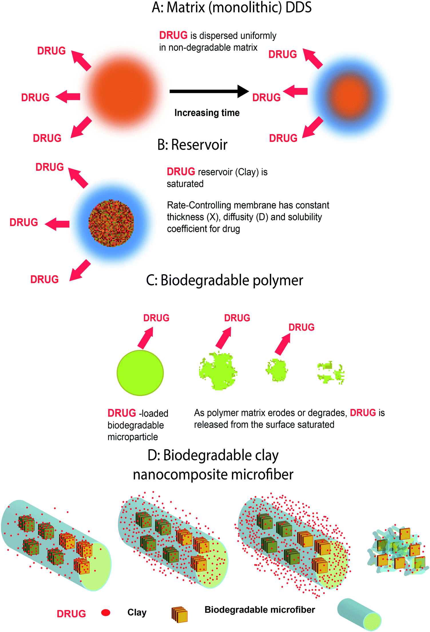 | ||
| Fig. 5 Different method of drug release based on: (a) monolithic, (b) reservoir, (c) degradation and (d) combined a, b and c drug delivery systems. | ||
In membrane-controlled reservoir devices like clay, the drugs are contained in cores and canals, which are surrounded by a polymer membrane, and released by diffusion through this rate-controlling membrane or layers (Fig. 5b).
In biodegradable drug delivery systems, the polymers used in the formulation and fabrication of biodegradable drug delivery devices erode (with or without changes to the chemical structure) or degrade (breakdown of the main chain bonds) as a result of the exposure to chemicals (water) or biological agents (enzymes).1 The drug molecules, which are initially dispersed in the polymer, are released as the polymer starts eroding or degrading as shown in Fig. 5c.
In biodegradable clay nano and microfiber nanocomposites, it is expected that the fourth mechanism as shown in Fig. 5d may be a combination of the a, b and c systems.
6. Characterization
The advanced devices used in most studies to understand and investigate nanoscale phenomena in clay nanocomposites for drug delivery systems are listed in Table 2.60,87,88,92–94| Loaded drug | Pore volume and surface area | Size and morphology | Analyzing methods | Ref. | ||||||||
|---|---|---|---|---|---|---|---|---|---|---|---|---|
| BJH | BET | SEM | TEM | EDX | AFM | CLSMa | X-ray XRD | FTIR | Thermal analysis | |||
| a CLSM (confocal laser scanning microscopy). | ||||||||||||
| 1 | Isoniazid | * | * | * | * | * | * | 78 | ||||
| 2 | Propranolol HCl | * | * | * | * | 93 | ||||||
| 3 | 1,3,4-Oxa (thia) diazoles | * | * | * | 72 | |||||||
| 4 | Dexamethasone | * | * | * | * | * | * | 61 | ||||
| 5 | Diclofenac sodium | * | * | * | * | 13 | ||||||
| 6 | Paclitaxel | * | * | * | * | * | 76 | |||||
| 7 | Nortriptyline and venlafaxine | * | * | * | * | 77 | ||||||
| 8 | Propranolol HCl | * | * | * | * | * | 80 | |||||
| Loaded drug | Entrapment efficiency | Release | In vitro | In vivo | Charge density | Water & dye uptake | Culture | Toxicity | Ref. | |||
|---|---|---|---|---|---|---|---|---|---|---|---|---|
| Dialysis | Centrifugation | Chromatography | ||||||||||
| 1 | Isoniazid | * | * | * | * | * | * | * | * | 78 | ||
| 2 | Propranolol HCl | * | * | * | * | * | 93 | |||||
| 3 | 1,3,4-Oxa (thia)diazoles | * | * | * | * | * | * | 72 | ||||
| 4 | Dexamethasone | * | * | * | * | 61 | ||||||
| 5 | Diclofenac sodium | * | 13 | |||||||||
| 6 | Paclitaxel | * | * | * | * | * | * | * | 76 | |||
| 7 | Nortriptyline and venlafaxine | * | * | * | * | 77 | ||||||
| 8 | Propranolol HCl | * | * | * | * | * | 80 | |||||
6.1. Average size, morphology and size distribution
Clays are in platy, film-like, flaky or tubular shapes with a thickness of 10 Å to 2 μm and their size can be determined using several techniques such as light microscopy, dynamic light scattering (DLS) analysis, electron microscopic analysis (scanning electron microscopy (SEM), transmission electron microscopy (TEM) and zetasizing. Also, clay size distribution and dispersion in polymers can be measured by using energy dispersive X-ray analysis (EDX), and exfoliation of clays and nanoclays can be determined by small angle X-ray diffraction (SAXRD) spectroscopy.64,786.2. Pore size analysis
The surface areas can be determined by nitrogen adsorption using a Quantachrome instrument using the Brunauer–Emmett–Teller (BET) equation. The pore size and pore volume can determined by the Barrett–Joyner–Halenda (BJH) method in the same instruments at a relative pressure using the (BJH) equation.42,78,95,966.3. Charge density and zeta potential
The zeta potential has an important role in the measurement of charge density in clay types. The surface charge was predominantly negative over most of the physiologically relevant pH range (>2) and the specific surface area of the l was very large (∼57 m2 g−1), indicating that the material has significant potential for extensive binding of cationic drugs.97 Specifically, the propranolol HCl (drug)–magnesium aluminum silicate complexes were broken into smaller particles because the negative charge of sodium alginate could interact molecularly not only with magnesium aluminum silicate (clay) but also with the positive charge of propranolol HCl.806.4. Thermal and mechanical behavior
The addition of nanoclay to the composite enhances the mechanical, thermal and other requisite properties.78 It is also reported that the thermal stability of the interlamellar organic species up to 750 °C also signifies the presence of interactions between the oxygen planes of the clay sheet and aromatic ring of the molecule and the shielding effect of the alumino-silicate layers.98 Bentonite too proved useful in improving the thermal stability, and can be used in the controlled release of naproxen.29 Increasing the magnesium aluminum silicate content of calcium alginate beads resulted in an increase in propranolol HCl thermal stability too.80 The mechanical and thermal properties of the electrospun nanocomposite nanofibers could be characterized by tensile strength, dynamic mechanical thermal analysis (DMTA), differential scanning calorimetry (DSC) and thermogravimetric analysis (TGA) experiments.42,64,88,93,94,986.5. Entrapment efficiency
For a therapeutic system to be used in a pharmaceutical application, the most important parameter of clay type composites is entrapment efficiency (EE%).More extensive loading should be achievable by entrapping agents within the lumen of tubules using retardant polymers, cationic coating and other approaches to moderate the release rate, or possibly by swapping intercalated water if present for low molecular weight agents.97 A low entrapment efficiency of water-soluble drugs in the polymers was a problem for developing a drug delivery system. This was due to the leakage of drug molecules, during the cross-linking process. Other major disadvantages of some biodegradable and hydrophilic polymers like alginate and poly(L lactide) (PLLA) were their fast disintegration and their high porosity, which resulted in a rapid drug release.77,79 An alternative approach to improve drug entrapment efficiency and modulate drug release involved the incorporation of water insoluble materials, like clays.80 Entrapment methods differ according to the kind of clay and polymers containing the nanocomposites. Several methods are reported to measure entrapment efficiency.77,80,97
Drug entrapment or encapsulation efficiency and drug loading are calculated using eqn (1) and (2):
| Drug loading% = (drug amount within the nanoparticles/total weight of nanoparticles) × 100 | (1) |
| Entrapment efficiency% = (drug amount within the nanoparticles/initial drug amount) × 100 | (2) |
6.6. In vitro and in vivo studies
According the route of administration, in vitro release can be determined by: the dialysis bag technique – suspension of the drug in a buffer in a constant temperature bath;37 SIF (simulated intestinal fluid) using UV-Vis spectroscopy;79 a dissolution method estimated by fitting the experimental drug release data into both models and analyzing it by linear regression analysis;77,99,100 or by a hemolysis assay based on hemoglobin absorbance (Abs) in the supernatant measured at 540 nm, with 655 nm as a reference, in a microplate spectrophotometer,61 which could be calculated using the following equation:| % hemolysis = (sample Abs540_655 nm − negative control Abs540_655 nm)/(positive control Abs540_655 nm − negative control Abs540_655 nm) × 100 | (3) |
In vivo studies of clay nanocomposite drug release depended on the route of administration investigated, the concentration, the effect and the time the drug was present in the tissues.
Experimental methods based on application have been developed on animals or physiologic liquids, like platelet aggregation and adhesion studies.61,72
Some characterization techniques for analyzing clay nanocomposite formulation, identified in the published literature, have been listed in Table 2.
6.7. Biocompatibility aspects
Clays in platelet and halloysite forms are biocompatible and environmentally friendly materials.61,65 It has been reported that nanocomposites containing montmorillonite clay and polyurethane can be applied to control the release of dexamethasone acetate, and biocompatibility studies demonstrated adequate interfacial interaction between polyurethane and subcutaneous tissue and a discreet inflammatory response which was completely resolved in 14 days.101 Also a nanocomposite of montmorillonite-chitosan has good biocompatibility and stimulates cell proliferation, and in addition shows mucoadhesive properties combined with low solubility in acid media, beyond retarding drug release in gastric environments for controlled drug release.102 Synthesis of novel nanohybrid materials by the incorporation of 2-D nanoparticles results in improved physical properties, notably biocompatibility and biodegradation, which are explicitly required for tissue engineering and drug delivery applications. Biocompatibility of these nanocomposites has been extensively verified through platelet adhesion, aggregation and hemolysis assay.616.8. Toxicity
Natural clay minerals are suitable to effectively modulate drug release. High specific surface area, adsorptive capacity, rheological properties, chemical inertness and low or null toxicity make clay minerals futuristic drug delivery carriers.103 The beneficial effects of a drug for human health are closely related to targeting selected body parts at a desired concentration level of the drug for a prolonged period of time. This concentration level for therapeutic applications can be achieved without reaching a higher toxic level or dropping below the minimum effective level.104 A study on mice shows that no significant effects of oral LDH nanoparticles on behavior, body weight gain, survival rate, or organosomatic index were observed up to a dose of 2000 mg kg−1 for 14 days. Plasma concentration of LDH nanoparticles rapidly decreased within 30 min depending on exposure doses, and their excretion via urine and feces was observed within 24 h. There is no significant increase in serum biochemical parameters and also acute hepatotoxicity, nephrotoxicity and organ accumulation were not found.105 High dose (5 mg kg−1) pulmonary exposures to sepiolite nanoclay particulates in male rats produced minimal alveolar macrophage aggregates, including occasional multinucleated cells in centriacinar air spaces, and minimal focal septal thickening of alveolar ducts and adjacent alveoli. Lesions regressed with increased post exposure duration and were very slight and focal by 3 months. Some evidence of particle-associated macrophage aggregates in draining lymphoid tissue was observed. However, no extrapulmonary target organ effects were observed in the particle-exposed at 3 months postinstillation exposures.106 Some findings suggest that the genotoxicity of organo-modified montmorillonite was caused by the organo-modifier.107 Serum supplementation is an important consideration in the toxicological assessments of nanomaterials on cells, which needs to be addressed in the standardization of in vitro testing methods. Clay particles, therefore, have cytotoxic properties that may be linked to their dispersion pattern. These adverse effects seem to be masked by 5% fetal calf serum (FCS).108,109 In another study concerned with the toxicity of nanosilicate platelets (NSP) derived from natural montmorillonite clay for feeding to rats, the acute oral toxicity was shown to have a low lethal dose (LD50) of greater than 5700 mg kg−1 body weight for both male and female Sprague–Dawley rats. The Comet assay showed no DNA damage after 24 h of incubation with an NSP of 1000 μg mL−1. Overall, the study demonstrated the safety of the NSP for potential uses in biomedical areas.110 It was also observed that a polyurethane bio-nanocomposite having a 2% bentonite nanoclay content is the ideal amount for surgical threads in investigations into their in vitro biocompatibility and non-toxicity. Cell culture tests were used to evaluate both cyto-toxicity and cyto-compatibility of the specimens.111 No data was found about carcinogenicity, reproductive toxicity, irritation and sensitization. So far, there is very limited data available on the toxicity of nanoclays. It seems important to elucidate if the modifiers are causing the toxic events or if the nanoclay itself can induce harmful effects.7. Administration
Not only have polymeric clay nanocomposites conjugated with drugs been reported to be used for oral drug delivery of 1, 3, 4-oxa(thia)diazoles, paclitaxel, nortriptyline, venlafaxine and propranolol HCl,11,16,72,76,77,80 they have good potential in chemotherapy with local administration of the drug at the site of resected cancerous cells.79 Also transdermal routes of administration for this system have been studied.16,72,77,90,918. Application
Clays were first used as fillers for oral use in tablet form and then came to the attention of the agricultural sector and cosmetic, nutrient pharmacological and pharmaceutical companies for smart drug delivery. They have enormous potential for therapeutic applications, being the subject of intensive research studies. Clays can reserve various drugs and nutrients and can have various applications, such as antioxidant, anticancer, anti-inflammatory, antimicrobial, anti-depressant, anti-bacterial, and blood pressure reduction. They are mostly used in oral routes and have been developed for transdermal and injection routes. Clay nanocomposites have been developed for wound healing because of their effectiveness in sustained drug releases.112,113 Also, because the surface characteristics of clays are biologically compatible and can support bone ingrowth, they can be applied for bone tissue healing by grafting.114–1169. Conclusion
The ion exchange nature and biocompatibility of clays make them versatile carriers for many vital drugs. In recent decades, polymer layered silicate nanocomposites have received more attention in controlled drug release and regenerative medicine. It is obvious that clays appear to be well preferred as fillers and drug delivery systems, because of their low toxicity aspect and economic benefits. Clay composites go to develop a convenient, prolonged, targeted and effective drug delivery system with the ability of loading both hydrophilic and hipophilic drugs. The efficiency of clay can be enhanced by using novel synthesis, loading, and modification and combination with polymers by controlling the nanometric phenomena and their effects on clay nanocomposites and they can subsequently prolong drug release. Thus, these fields need further exploration and research to obtain commercially available and economical clay nanocomposites in pharmaceutical industries.References
- R. A. Siegel and M. J. Rathbone, in Fundamentals and Applications of Controlled Release Drug Delivery, Springer, 2012, pp. 19–43 Search PubMed.
- A. Mostafavi, J. Emami, J. Varshosaz, N. M. Davies and M. Rezazadeh, Int. J. Pharm., 2011, 409, 128–136 CrossRef CAS PubMed.
- S. Yi, Y.-j. Chung, T.-E. Kim, H.-S. Shin, S. H. Yoon, J.-Y. Cho, I.-J. Jang, S.-G. Shin and K.-S. Yu, Clin. Ther., 2011, 33, 728–737 CrossRef CAS PubMed.
- C. Chakraborty, K. Dana and S. Malik, J. Colloid Interface Sci., 2012, 368, 172–180 CrossRef CAS PubMed.
- K. Zhang, Z. P. Xu, J. Lu, Z. Y. Tang, H. J. Zhao, D. A. Good and M. Q. Wei, Int. J. Mol. Sci., 2014, 15, 7409–7428 CrossRef CAS PubMed.
- L. A. de Sousa Rodrigues, A. Figueiras, F. Veiga, R. M. de Freitas, L. C. C. Nunes, E. C. da Silva Filho and C. M. da Silva Leite, Colloids Surf., B, 2013, 103, 642–651 CrossRef PubMed.
- G. Sposito, N. T. Skipper, R. Sutton, S.-h. Park, A. K. Soper and J. A. Greathouse, Proc. Natl. Acad. Sci. U. S. A., 1999, 96, 3358–3364 CrossRef CAS.
- T. Takahashi and M. Yamaguchi, J. Inclusion Phenom. Mol. Recognit. Chem., 1991, 10, 283–297 CrossRef CAS.
- T. Takahashi and M. Yamaguchi, J. Colloid Interface Sci., 1991, 146, 556–564 CrossRef CAS.
- T. Bandosz, J. Jagiello, K. Amankwah and J. Schwarz, Clay Miner., 1992, 27, 435–444 CAS.
- T. Takahashi, Y. Yamada, K. Kataoka and Y. Nagasaki, J. Controlled Release, 2005, 107, 408–416 CrossRef CAS PubMed.
- J. U. Ha, Study of controlled release of active pharmaceutical ingredients from functionalized nanoclays and polymer matrices, New Jersey Institute Of Technology, 2011 Search PubMed.
- J. U. Ha and M. Xanthos, Int. J. Pharm., 2011, 414, 321–331 CrossRef CAS PubMed.
- Y.-R. K. Soo-Jin Choi, Bioinspired layered nanoclays for nutraceutical delivery system, Washington, DC, 2013 Search PubMed.
- J. N. Israelachvili, Intermolecular and surface forces: revised third edition, Academic press, 2011 Search PubMed.
- C. Aguzzi, P. Cerezo, C. Viseras and C. Caramella, Appl. Clay Sci., 2007, 36, 22–36 CrossRef CAS.
- R. K. Gupta, E. Kennel and K.-J. Kim, Polymer nanocomposites handbook, CRC press, 2009, ch. 4 Search PubMed.
- J.-H. Choy, S.-J. Choi, J.-M. Oh and T. Park, Appl. Clay Sci., 2007, 36, 122–132 CrossRef CAS.
- F.-H. Lin, Y.-H. Lee, C.-H. Jian, J.-M. Wong, M.-J. Shieh and C.-Y. Wang, Biomaterials, 2002, 23, 1981–1987 CrossRef CAS PubMed.
- G. V. Joshi, H. A. Patel, B. D. Kevadiya and H. C. Bajaj, Appl. Clay Sci., 2009, 45, 248–253 CrossRef CAS.
- F. Bonina, M. Giannossi, L. Medici, C. Puglia, V. Summa and F. Tateo, Appl. Clay Sci., 2007, 36, 77–85 CrossRef CAS.
- V. Ambrogi, G. Fardella, G. Grandolini and L. Perioli, Int. J. Pharm., 2001, 220, 23–32 CrossRef CAS PubMed.
- R. R. Price, B. P. Gaber and Y. Lvov, J. Microencapsulation, 2001, 18, 713–722 CrossRef CAS PubMed.
- V. Ambrogi, G. Fardella, G. Grandolini, L. Perioli and M. C. Tiralti, AAPS PharmSciTech, 2002, 3, 77–82 CrossRef.
- V. Ambrogi, G. Fardella, G. Grandolini, M. Nocchetti and L. Perioli, J. Pharm. Sci., 2003, 92, 1407–1418 CrossRef CAS PubMed.
- M. Del Arco, E. Cebadera, S. Gutierrez, C. Martin, M. Montero, V. Rives, J. Rocha and M. Sevilla, J. Pharm. Sci., 2004, 93, 1649–1658 CrossRef CAS PubMed.
- J.-C. Dupin, H. Martinez, C. Guimon, E. Dumitriu and I. Fechete, Appl. Clay Sci., 2004, 27, 95–106 CrossRef CAS.
- B. Li, J. He, D. G. Evans and X. Duan, Appl. Clay Sci., 2004, 27, 199–207 CrossRef CAS.
- M. Wei, S. Shi, J. Wang, Y. Li and X. Duan, J. Solid State Chem., 2004, 177, 2534–2541 CrossRef CAS.
- L. Mohanambe and S. Vasudevan, J. Phys. Chem. B, 2005, 109, 15651–15658 CrossRef CAS PubMed.
- C. D. Nunes, P. D. Vaz, A. C. Fernandes, P. Ferreira, C. C. Romao and M. J. Calhorda, Eur. J. Pharm. Biopharm., 2007, 66, 357–365 CrossRef CAS PubMed.
- N. G. Veerabadran, R. R. Price and Y. M. Lvov, Nano, 2007, 2, 115–120 CrossRef CAS.
- F. P. Bonina, M. L. Giannossi, L. Medici, C. Puglia, V. Summa and F. Tateo, Appl. Clay Sci., 2008, 41, 165–171 CrossRef CAS.
- M. Del Arco, A. Fernandez, C. Martin, M. Sayalero and V. Rives, Clay Miner., 2008, 43, 255–265 CrossRef CAS.
- Y. M. Lvov, D. G. Shchukin, H. Mohwald and R. R. Price, ACS Nano, 2008, 2, 814–820 CrossRef CAS PubMed.
- M. Del Arco, A. Fernández, C. Martín and V. Rives, Appl. Clay Sci., 2009, 42, 538–544 CrossRef CAS.
- G. V. Joshi, B. D. Kevadiya, H. A. Patel, H. C. Bajaj and R. V. Jasra, Int. J. Pharm., 2009, 374, 53–57 CrossRef CAS PubMed.
- T. Pongjanyakul, W. Khunawattanakul and S. Puttipipatkhachorn, Appl. Clay Sci., 2009, 44, 242–250 CrossRef CAS.
- M. Del Arco, A. Fernandez, C. Martin and V. Rives, J. Solid State Chem., 2010, 183, 3002–3009 CrossRef CAS.
- Y. Chen, A. Zhou, B. Liu and J. Liang, Appl. Clay Sci., 2010, 49, 108–112 CrossRef CAS.
- F. Kovanda, Z. Maryšková and P. Kovář, J. Solid State Chem., 2011, 184, 3329–3335 CrossRef CAS.
- A. Tabak, N. Yilmaz, E. Eren, B. Caglar, B. Afsin and A. Sarihan, Chem. Eng. J., 2011, 174, 281–288 CrossRef CAS.
- R. I. Iliescu, E. Andronescu, G. Voicu, A. Ficai and C. I. Covaliu, Appl. Clay Sci., 2011, 52, 62–68 CrossRef CAS.
- I. Teasdale and O. Brüggemann, Polymers, 2013, 5, 161–187 CrossRef PubMed.
- F. C. H. Pinto, A. Silva-Cunha, G. A. Pianetti, E. Ayres, R. L. Oréfice and G. R. Da Silva, J. Nanomater., 2011, 2011, 15 Search PubMed.
- A. Ainurofiq and S. Choiri, Trop. J. Pharm. Res., 2015, 14, 1129–1135 CrossRef CAS.
- V. Ambrogi, L. Perioli, M. Ricci, L. Pulcini, M. Nocchetti, S. Giovagnoli and C. Rossi, Microporous Mesoporous Mater., 2008, 115, 405–415 CrossRef CAS.
- D. R. Katti, K. S. Katti, M. Raviprasad and C. Gu, J. Nanomater., 2012, 2012, 28 Search PubMed.
- Y. Wang, M. Li, J. Rong, G. Nie, J. Qiao, H. Wang, D. Wu, Z. Su, Z. Niu and Y. Huang, Colloid Polym. Sci., 2013, 291, 1541–1546 CAS.
- V. Mittal, Materials, 2009, 2, 992–1057 CrossRef CAS.
- S. Pavlidou and C. Papaspyrides, Prog. Polym. Sci., 2008, 33, 1119–1198 CrossRef CAS.
- M. Segad, B. Jönsson and B. Cabane, J. Phys. Chem. C, 2012, 116, 25425–25433 CAS.
- M. Alexandre and P. Dubois, Mater. Sci. Eng., R, 2000, 28, 1–63 CrossRef.
- S. Abedi and M. Abdouss, Appl. Catal., A, 2014, 475, 386–409 CrossRef CAS.
- K. T. Campbell, D. Q. Craig and T. McNally, J. Appl. Polym. Sci., 2014, 131 DOI:10.1002/app.40284.
- H. Mahdavi, H. Mirzadeh, M. J. Zohuriaan-Mehr and F. Talebnezhad, Am. J. Sci., 2013, 9, 203–214 Search PubMed.
- K. A. Carrado and L. Xu, Chem. Mater., 1998, 10, 1440–1445 CrossRef CAS.
- S. S. Ray, Clay-containing polymer nanocomposites: from fundamentals to real applications, Newnes, 2013 Search PubMed.
- G. Vilar, J. Tulla-Puche and F. Albericio, Curr. Drug Delivery, 2012, 9, 367–394 CrossRef CAS.
- A. U. Liyanage, E. U. Ikhuoria, A. A. Adenuga, V. T. Remcho and M. M. Lerner, Inorg. Chem., 2013, 52, 4603–4610 CrossRef CAS PubMed.
- A. Mishra, S. K. Singh, D. Dash, V. K. Aswal, B. Maiti, M. Misra and P. Maiti, Acta Biomater., 2014, 10, 2133–2146 CrossRef CAS PubMed.
- W. Li, L. Sun, L. Pan, Z. Lan, T. Jiang, X. Yang, J. Luo, R. Li, L. Tan and S. Zhang, Eur. J. Pharm. Biopharm., 2014, 88, 706–717 CrossRef CAS PubMed.
- J. Xue, Y. Niu, M. Gong, R. Shi, D. Chen, L. Zhang and Y. Lvov, ACS Nano, 2015, 9, 1600–1612 CrossRef CAS PubMed.
- M. Koosha, H. Mirzadeh, M. A. Shokrgozar and M. Farokhi, RSC Adv., 2015, 5, 10479–10487 RSC.
- E. Abdullayev and Y. Lvov, J. Mater. Chem. B, 2013, 1, 2894–2903 RSC.
- D. Q. Craig and J. A. McNally, WO2004098574 A1, 2005.
- A. R. Menjoge, R. M. Kannan and D. A. Tomalia, Drug Discovery Today, 2010, 15, 171–185 CrossRef CAS PubMed.
- P. Kesharwani, K. Jain and N. K. Jain, Prog. Polym. Sci., 2014, 39, 268–307 CrossRef CAS.
- A. H. Najafabadi, S. Azodi-Deilami, M. Abdouss, H. Payravand and S. Farzaneh, J. Mater. Sci.: Mater. Med., 2015, 26, 1–11 Search PubMed.
- S. Vakilian, S. Mashayekhan, I. Shabani, M. Khorashadizadeh, A. Fallah and M. Soleimani, Int. J. Biol. Macromol., 2015, 75, 248–257 CrossRef CAS PubMed.
- S. Moghassemi and A. Hadjizadeh, J. Controlled Release, 2014, 185, 22–36 CrossRef CAS PubMed.
- N. Salahuddin, A. Elbarbary, N. G. Allam and A. F. Hashim, J. Appl. Polym. Sci., 2014, 131 DOI:10.1002/app.41177.
- M. Mahkam, A. Latifpour, A. A. Rafi, L. M. Gheshlaghi and A. Takfallah, Int. J. Polym. Mater. Polym. Biomater., 2015, 64, 32–37 CrossRef CAS.
- W. Mohamed, A. Mostafa and H. Nasr, Polym.-Plast. Technol. Eng., 2014, 53, 1425–1433 CrossRef CAS.
- S. Barkhordari, M. Yadollahi and H. Namazi, J. Polym. Res., 2014, 21, 1–9 CAS.
- Y. Dong and S.-S. Feng, Biomaterials, 2005, 26, 6068–6076 CrossRef CAS PubMed.
- S. Rajkumar, B. D. Kevadiya and H. C. Bajaj, Asian J. Pharm. Sci., 2015, 10, 452–458 CrossRef.
- N. Banik, M. Iman, A. Hussain, A. Ramteke, R. Boruah and T. K. Maji, New J. Chem., 2013, 37, 3981–3990 RSC.
- R. I. Iliescu, E. Andronescu, C. D. Ghitulica, G. Voicu, A. Ficai and M. Hoteteu, Int. J. Pharm., 2014, 463, 184–192 CrossRef CAS PubMed.
- T. Pongjanyakul and T. Rongthong, Carbohydr. Polym., 2010, 81, 409–419 CrossRef CAS.
- Z. S. Petrović and D. Fajnik, J. Appl. Polym. Sci., 1984, 29, 1031–1040 CrossRef.
- C. Lyon and V. Garrett, J. Am. Oil Chem. Soc., 1973, 50, 112–114 CrossRef CAS.
- Z. S. Petrović, Polym. Rev., 2008, 48, 109–155 CrossRef.
- D. P. Pfister and R. C. Larock, Composites, Part A, 2010, 41, 1279–1288 CrossRef.
- S. Husić, I. Javni and Z. S. Petrović, Compos. Sci. Technol., 2005, 65, 19–25 CrossRef.
- I. Gupta, M. Singh, P. Singh and K. Singh, Research and industry, 1975 Search PubMed.
- A. Kaushik, D. Ahuja and V. Salwani, Composites, Part A, 2011, 42, 1534–1541 CrossRef.
- M. Iman, A. K. Manhar, M. Mandal and T. K. Maji, RSC Adv., 2014, 4, 33826–33839 RSC.
- R. Kuktaite, H. Türe, M. S. Hedenqvist, M. Gällstedt and T. S. S. Plivelic, ACS Sustainable Chem. Eng., 2014, 2, 1439–1445 CrossRef CAS.
- K. Saha, B. Butola and M. Joshi, J. Appl. Polym. Sci., 2014, 131 DOI:10.1002/app.40824.
- K. Saha, B. S. Butola and M. Joshi, J. Appl. Polym. Sci., 2014, 131 DOI:10.1002/app.40230.
- S. W. Kim, S. O. Han, I. N. Sim, J. Y. Cheon and W. H. Park, J. Nanomater., 2015, 2015, 3 Search PubMed.
- M. Datta, Appl. Clay Sci., 2013, 80, 85–92 Search PubMed.
- Y. Jiang, B. Li, X. Chen and M. Zhu, J. Appl. Polym. Sci., 2012, 125 DOI:10.1002/app.35542.
- G. Yang, H. Gong, X. Qian, P. Tan, Z. Li, T. Liu, J. Liu, Y. Li and Z. Liu, Nano Res., 2015, 8, 751–764 CrossRef CAS.
- X. Huang, N. P. Young and H. E. Townley, Nanomater. Nanotechnol., 2014, 4 DOI:10.5772/58290.
- S. Levis and P. Deasy, Int. J. Pharm., 2002, 243, 125–134 CrossRef CAS PubMed.
- A. Tabak, B. Afsin, S. Aygun and E. Koksal, J. Therm. Anal. Calorim., 2007, 87, 377–382 CrossRef.
- P. Costa and J. M. S. Lobo, Eur. J. Pharm. Sci., 2001, 13, 123–133 CrossRef CAS PubMed.
- R. Bhaskar, R. Murthy, B. Miglani and K. Viswanathan, Int. J. Pharm., 1986, 28, 59–66 CrossRef CAS.
- G. R. Da Silva, E. Ayres, R. L. Orefice, S. A. L. Moura, D. C. Cara and A. D. S. Cunha Jr, J. Drug Targeting, 2009, 17, 374–383 CrossRef PubMed.
- B. D. Kevadiya, G. V. Joshi and H. C. Bajaj, Int. J. Pharm., 2010, 388, 280–286 CrossRef CAS PubMed.
- M. I. Carretero, Appl. Clay Sci., 2002, 21, 155–163 CrossRef CAS.
- D. Depan, A. P. Kumar and R. P. Singh, Acta Biomater., 2009, 5, 93–100 CrossRef CAS PubMed.
- J. Yu, H.-E. Chung and S.-J. Choi, J. Nanomater., 2013, 2013, 12 Search PubMed.
- D. B. Warheit, C. M. Sayes, S. R. Frame and K. L. Reed, Toxicol. Lett., 2010, 192, 286–293 CrossRef CAS PubMed.
- A. K. Sharma, B. Schmidt, H. Frandsen, N. R. Jacobsen, E. H. Larsen and M.-L. Binderup, Mutat. Res., Genet. Toxicol. Environ. Mutagen., 2010, 700, 18–25 CrossRef CAS PubMed.
- S. Lordan and C. L. Higginbotham, Cell Biol. Int., 2012, 36, 57–61 CrossRef CAS PubMed.
- S. Lordan, J. E. Kennedy and C. L. Higginbotham, J. Appl. Toxicol., 2011, 31, 27–35 CrossRef CAS PubMed.
- P.-R. Li, J.-C. Wei, Y.-F. Chiu, H.-L. Su, F.-C. Peng and J.-J. Lin, ACS Appl. Mater. Interfaces, 2010, 2, 1608–1613 CAS.
- K. M. Zia, M. Zuber, M. Barikani, R. Hussain, T. Jamil and S. Anjum, Int. J. Biol. Macromol., 2011, 49, 1131–1136 CrossRef CAS PubMed.
- M. Ghadiri, W. Chrzanowski and R. Rohanizadeh, J. Mater. Sci.: Mater. Med., 2014, 25, 2513–2526 CrossRef CAS PubMed.
- M. Kokabi, M. Sirousazar and Z. M. Hassan, Eur. Polym. J., 2007, 43, 773–781 CrossRef CAS.
- A. Berent, K. Tobias, K. Tobias and S. Johnston, Hepatic Vascular Anomalies, ed. K. M. J. S. Tobias, 2012, vol. 2, pp. 1624–1658 Search PubMed.
- T. W. Fossum, Small animal surgery textbook, Elsevier Health Sciences, 2013 Search PubMed.
- A. K. Gaharwar, S. Mukundan, E. Karaca, A. Dolatshahi-Pirouz, A. Patel, K. Rangarajan, S. M. Mihaila, G. Iviglia, H. Zhang and A. Khademhosseini, Tissue Eng., Part A, 2014, 20, 2088–2101 CrossRef CAS PubMed.
| This journal is © The Royal Society of Chemistry 2016 |