
Naphthothiazole-based highly selective and sensitive fluorescent and colorimetric chemosensor for detection of pollutant metal ions†
Mohammad Zareh Jonaghania,
Hassan Zali-Boeini*a,
Reza Taherib,
Hadi Amiri Rudbaria and
Banafshe Askaria
aDepartment of Chemistry, University of Isfahan, 81746-73441, Isfahan, Iran. E-mail: h.zali@chem.ui.ac.ir
bEnvironmental Research Institute, University of Isfahan, Isfahan, Iran
First published on 4th April 2016
Abstract
A new fluorescent and colorimetric chemosensor based on naphthothiazole S1 was synthesized and its applications as chemosensor for the selective sensing of pollutant metal ions in water resources and/or biological systems were investigated. Sensor S1 showed great selectivity for Zn2+ and Sn2+ over other metal ions. The Job's plot showed a 1![[thin space (1/6-em)]](https://www.rsc.org/images/entities/char_2009.gif) :1 stoichiometry between S1 with Zn2+ and Sn2+. Interaction mode of sensor S1 with Zn2+ ion was further approved by single crystals X-ray crystallography method. The detection limit for the fluorescent chemosensor S1 toward Zn2+ and Sn2+ were respectively 2.60 × 10⁻⁸ M and 8.21 × 10⁻⁸ M.
:1 stoichiometry between S1 with Zn2+ and Sn2+. Interaction mode of sensor S1 with Zn2+ ion was further approved by single crystals X-ray crystallography method. The detection limit for the fluorescent chemosensor S1 toward Zn2+ and Sn2+ were respectively 2.60 × 10⁻⁸ M and 8.21 × 10⁻⁸ M.
Introduction
Due to the important role of cations in biological, environmental, and chemical processes, design and synthesis of receptors for selective detection of a specific cation is a rapidly growing area in supramolecular chemistry in recent years.1 Among them, sensors targeting metal ions such as zinc,2 mercury,3 tin4 and copper5 cations have gained considerable attention due to important environmental issues.After iron, zinc is the most abundant transition metal ion in the human body and plays vital roles as an essential micronutrient in biological processes such as gene expression6 and regulation of enzymes and other proteins.7 Also aberration of its concentration is related to many diseases such as Alzheimer's syndrome.8 Tin is commonly used as heavy metals in industry as catalyst in production of biodiesel and allylation of carbonyl compounds.9 It is also involved in several biochemical processes occurring at the cellular level.10
Therefore, significant efforts have been made to develop selective chemosensors and/or chemodosimeters for detecting such environmentally important cations.11
Amid the chemosensors for detection of cations, colorimetric and fluorescent chemosensors are of great concerns among the chemist and biologists because of their high selectivity, high sensitivity, and simplicity.12
Although numerous sensors are available for sensing of metal ions, the design of new chemical sensors that are simpler, easier to synthesize, and can selectively recognize multiple analytics, especially species with similar chemical properties continues to develop at an unceasing pace. Meanwhile, it is a nice challenge to develop a small molecule probe capable of discriminating Zn2+ from Cd2+. Intrigued by this, we were interested in studying heterocyclic ligands for use as fluorescent and colorimetric chemosensors.
Results and discussion
Initially, our researches commenced by synthesis of quinoline-substituted naphthothiazole S1 in two simple steps via modified Willgerodt–Kindler reaction of quinoline, 1-naphthylamine and sulfur under MW irradiation followed by oxidative cyclization reaction of the corresponding thioamide (Scheme 1). After that, application of this chemosensors for detection of zinc, tin, and many other cations were investigated.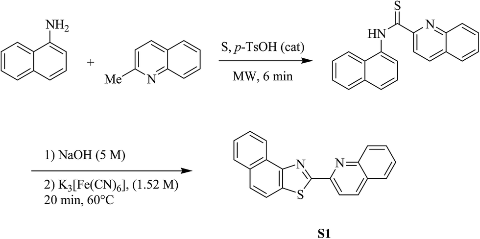 | ||
| Scheme 1 A simple two step synthesis of S1 starting from 1-naphthylamine. | ||
At the onset of our investigations, the UV absorption spectra of sensor S1 in aqueous solution of acetonitrile (CH3CN–H2O 80/20, v/v) was taken alone and in the course of titration with Sn2+, Cd2+, Zn2+, Mn2+, Cu2+, Ba2+, Co2+, Ca2+, Ni2+, Pb2+, Hg2+, Fe3+, Al3+, Cr3+, and Ag+ (Fig. 1A).
 | ||
| Fig. 1 (A) UV-vis absorption spectra of the chemosensor S1 (30 μM) with various metal ions in CH3CN/H2O (80:20). [Mn+] = 100 μM; (B) fluorescence spectra of S1 (0.1 μM) in the presence of various metal ions in CH3CN/H2O (80:20, v/v) at room temperature. [Mn+] = 5 μM. | ||
The absorption spectrum of free S1 exhibited a maximum centered at 360 nm (ε = 26700 M−1 cm−1) with a shoulder at 378 nm. When 5 μM solution of Cd2+, Mn2+, Cu2+, Ba2+, Co2+, Ca2+, Ni2+, Pb2+, Hg2+, Fe3+, Al3+, Cr3+, and Ag+ metal ions were added, the absorption maxima of S1 did not move, whereas the intensity of the maxima exhibited a little decrease during addition of Sn2+, Zn2+, alongside with small red shifts.
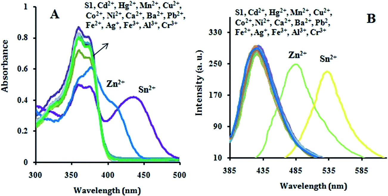
The selectivity of the fluorescence response of S1 to various metal ions in aqueous solution of acetonitrile (CH3CN–H2O 80/20, v/v) were examined. Fig. 1B shows the fluorescence response of S1 (428 nm, blue fluorescence, ε = 32000 M−1 cm−1) to diverse metal ions. Selective and large red shifts in the fluorescent spectra of the S1 were observed upon addition of Zn2+ (486 nm, green fluorescence, ε = 32000 M−1 cm−1) and Sn2+ (533 nm, yellow fluorescence, ε = 32000 M−1 cm−1) solutions to the solution of S1 in aqueous CH3CN. Investigation of other metal ions such as Cd2+, Ca2+, Mn2+, Ni2+, Cu2+, Co2+, Ba2+, Pb2+, Hg2+, Fe3+, Al3+, Cr3+, and Ag+ revealed that they did not affect the fluorescence emission of S1. Fig. 2 shows the images of color (A) and fluorescence (B) response changes of S1 (10 μM) upon addition of Sn2+, Zn2+, Cd2+, Hg2+, Mn2+, Cu2+, Co2+, Ni2+, Ca2+, Ba2+, Pb2+, Fe3+, Al3+, Cr3+ and Ag+ (50 μM) cations in CH3CN–H2O (80/20, v/v).
 | ||
| Fig. 2 Color (A) and fluorescence (B) change upon addition of various metal cations to S1 in CH3CN–water under visible and UV light respectively. | ||
Moreover, the 1H NMR spectra of S1 and S1–Zn2+ were recorded and the observed significant chemical shift show intense interaction between S1 and Zn2+ (Fig. 3).
 | ||
| Fig. 3 1H NMR spectra of S1 with 1.0 equiv. of ZnCl2 in DMSO-d6. | ||
The relationship between S1 fluorescence intensity and concentrations of Zn2+ and Sn2+ were examined in CH3CN–H2O (80/20, v/v) and is shown respectively in Fig. 4A and B. A good linear relationship between the fluorescence intensity at 486 nm and concentration of Zn2+ (0–5 μM) (Fig. 4C) and fluorescence intensity at 533 nm and concentration of Sn2+ (0–5 μM) (Fig. 4D) were also found with a detection limit of 2.60 × 10⁻⁸ M and 8.21 × 10⁻⁸ M respectively for Zn2+ and Sn2+ (S/N = 3).
 | ||
| Fig. 4 (A) Fluorescence emission spectra of S1 (0.1 μM) upon the addition of Zn2+ (0–5 μM) and (B) Sn2+ (0–5 μM) in CH3CN/H2O (80/20, v/v); (C) linear relationship between the fluorescence intensity at 486 nm and concentration of Zn2+ (0–5 μM) and (D) fluorescence intensity at 533 nm and concentration of Sn2+ (0–5 μM). | ||
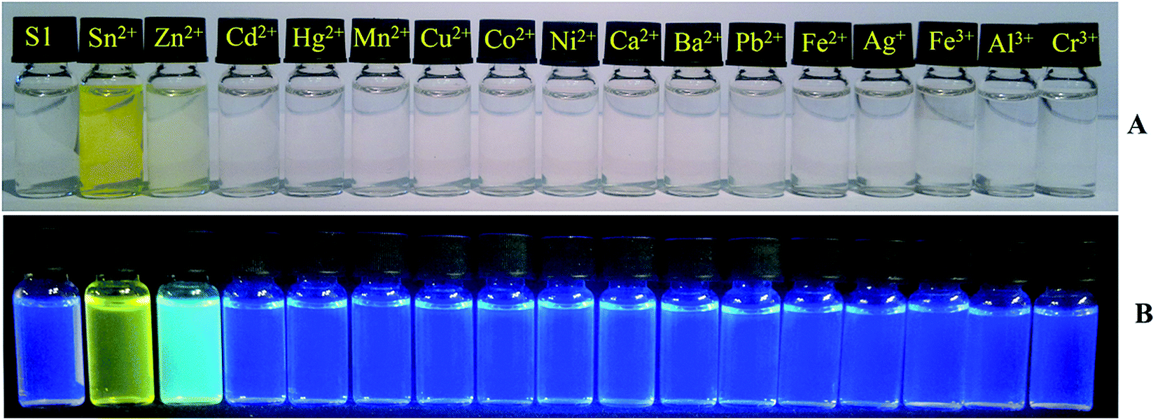
In order to examine selectivity of the sensor S1 for detection of Zn2+ and Sn2+ over other metal ions, competition experiments were conducted. Hence, fluorescent intensity for Zn2+ and Sn2+ were investigated respectively at 486 nm and 533 nm in the presence of other metal ions (Fig. 5). As shown in Fig. 5A all other metal ions (including Sn2+, and Cd2+) have no obvious effect on the emission spectra of S1–Zn2+ but emission intensity for the interaction of S1–Sn2+ decreases in the presence of Zn2+ (Fig. 5B). These data obviously demonstrate that S1 tends to bind stronger with Zn2+ compare to Sn2+ and sensor S1 can be used successfully for selective detection of Zn2+ ions in the presence of Sn2+ ions in aqueous media.
 | ||
| Fig. 5 Fluorescence response of S1 (0.1 μM) to 5 μM of (A) Zn2+ and (B) Sn2+ in CH3CN–H2O (80/20, v/v) in the presence of 100 μM of various mental ions. | ||
The efficiency of the sensor S1 for the selective detection of some environmentally important and toxic pollutant metal ions in real samples was also investigated. For instance, S1 was successfully utilized for detection of Sn2+ ion (in the absence of Zn2+) contamination in a plant cell. In this study, micro alga which are unicellular photosynthetic unites were chosen. These autotrophic cells can live in wide environmental conditions; from fresh clear water (such as Micrasterias furcata) to high contaminated waste water (such as Scenedesmus obliquus). In the case of Scenedesmus obliquus the cell walls behave like as a faintly acidic cation exchanger capable of removing heavy metal pollutants such as cadmium, mercury, and tin ions from the waste water.13 This feature intrigued us to show ability of Scenedesmus obliquus in absorption of tin ions from aqueous solutions followed by sensing the absorbed Sn2+ ion by chemosensor S1. In this experiment, the plant cell was incubated for 60 minutes in a solution of Sn2+ ion in water (10 μM) and then treated with a solution of S1 in CH3CN/H2O (80/20, 5 μM). Thereafter, the fluorescence image of the treated cells were acquired using a confocal fluorescence microscope showing a bright yellow-green fluorescence for the Sn2+ absorbed cells (Fig. 6).
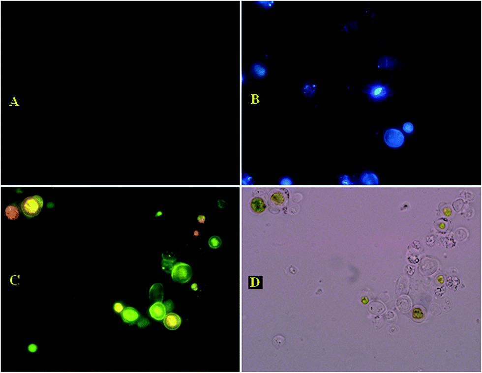 | ||
| Fig. 6 Fluorescence image of live Scenedesmus obliquus cells (A) cells incubated with 10 μM SnCl2 for 1 h at 25 °C. (B) Cells incubated with 5 μM S1 for 5 min at 25 °C. (C) Cells supplemented with 10 μM SnCl2 for 1 h at 25 °C and stained with 5 μM S1 at 25 °C for 5 min. (D) Brightfield image of the Scenedesmus obliquus cells shown in panel C. | ||
In order to investigate the mode of interaction of the receptor S1 with metal ions, single crystal structure of compound made from the interaction of S1 and Zn2+ ion was prepared (Fig. 7, CCDC 1426152†). The suitable single crystals for X-ray structure analysis were obtained by slow evaporation of a solution of the complex compound formed between S1 and Zn2+ ion in acetonitrile.
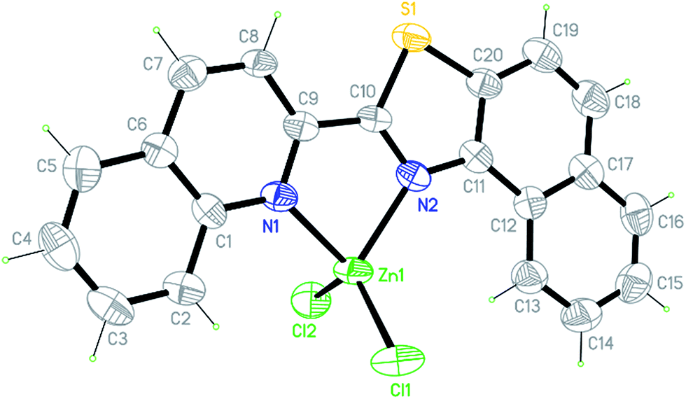 | ||
| Fig. 7 ORTEP representation of S1 + ZnCl2. Displacement ellipsoids are drawn at the 50% probability level and H atoms are shown as small spheres of arbitrary radii. | ||
As it is obvious from Fig. 7, a 1:1 ratio is observed for the mode of interaction of Zn2+ and probably all other two-valent metal ions with sensor S1.
In order to determine the stoichiometry in the interaction of Zn2+ and Sn2+ ions with S1 different mole ratios of S1 to Zn2+ and Sn2+ were investigated. The Job's plot showed 1:1 stoichiometry between S1 with Zn2+ and Sn2+ (Fig. 8A and B). The association constant Ka for the interaction of S1 with Zn2+ and Sn2+ was evaluated graphically by plotting 1/(F − F0) against 1/[Zn2+] and 1/[Sn2+] respectively (Fig. 8C and D). The data were linearly fitted according to the Benesi–Hilderbrand equation and the association constants were obtained from the intercept and slope of the line. The association constant for Zn2+ and Sn2+ was estimated to be respectively 15 × 105 M−1 and 7.5 × 10⁵ M−1.
 | ||
| Fig. 8 Job's plot diagram of sensor S1 for Zn2+ (A) and Sn2+ (B) respectively. Estimation of association constants for the interaction of S1 and Zn2+ (C) and Sn2+ (D) using the Benesi–Hilderbrand equation. | ||
Experimental
General information
Reagents and solvents were obtained from commercial suppliers and used as received unless otherwise indicated. Melting points were determined with a Stuart Scientific SMP-2 apparatus and are uncorrected. Reactions were monitored by thin-layer chromatography (TLC Silica gel 60 F254). 1H NMR (400 MHz) and 13C NMR (100 MHz) spectra were recorded on a Bruker Avance 400 MHz NMR spectrometer referenced to residual solvent protons and signals are reported in ppm (δ). The singlet peak at 3.35 ppm in 1H NMR spectra corresponds to protons of H2O. X-ray crystallographic data was obtained using a Bruker AXS Kappa APEX II single crystal X-ray diffraction instrument.Preparation of N-(naphthalen-1-yl)quinoline-2-carbothioamide
In a round bottom flask equipped with magnetic stirrer and a condenser, naphthyl amine (10 mmol, 1.43 g), sulfur (11 mmol, 0.36 g), quinaldine (14 mmol, 2.00 g), and catalytic amount of p-TsOH was added and the mixture was stirred and heated under MW irradiation (400 W) at 135 °C for 6 min. After cooling to 75 °C, the reaction mixture was poured in EtOH (20 mL) and left in a refrigerator for 2 h to deposit the corresponding thioamide as fine crystals. Then, the precipitated crystal was filtered, washed with cold MeOH (2 × 10 mL, 0 °C), and air dried to obtain the final pure product as yellow crystals in good yield (76%).Preparation of 2-(quinolin-2-yl)naphtho[1,2-d]thiazole (S1)
In a round bottom flask, a mixture of N-(naphthalen-1-yl)quinoline-2-carbothioamide (5 mmol, 629 mg) and NaOH solution (6 mmol, 1 M, 6 mL) was stirred and heated at 70 °C for 2 min. Thereafter, a solution of K3[Fe(CN)6] (7 mmol, 1 M, 7 mL) was added dropwise under vigorous stirring at the same temperature during 20 min. Then, the precipitated product was filtered, washed with distilled warm water (2 × 10 mL), and cold MeOH–water (2 × 5 mL). The product was dried in an oven (80 °C) to obtain 2-(quinolin-2-yl)naphtho[1,2-d]thiazole S1 as a fine yellow powder in good yield (505 mg, 81%).Conclusions
To close, we have described the synthesis, properties, and applications of S1 as new fluorescent and/or colorimetric chemosensors for imaging various toxic metal ions in living biological samples and water resources. S1 is promising as an effective fluorescent probe for detection of Zn2+ and Sn2+ with strong visible emission profiles. Additionally, confocal fluorescence microscopy experiments approve that S1 can be used for detecting Sn2+ (or Zn2+) levels within living cells. Applications of S1 and related chemical tools to tin bio-accumulations in animal and plant tissues are underway.Acknowledgements
We are grateful to the University of Isfahan research council for partial support of this work.Notes and references
- (a) K. P. Carter, A. M. Young and A. E. Palmer, Chem. Rev., 2014, 114, 4564–4601 CrossRef CAS PubMed; (b) H. Zhu, J. Fan, B. Wang and X. Peng, Chem. Soc. Rev., 2015, 44, 4337–4366 RSC; (c) H. N. Kim, W. X. Ren, J. S. Kim and J. Yoon, Chem. Soc. Rev., 2012, 41, 3210–3244 RSC.
- (a) Y. Zhang, X. Guo, W. Si, L. Jia and X. Qian, Org. Lett., 2008, 10, 473–476 CrossRef CAS PubMed; (b) L. Xue, C. Liu and H. Jiang, Org. Lett., 2009, 11, 1655–1658 CrossRef CAS PubMed; (c) Z. Xu, K. H. Baek, H. N. Kim, J. Cui, X. Qian, D. R. Spring, I. Shin and J. Yoon, J. Am. Chem. Soc., 2010, 132, 601–610 CrossRef CAS PubMed; (d) L. Xue, Q. Liu and H. Jiang, Org. Lett., 2009, 11, 3454–3457 CrossRef CAS PubMed; (e) A. E. Majzoub, C. Cadiou, L. Dechamps-Olivier, B. Tinant and F. Chuburu, Inorg. Chem., 2011, 50, 4029–4038 CrossRef PubMed; (f) C. Lu, Z. Xu, J. Cui, R. Zhang and X. Qian, J. Org. Chem., 2007, 72, 3554–3557 CrossRef CAS PubMed; (g) M. Li, H. Y. Lu, R. L. Liu, J. D. Chen and C. F. Chen, J. Org. Chem., 2012, 77, 3670–3673 CrossRef CAS PubMed; (h) F. Qian, C. Zhang, Y. Zhang, W. He, X. Gao, P. Hu and Z. Guo, J. Am. Chem. Soc., 2009, 131, 1460–1468 CrossRef CAS PubMed.
- (a) S. Yoon, A. E. Albers, A. P. Wong and C. J. Chang, J. Am. Chem. Soc., 2005, 127, 16030–16031 CrossRef CAS PubMed; (b) Y. K. Yang, S. K. Ko, I. Shin and J. Tae, Nat. Protoc., 2007, 2, 1740–1745 CrossRef CAS PubMed; (c) M. Suresh, S. Mishra, S. K. Mishra, E. Suresh, A. K. Mandal, A. Shrivastav and A. Das, Org. Lett., 2009, 11, 2740–2743 CrossRef CAS PubMed; (d) M. Matsushita, M. M. Meijler, P. Wirsching, R. A. Lerner and K. D. Janda, Org. Lett., 2005, 7, 4943–4946 CrossRef CAS PubMed; (e) J. Huang, Y. Xu and X. Qian, J. Org. Chem., 2009, 74, 2167–2170 CrossRef CAS PubMed; (f) J. Hatai, S. Pal, G. P. Jose and S. Bandyopadhyay, Inorg. Chem., 2012, 51, 10129–10135 CrossRef CAS PubMed; (g) Y. K. Yang, K. J. Yook and J. Tae, J. Am. Chem. Soc., 2005, 127, 16760–16761 CrossRef CAS PubMed; (h) X. Guo, X. Qian and L. Jia, J. Am. Chem. Soc., 2004, 126, 2272–2273 CrossRef CAS PubMed; (i) Y. Wu, Y. Dong, J. Li, X. Huang, Y. Cheng and C. Zhu, Chem.–Asian J., 2011, 6, 2725–2729 CrossRef CAS PubMed.
- A. K. Mahapatra, S. K. Manna, D. Mandal and C. D. Mukhopadhyay, Inorg. Chem., 2013, 52, 10825–10834 CrossRef CAS PubMed.
- (a) L. Zeng, E. W. Miller, A. Pralle, E. Y. Isacoff and C. J. Chang, J. Am. Chem. Soc., 2006, 128, 10–11 CrossRef CAS PubMed; (b) E. L. Que and C. J. Chang, J. Am. Chem. Soc., 2006, 128, 15942–15943 CrossRef CAS PubMed; (c) E. W. Miller, L. Zeng, D. W. Domaille and C. J. Chang, Nat. Protoc., 2006, 1, 824–827 CrossRef CAS PubMed; (d) W. Chen, X. Tu and X. Guo, Chem. Commun., 2009, 1736–1738 RSC; (e) S. Goswami, D. Sen and N. K. Das, Org. Lett., 2010, 12, 856–859 CrossRef CAS PubMed; (f) R. Martınez, F. Zapata, A. Caballero, A. Espinosa, A. Tarraga and P. Molina, Org. Lett., 2006, 8, 3235–3238 CrossRef PubMed.
- (a) Z. K. Wu, Y. F. Zhang, J. S. Ma and G. Q. Yang, Inorg. Chem., 2006, 45, 3140–3142 CrossRef CAS PubMed; (b) S. Y. Assaf and S. H. Chung, Nature, 1984, 308, 734–736 CrossRef CAS PubMed; (c) J. M. Berg and Y. Shi, Science, 1996, 271, 1081–1085 CAS; (d) G. K. Andrews, BioMetals, 2001, 14, 223–237 CrossRef CAS PubMed.
- (a) C. J. Frederickson, J. Y. Koh and A. I. Bush, Nat. Rev. Neurosci., 2005, 6, 449–462 CrossRef CAS PubMed; (b) J. M. Berg, J. Biol. Chem., 1990, 265, 6513–6516 CAS.
- (a) Z. H. Dai, X. D. Xu and J. W. Canary, Chem. Commun., 2002, 1414 RSC; (b) E. Andrasi, E. Farkas, D. Gawlik, U. Rosick and P. Bratter, J. Alzheimer's Dis., 2000, 2, 17–26 CAS.
- (a) A. L. Cardoso, S. C. Gonzaga Neves and M. J. da Silva, Energies, 2008, 1, 79–92 CrossRef CAS; (b) X. H. Tan, B. Shen, L. Liu and Q. X. Guo, Tetrahedron Lett., 2002, 43, 9373–9376 CrossRef CAS.
- N. J. Snoeij, A. H. Penninks and W. Seinen, Environ. Res., 1987, 44, 335–353 CrossRef CAS PubMed.
- (a) R. Parkesh, T. C. Lee and T. Gunnlaugsson, Org. Biomol. Chem., 2007, 5, 310–317 RSC; (b) N. C. Lim, J. V. Schuster, M. C. Porto, M. A. Tanudra, L. Yao, H. C. Freake and C. Bruckner, Inorg. Chem., 2005, 44, 2018–2030 CrossRef CAS PubMed; (c) E. M. Nolan, S. C. Burdette, J. H. Hervey, S. A. Hilderbrand and S. J. Lippard, Inorg. Chem., 2004, 43, 2624–2635 CrossRef CAS PubMed; (d) S. Aoki, D. Kagata, M. Shiro, K. Takeda and E. Kimura, J. Am. Chem. Soc., 2004, 126, 13377–13390 CrossRef CAS PubMed.
- (a) J. Liu, K. Wu, X. Li, Y. Han and M. Xia, RSC Adv., 2013, 3, 8924 RSC; (b) X. Chen, T. Pradhan, F. Wang, J. S. Kim and J. Yoon, Chem. Rev., 2012, 112, 1910–1956 CrossRef CAS PubMed.
- C. M. Monteiro, P. M. L. Castro and F. X. Malcata, World J. Microbiol. Biotechnol., 2009, 25, 1573–1578 CrossRef CAS.
Footnote |
| † Electronic supplementary information (ESI) available. CCDC 1426152. For ESI and crystallographic data in CIF or other electronic format see DOI: 10.1039/c6ra03689a |
| This journal is © The Royal Society of Chemistry 2016 |