O. V. Dement'eva and
V. M. Rudoy
A. N. Frumkin Institute of Physical Chemistry and Electrochemistry of Russian Academy of Sciences, Leninsky pr. 31-4, Moscow, 119071, Russia. E-mail: dema_ol@mail.ru
First published on 30th March 2016
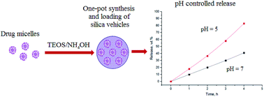
A new approach is proposed for creating of pH-sensitive mesoporous silica nanocontainers with ultrahigh capacity for amphiphilic functional compounds. It relies on the use of micelles of such compounds as templates for the sol–gel synthesis of SiO2 nanoparticles. The possibility of such one-stage synthesis and loading of SiO2 nanocontainers has been demonstrated using the micelles of the bactericidal drug Myramistin as a template.
We propose to use the micelles of the functional amphiphilic compound which then must be loaded (instead of inert surfactant ones) as a template in the sol–gel synthesis of silica nanocontainers. The results of our studies, which were carried out using the micelles of cetyltrimethylammonium bromide (CTAB) as a model functional template, suggest that this approach paves the way to a one-pot synthesis and loading of SiO2 nanocontainers with ultrahigh capacity (up to about 1 g per 1 g of SiO2) for the functional compound.9,10 Herewith the rate of functional compound release from the particles into an aqueous medium is determined by two main processes: the gradual dissolution of the silica matrix by the penetrating water and the interaction of the molecules/ions of the templating surfactant with the pores' surface. The relative contributions of these processes strongly depend on the pH of an aqueous medium.10
In this study we first demonstrate the prospects and advantages of the proposed approach using the micelle-forming, broad-spectrum bactericidal drug benzyldimethyl[3-(miristoilamino)propyl]ammonium chloride, known under the trademark Myramistin (its structural formula is shown below in Scheme 1).11 This compound can be referred to both the asymmetric cationic gemini surfactants12 and to ionic liquids.13 It should be emphasized that there are currently a rather limited number of studies aimed at the template synthesis of mesoporous silica using gemini surfactants.1,14–20 Moreover, as a rule, these studies concern the bulk materials1,14–19 synthesized mainly using the micelles of symmetric gemini surfactants as templates.14,15,17,19 To the best of our knowledge there is only one publication reporting the possibility of producing submicron mesoporous silica particles with a given size by sol–gel synthesis with the micelles of asymmetric gemini surfactant [C18H37N+(CH3)2(CH2)3N+(CH3)3]Br2.20
The critical micelle concentration of Myramistin is equal to approximately 1 mM.12 According to the dynamic light scattering data, in 1–5 mM aqueous solutions, it forms spherical micelles with an average diameter of about 5 nm. The UV spectrum of Myramistin aqueous solution is characterized by the pronounced absorption band at 263 nm (see Fig. 1). This band may be used to control both the degree of the drug incorporation into the silica nanocontainers during their synthesis and the kinetics of its subsequent release into the environment.
Fig. 1 Absorption spectra of (1) aqueous Myramistin solution with a concentration of 3 mM and (2) supernatant, sampled after precipitation of synthesized SiO2 nanocontainers. |
Mesoporous silica nanocontainers were synthesized at room temperature as follows. A weighed portion of Myramistin‡ (110 mg) was put into 75 mL of the ammonium hydroxide (Sigma-Aldrich) aqueous solution (0.33 M) under vigorous stirring. After complete dissolution of the drug, 3 mL of tetraethoxysilane (TEOS) (Sigma-Aldrich) solution in absolute ethanol (0.6 M) was added, i.e., the Myramistin/NH4OH/TEOS/C2H5OH molar ratio was equal to 3/330/23/570. The reaction system was stirred for 6 h and then left to stand for an additional 24 h. Then SiO2 particles were precipitated by centrifugation. The supernatant was removed, and the nanoparticles were redispersed in deionized water. The precipitation/redispersion procedure was repeated three times. For the thermal and IR spectral analyses, the obtained dispersion was lyophilized.
As follows from the high resolution transmission electron microscopy (HRTEM) data (Fig. 2), the spherical SiO2 particles with average diameter of 160–170 nm are formed as a result of the sol–gel synthesis. These particles are characterized by a monomodal size distribution and a clearly discernible ordered porous structure. The pores' diameter is equal to ∼3 nm.
Fig. 2 HRTEM image of the silica nanocontainers synthesized using the Myramistin micelles as a template. |
Two groups of the absorption bands are present in the FTIR spectrum of synthesized SiO2 nanocontainers (Fig. 3). The first one is inherent to Myramistin and the second one indicates the formation of the Si–O–Si network (see Table 1). Comparison of UV spectra of Myramistin stock solution and the supernatant, sampled after precipitation of synthesized SiO2 nanocontainers, suggests (Fig. 1) that of about 95 wt% of the drug introduced into the reaction system is encapsulated in the silica matrix. This result is quite understandable, taking into account that both micelles and monomeric molecules of the templating surfactant are involved in the formation of a liquid-crystalline mesophase at the early stages of the sol–gel synthesis of SiO2 nanoparticles10 (see also review1 and references therein).
Fig. 3 FTIR spectra of (1) Myramistin and (2) mesoporous silica nanocontainers synthesized using its micelles. |
Wave number, cm−1 | The assignment of the band21 | Myramistin | SiO2-Myramistin |
---|---|---|---|
2920, 2860 | Stretching vibrations of C–H bonds in alkanes | + | + |
1470 | Bending vibrations of C–H bonds in alkanes | + | + |
3030 | Stretching vibrations of C–H in benzene ring | + | + |
1660 | In-plane skeletal vibrations of the benzene ring | + | + |
3280, 3460 | Stretching vibrations of N–H bonds in secondary amides | + | + |
1040, 1200 | Stretching vibrations of Si–O–Si bonds | + | |
960 | Bending vibrations of Si–OH bonds | + |
The results of thermogravimetric analysis (TGA) indicate (Fig. 4), that the weight losses by the synthesized particles in the range, corresponding to the thermal degradation of the templating drug (150–350 °C), are about 45 wt%. Thus, the Myramistin-to-SiO2 weight ratio in the prepared mesoporous nanocontainers (i.e., their capacity for this targeted substance) is about 0.9 g g−1. This value, obviously, is significantly greater than the one achieved upon sorption of this surfactant from its solution by MSNs synthesized by the traditional scheme which implies the template's removal from the silica matrix.1–3 Thus, as was shown earlier, the amount of CTAB encapsulated in MSNs during their synthesis is 0.9 g per 1 g of SiO2.10 At the same time, the amount of CTAB sorbed from its solution by the same MSNs after removing the template does not exceed 0.3 g per 1 g of SiO2.10
Fig. 4 TGA curves for (1) pristine Myramistin and (2) SiO2 nanocontainers, synthesized using its micelles. |
Analysis of the TGA data, presented in Fig. 4, suggests that the mechanisms of thermal degradation of pristine and encapsulated into silica nanocontainers Myramistin differ significantly; a similar pattern was observed in the case of silica nanoparticles synthesized using CTAB micelles.5,22,23 One reason for this difference could be partial incorporation of head groups of Myramistin molecules in the pore walls of the growing silica nanoparticles. As well, one cannot exclude that the ions, captured by SiO2 matrix from the reaction system during sol–gel synthesis, could impact to the process of thermal degradation of the template molecules.10
A kinetic study of Myramistin release from SiO2 nanocontainers into an aqueous medium in quasi-dynamic conditions§ showed that the rate of this process depends strongly on the pH value (experiments were carried out at room temperature of 23 °C). Thus, of about 40 wt% encapsulated Myramistin desorbs in deionized water (pH ≈ 6.8) for 4 h (curve 1 in Fig. 5). At pH value of aqueous medium decreased to about 5, the rate of templating drug release increases significantly. As a result, over the same period of time, Myramistin almost completely desorbs from the nanoparticles (curve 2 in Fig. 5). These results are of considerable interest from the viewpoint of controlling the release of drugs in the body, including directly into the cells. Indeed, it is known that the intercellular pH value in healthy human tissues is 6.8–7.2, while in tumors or places of inflammation it decreases down to 5.24 Still lower pH values are typical for the stomach25 and some cell compartments, i.e. endosomes and lysosomes.26
Fig. 5 Kinetics of Myramistin release from SiO2 nanocontainers into (1) water and (2) aqueous medium with pH = 5. |
Myramistin desorption is well described by the zero-order kinetics.27,28 It means that the rate-controlling stage for release of templating molecules is the water penetration into silica nanocontainers, i.e. their swelling.28 Note that, according to our preliminary data, such penetration occurs much faster than in nanocontainers synthesized using the CTAB micelles as a template.10 It was also found that increasing the temperature to 37 °C leads to some increase in the rate of Myramistin desorption, and this effect is more pronounced in the case of the dispersion medium with pH ≈ 5 (both of these effects will be discussed in our next publication in more detail).
Footnotes |
† Electronic supplementary information (ESI) available. See DOI: 10.1039/c6ra03602c |
‡ The substance was kindly provided by Dr V. N. Barvinchenko, Institute of Surface Chemistry, Kiev, Ukraine. |
§ The term “quasi-dynamic conditions” means the periodic replacement of the aqueous medium, in which silica nanocontainers are dispersed, by its new portion, as was done earlier.10 |
This journal is © The Royal Society of Chemistry 2016 |