DOI:
10.1039/C6RA03578G
(Paper)
RSC Adv., 2016,
6, 57863-57871
Design and development of a piscine collagen blended pullulan hydrogel for skin tissue engineering
Received
8th February 2016
, Accepted 31st May 2016
First published on 7th June 2016
Abstract
This present study was designed to prepare a super-absorbent tailor-made collagen–pullulan hydrogel with improved mechanical stability and well-defined biocompatibility for skin tissue engineering. Three-dimensional (3D) scaffolds were fabricated using natural polysaccharide pullulan, cross-linked with sodium trimetaphosphate (STMP) and blended with collagen to form a polymeric network. Collagen was extracted from the skin of unexplored puffer fish (Lagocephalus inermis). The cross-linking occurred at alkaline pH at room temperature to give translucent, clear and soft hydrogels. Swelling studies revealed a remarkable water absorption property with a swelling ratio up to 320%, an ideal characteristic for the hydrogel to provide a moist wound healing environment. SEM analysis revealed the highly interconnected porous structure of the collagen blended pullulan hydrogels. MTT assay performed on NIH3T3 fibroblast cell lines revealed that the prepared hydrogels were 100% biocompatible with enhanced cell adhesion and proliferation. The hydrogels promote angiogenesis in the chick chorioallantoic membrane, which was investigated by the CAM assay. Wound healing studies exhibit a statistically significant (<0.05) response to wound contraction and re-epithelialization in an excision wound model in rats. Compared with 49% wound closure in 11 ± 2 days in the controls, 96% wound closure was observed in rats treated with the collagen–pullulan hydrogel (CGPNH). The highly porous collagen–pullulan hydrogels were successfully developed with a significant in vitro and in vivo biological performance, and are a promising biomaterial for wound healing applications.
Introduction
Hydrogels are super-absorbent three-dimensional (3D) macromolecular networks of hydrophilic polymers which play significant roles in biomedical applications, especially in skin repairs and tissue engineering. The 3D integrity of hydrogels in their swollen state is maintained by either physical or chemical cross-linking.1 The remarkable swelling property of a hydrogel is mainly due to its network elasticity and porosity. Hydrogels can absorb and release water in a reversible manner, in response to specific environmental stimuli viz. temperature, pH, ionic strength etc.2 It is possible to change the chemistry of hydrogels by controlling their polarity, swelling behaviours, surface, and mechanical properties. Such a smart behaviour towards the adjustment of physiological variations promotes them as a potential candidate for wound healing applications.3 Natural biodegradable polymers are generally hydrophilic in nature with excellent biocompatibility and weak immunogenicity.4 Since studies on natural polymers are always fascinating and are an interesting area of research for the development of newer products, and for this purpose polysaccharides and proteins have been extensively investigated for dermal reconstructions. The blending of natural polymers will produce a potentially active biomaterial with tailored properties without destroying the original properties.
Pullulan, an excellent polysaccharide produced from the yeast-like fungi Aureobasidium pullulans is composed of linear α-(1 → 6) linked maltotriose units and a small number of α-(1 → 4) linked maltotetraose units. It is biodegradable in biologically active environments, and it forms easily thermo-stable, transparent, flexible films with an extremely low oxygen permeability.5 It has good adhesive and binding properties. As a hydrophilic polymer, it has the unique characteristic of excellent swelling properties, which makes them a suitable candidate for various biomedical applications, especially in dermal tissue engineering.6
Collagen, an interesting polymer with a unique structure, size and amino acid sequence has a wide range of applications in biomedical, pharmaceutical and cosmetic industries. It is a major component of extracellular matrix (ECM) and one of the most abundant proteins in mammalian tissues. Collagen can also form fibres with extra strength and stability through its cell aggregation and cross-linking properties.7 However, its applicability is limited owing to its high cost and the probability of disease transmission from the bovine sources.8 Recent studies proved that the collagen derived from marine sources was much safer and did not produce any immunological response or allergic reactions when compared to mammalian tissue collagen.9 Thus, in the present study, collagen extracted from the skin of puffer fish Lagocephalus inermis (a novel marine source) was studied for the preparation of a hydrogel-based system. This is the first paper reporting the extraction of collagen from this under-utilized fish of Indian origin belonging to the family Tetraodontidae. In this study, we aimed to develop an effective porous biomimetic hydrogel from fish skin collagen blended with pullulan for wound healing applications.
Materials and methods
Collection of fish skin
The smooth golden puffer fish Lagocephalus inermis were collected in July 2014, from Mallipatinum seashore, Tamil Nadu, India and were brought to the laboratory at 4 °C. The skin was removed from the fish and immediately washed with distilled water to remove the blood stains and debris and cut into small pieces approximately 5 × 5 cm and stored at −20 °C until further use.
Chemicals
Pullulan (molecular weight 200![[thin space (1/6-em)]](https://www.rsc.org/images/entities/char_2009.gif) 000), sodium trimetaphosphate (STMP), collagenase A (from Clostridium histolyticum) and pullulanase were purchased from Sigma-Aldrich, India. Until and unless mentioned all the chemicals and reagents used in this experiment were obtained from Sigma-Aldrich, India. Deionized water was used throughout the studies. NIH3T3 fibroblast cell lines were procured from National Centre for Cell Sciences (NCCS), Pune, India.
000), sodium trimetaphosphate (STMP), collagenase A (from Clostridium histolyticum) and pullulanase were purchased from Sigma-Aldrich, India. Until and unless mentioned all the chemicals and reagents used in this experiment were obtained from Sigma-Aldrich, India. Deionized water was used throughout the studies. NIH3T3 fibroblast cell lines were procured from National Centre for Cell Sciences (NCCS), Pune, India.
Extraction of collagen from fish skin
Acid-soluble collagen was extracted from the puffer fish skin as per the method of Nagai and Suzuki10 with slight modification. To remove non-collagenous proteins, the fish skin was mixed with 0.1 M NaOH at a ratio of 1:10 (w/v). The mixture was then continuously stirred for 6 h at 4 °C and then washed with cold water until neutral pH was obtained. Pre-treated skins were de-fatted with 10% butyl alcohol for 48 h and the solvent was changed every 6 h. The de-fatted skin was treated with 0.5 M acetic acid for 24 h. The mixture was filtered through two layers of cheese cloth and the supernatant was separated out. Salting out of the collagen from the supernatant was achieved using 0.9 M NaCl and it was kept undisturbed for 24 h at 4 °C. Then the suspension was centrifuged at 8000 rpm for 1 h at 4 °C. Then the precipitate was dialyzed against 0.1 M acetic acid and distilled water for 24 h each, respectively. The final extracted product was acid-soluble collagen.
Hydrogel fabrication
Hydrogel fabrication was done according to the method of Dulong, with slight modifications.11 For this study, five hydrogel formulations were prepared, all of which contained a constant pullulan concentration (2 g). The polymer and cross-linking concentrations of different blends are shown in Table 1. For the synthesis of pullulan hydrogels (PNG, PNS, and PNH), 2 g of pullulan was mixed with different concentrations (1, 1.5, 2 g, respectively) of STMP in 10 mL of deionized water. Finally, 50 μL of 0.1 M NaOH was added to the mixture and stirred well for 90 min at room temperature until a transparent viscous liquid was produced. This transparent viscous liquid then underwent a sol–gel transition when incubated at 50 °C for 30 min.
Table 1 Pullulan and collagen–pullulan hydrogel compositions
| Hydrogels |
Sample |
PN (g) |
STMP (g) |
CG (g) |
NaOH (μL) |
| Pullulan hydrogels |
PNG |
2 |
1 |
0 |
50 |
| PNS |
2 |
1.5 |
0 |
50 |
| PNH |
2 |
2 |
0 |
50 |
| Collagen–pullulan |
CGPNH |
2 |
2 |
0.1 |
50 |
| Hydrogels |
CGPNH1 |
2 |
2 |
0.2 |
50 |
For the synthesis of collagen–pullulan hydrogels (CGPNH, CGPNH1), different concentrations of collagen (0, 0.1 and 0.2 g) were mixed with 2 g of pullulan and 2 g of STMP in 10 mL of deionized water with constant stirring, respectively. The pH was adjusted to 9.0 using 0.1 M NaOH and the composite mixture was stirred well for 90 min at room temperature to promote the homogenous distribution of polymers within the hydrogel. The mixture was poured into Petri dishes and incubated at 50 °C for 30 min for the formation of hydrogels. A circular punch was used to cut 6 mm diameter gels. The prepared hydrogels were washed extensively with phosphate buffered saline until neutral pH was obtained and stored at 4 °C until further use. The hydrogels were sterilized overnight under UV light prior to all experiments.
Evaluation of formulated hydrogels
Appearance. The hydrogels formulated were observed for their visual appearance, color, texture feel upon applications such as smoothness, stiffness, and stickiness.
pH. The pH of the hydrogels has also played an important role in the formation of a gel. The pH of the hydrogels was determined by a pH meter. The determinations were carried out in triplicate and the average of three readings was recorded.
Viscosity. The viscosities of the formulated hydrogels were determined using a Brook-field viscometer (spindle number LV-16) in triplicate and the average of three readings was recorded.
Physicochemical characterization of hydrogels
Fourier transform infrared spectroscopy (FTIR). FTIR analysis was done in the range of 500–4000 cm−1 with resolution 4 cm−1 using Nicolet Magna-IR 560, USA.
Thermogravimetric analysis (TGA). Thermogravimetric analysis was performed using a high resolution 2950 TGA thermogravimetric analyzer (TA instruments Co., Austria). Samples weighing between 10 and 20 mg were placed in a platinum pan and the test was carried out in a programmed temperature range of 0–800 °C at a heating rate of 5 °C min−1 under a nitrogen atmosphere at a flow rate of 50 mL min−1.
Differential scanning calorimetry (DSC). The thermal characteristics of the hydrogels were investigated using a differential scanning calorimeter (TA instruments Co., Austria) in order to measure their crystallization temperature (TC) and melting temperature (Tm). The analysis was performed at a heating rate of 20 °C min−1, from 30 °C to 200 °C, under nitrogen gas at a flow rate of 10 mL min−1.
Swelling studies. The degree of swelling could be characterized as the water retention ability of the hydrogel. Small pieces (2 cm2) of PNH and CGPNH hydrogels were weighed and soaked in phosphate buffered saline (pH 7.4) at 37 °C. Excess liquid was gently shaken off and the weights of the swollen gels were obtained. The hydrogel weights were measured at 6, 12, 18, 24, 48, and 72 h. Six samples were tested for each condition. Then, the swollen products were dried at 37 °C under vacuum to a constant weight. The equilibrium percentage of swelling (% swelling) of the hydrogels was calculated as per eqn (1):12| | |
% of swelling = [(Ws − Wd)/Wd] × 100
| (1) |
where Ws is the weight of the product after hydration for 72 h and Wd is the weight of the dried product.13
In vitro biodegradation studies. The in vitro biodegradation of the PNH and CGPNH hydrogels was carried out according to previously described procedures.14–16 The PNH and CGPNH hydrogels were incubated with collagenase A (2 mg mL−1), pullulanase (4 U mL−1) in PBS and weighed every 12 h for 7 days. Combination degradation studies using both the enzymes were conducted in PBS using the same concentration as above. The initial weight at time 0 was the dry weight of the hydrogels (W0), and after being degraded by the enzymes, the wet hydrogels were washed three times with water (W1). These hydrogels were used for subsequent measurements. The experiments were performed in triplicate under each condition at 37 °C. The weight loss (%) was calculated as follows (eqn (2)):| | |
% of weight loss = (W0 − W1)/W0 × 100
| (2) |
Morphology observation. The internal structure of the hydrogels was investigated by scanning electron microscopy (JSM-5600LV, JEOL). The hydrogels were dehydrated by a freeze dryer and sputter-coated with gold before SEM characterization.
Hemocompatibility test. The hemostatic potential of the hydrogels was determined using human blood according to Lu et al.,16 with slight modifications. The percentage hemolysis was calculated as per the following formula (eqn (3)):| |
 | (3) |
where OD is the optical density.
Biocompatibility. Biocompatibility was assessed using MTT [3-(4,5-dimethylthiazol-2-yl)-2,5-diphenyltetrazolium bromide] assay. The samples were placed on polystyrene Petri plates and were sterilized with ethanol-UV treatment. This was followed by seeding with 20 μL of NIH3T3 fibroblast cell suspension (4 × 10⁵ cells per mL) on the samples and incubated at 37 °C for 1 h to allow cell adherence to the surface of the samples. Thereafter, the samples were incubated for another 2 days. The cell viability was measured after 24 h and 48 h using MTT assay. MTT was added at the end of the incubation period and the plates were incubated for 4 h at 37 °C. Following incubation, the media was aspirated completely and the MTT formazan crystals formed were dissolved by the addition of dimethylsulfoxide (DMSO), and the absorbance was read at 570 nm using a multi-well plate reader (Multiskan Ascent VI.24). Untreated wells served as the cell control. Data were normalized to the control to evaluate the biocompatible potential of the hydrogel on cells.17,18
In vitro chick chorioallantoic membrane assay. The angiogenic property of the hydrogels was assessed by the simple and inexpensive CAM assay method. Four-day old chick eggs were obtained and incubated at 37 °C and 60% humidity in an egg incubator for 72 h. On the eighth day, 2–3 mL of albumen from the egg was aspirated at the acute pole of the egg to create a false air sac, facilitating its dissociation from the egg shell. A square window of 1 cm2 was cut and a lyophilized hydrogel disc was loaded and placed in the junction between two big vessels. The window was re-sealed and incubated for 72 h, and a sterilized methyl cellulose disc was used as the control.19
In vivo studies: maintenance of animals. All experiments were performed in compliance with the relevant laws and institutional guidelines. The animal study was approved by the institutional ethical committee (Reg. no: GSP-IAEC-2013-04-06) for conducting experiments on animals according to the regulations of CPCSEA, at the department of pharmacology laboratory, GIET School of Pharmacy, Rajahmundry, Andra pradesh, India.The wound healing potential of the hydrogel was studied using Wistar albino rats (weighing 150–200 g) of either sex. The animals were housed in polyacrylic cages and maintained under standard laboratory conditions (25 ± 2 °C) with a dark and light cycle (14/10 h). They were allowed free access to a standard dry pellet diet (Hindustan Lever, Kolkata, India) and water ad libitum.
Excision wound model. All experiments were carried out upon getting approval from the institutional ethical committee. Rats were inflicted with excision wounds according to the method of Morton and Malone with slight modifications. Animals were divided into three groups of six animals in each. Group I served as the control. Animals in Group II and Group III were treated with only PNH and CPNHG hydrogels. An excision wound of 20 mm in diameter was made on the pre-shaved, sterile dorsal surfaces of the animal by means of a sterile surgical apparatus under ether anaesthesia in aseptic conditions. The wounds were of the full thickness type extending up to the subcutaneous tissue. The day of wound infection was considered as day zero of the experimental period.20,21
Healing assessment. The progressive changes in the wound area were measured in mm at intervals of every 3 days. The gradual reduction of the wounded area was measured on the 3rd, 6th, 9th, and 12th day after initiation of the wound. Histopathological observations were done using eosin-haematoxylin staining of the tissues and compared to the control tissues for fibroblast proliferation, angiogenesis and collagen synthesis.
Statistical analysis. All the results are given as a mean ± standard deviation of three individual experiments (n = 3). The comparison between means was done by one-way analysis of variance (P < 0.05) followed by Duncan’s multiple range analysis using statistical software package SPSS, version 13.0.
Results and discussion
Different hydrogels were prepared at different cross-linking densities by varying the STMP concentration (Table 1) at constant NaOH and pullulan concentration (PNG, PNS and PNH). For a low concentration of STMP (PNG and PNS), no cross-linking reaction occurred properly. The best result was obtained with the PNH hydrogel, where the polymer and STMP ratios are equal. Various concentrations (0, 5% and 10%) of extracted fish skin collagen were blended with constant pullulan (2 g) and STMP (2 g) concentration for the preparation of the collagen–pullulan hydrogel (CGPNH, CGPNH1) (Fig. 1 and 2). The prepared hydrogels were characterized by their appearance, pH, viscosity and swelling studies (Table 2). The prepared PNH and PNH1 gels were non-viscous and hard in texture due to the influence of pH and the cross-linking agent (STMP), whereas the CGPNH1 hydrogel showed a more viscous and flexible texture. The PNH and CGPNH hydrogels were clear, soft, flexible, homogeneous and translucent in nature with extra strength and stability through self-aggregation and cross-linking (Fig. 3 and 4). On the basis of the above studies the PNH and CGPNH formulations were found to be more optimum for further in vitro and in vivo studies.
 |
| | Fig. 1 Pullulan cross-linked with STMP under alkaline conditions. | |
 |
| | Fig. 2 Synthesis of collagen–pullulan hydrogel (CGPNH). | |
Table 2 Characterization of formulated hydrogels
| Hydrogels |
Appearance |
Viscosity |
pH |
Gelation time |
| PNG |
Yellowish color, semi-solid |
3000 cps |
8.2 |
60 min |
| PNS |
Glossy, yellowish color |
3200 cps |
8.6 |
45 min |
| PNH |
Glossy, clear, transparent gel |
4000 cps |
9.0 |
20 min |
| CGPNH |
Glossy, clear, transparent gel |
4600 cps |
9.0 |
20 min |
| CGPNH1 |
Glossy, clear, transparent gel |
4800 cps |
9.0 |
30 min |
 |
| | Fig. 3 Transparent and soft CGPNH hydrogels. | |
 |
| | Fig. 4 Different concentrations of pullulan and collagen–pullulan hydrogel formation: (a) PNG, hydrogel not formed, (b) PNS, (c) PNH, (d) CGPNH, and (e) CGPHN1. | |
Thermogravimetric analysis (TGA)
The thermal stability of the CGPNH hydrogel in comparison with that of the PNH hydrogel and puffer fish skin collagen was characterized using TGA as shown in Fig. 5. The thermogram of the pure collagen and pullulan hydrogel consists of three degradation stages, while that of the porous CGPNH hydrogel consists of four stages. From the graph, it can be analysed that below 100 °C, all the sample weight loss was mainly due to water loss from the protein and polysaccharide. For collagen and pullulan major weight loss occurred at 60% (in the ranges 175–380 °C and 255–384 °C) and 19% (in the ranges 400–503 °C and 451–580 °C), which signifies the breakdown or chain cleavage of the organic compounds leading to rapid weight loss and the formation of gaseous elements.16,22–24 Furthermore, these two peaks assigned to pullulan were observed in the thermogram of the synthesized porous CGPNH hydrogels in the range of 365–490 °C of 30% weight loss and 455–520 °C of 8%, respectively. These peaks could be assigned to the thermal degradation of polysaccharide rings. At the end of the experiment at 830 °C, only 10.11% of the cross-linked CGPNH hydrogel remains as residue. From the above result, it is confirmed that collagen blended with pullulan extensively improves the thermal stability of the CGPNH hydrogel when compared with the cross-linked pullulan hydrogel (PNH).
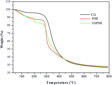 |
| | Fig. 5 TGA curve of fish skin collagen (CG), pullulan hydrogel (PNH), and collagen–pullulan hydrogel (CGPNH). | |
Differential scanning calorimetry (DSC)
Fig. 6 illustrates the DSC thermograms of the pure collagen, PNH and CGPNH hydrogels. Relatively large and sharp endothermic peaks observed in the DSC curves for each of the membranes are due to the glass transition (Tg) of molecules at the particular temperature. The glass transition temperature of extracted fish skin collagen was found to be at ∼89 °C, whereas the PNH hydrogel exhibits a glass transition temperature at ∼147 °C.2,25,26 The blended collagen–pullulan hydrogel showed two thermogram peaks of glass transition temperature at 94 °C and 197 °C. A possible cause of such a high glass transition temperature could be the existence of strong hydrogen bonding between the cross-linked pullulan blended with collagen24,26 (CGPNH).
 |
| | Fig. 6 DSC thermograms of fish skin collagen (CG), pullulan (PNH), and collagen–pullulan hydrogel (CGPNH). | |
Fourier transform infrared spectroscopy (FTIR)
FTIR peaks of fish skin collagen (Fig. 7) display bands at 1650, 1554 and 1253 cm−1, which are characteristic of amide I, II and III bands, respectively.27,28 The bands at 3421 and 2925 cm−1 represent the stretching vibrations of N–H and C–H bonds, respectively.10,29 The infrared spectrum of the cross-linked PNH hydrogel exhibits a strong absorption peak at 3315 cm−1 due to OH stretching and vibration and at 2940 cm−1 due to C–H stretching. It also displayed the typical absorption bands for the α-configuration of α-D-glucopyranose units in pullulan at 849 cm−1. The FTIR spectrum of the porous CGPNH hydrogel shows a broad peak around 3305 cm−1 indicating stretching of hydroxyl groups and an absorption band at 3164 cm−1 indicating the presence of a hydroxyl group with polymeric association.2,23,24 However, the most characteristic bands of collagen were observed in the spectrum of the hydrogel with two very sharp peaks at 1646 cm−1 (N–H) and 1543 cm−1 (C–O). At the same time, there was only a single peak at 1559 cm−1 assigned to the stretching vibration of C–O in pullulan. Other features of pullulan were also found from the spectrum, including a C–H bend (1307 cm−1), C–O stretch (1122 cm−1) and a very sharp peak at 1021 cm−1 belonging to the stretching of the C6–OH bonds. Moreover, the two main linkages of pullulan, α-(1,4) and α-(1,6)-D-glucosidic bonds, were observed at 741 cm−1 and 907 cm−1. From the above discussion, it is confirmed that the blending of collagen with pullulan has effectively occurred.
 |
| | Fig. 7 FTIR analysis of pullulan hydrogel (PNH), fish skin collagen (CG), and collagen–pullulan hydrogel (CGPNH). | |
Swelling studies
The swelling behaviour of fish skin collagen, cross-linked PNH and CGPNH hydrogels was investigated in PBS buffer at 37 °C for 72 h (Fig. 8). The CGPNH hydrogel indicated the highest water retention capacity (∼390%) compared to that of the PNH hydrogel (∼210%) and collagen (∼110%). The swollen hydrogels retained their original shape and did not degrade after overnight incubation, and due to these characteristic features it can be categorized as super-absorbent. The water binding ability of the hydrogel membrane could be mainly attributed to the hydrophilic nature of the pullulan. In general, the water uptake decreases as the cross-linking degree is increased because of a decrease in the number of hydrophilic groups as well as more difficulty in the structural expansion due to the more dense covalently linked network. This swelling property of the hydrogel combined with the porosity will greatly aid in the absorption of exudates in a wound environment.26 Hence the prepared CGPNH hydrogel may be an excellent wound dressing material.
 |
| | Fig. 8 Swelling studies of fish skin collagen, PNH and CGPNH hydrogels. | |
SEM analysis
Fig. 9 shows SEM photographs of the internal microstructure morphology of all 5 samples of the prepared hydrogel. The PNG and PNS hydrogels show a rough surface morphology whereas in the CGPNH hydrogel, a more ordered porous structure was produced with highly interconnected pores compared to the PNH hydrogel, which shows a less porous structure. The CGPNH1 hydrogel showed an irregular porous morphology, with increased pore size. It is assumed that these pores are the regions of water permeation and interaction sites of external stimuli with the hydrophilic groups of the hydrogels. Therefore the highly ordered porous structure and relatively increased cross-linking density of the CGPNH hydrogels are found to be more suitable for the application of wound dressing and targeted drug delivery applications.
 |
| | Fig. 9 SEM analysis of the surface morphology of the prepared hydrogels: (a) PNG, (b) PNS, (c) PNH, (d) CGPNH, and (e) CGPNH1 hydrogels. Among all the prepared hydrogels, the CGPNH hydrogel showed a more ordered and highly interconnected porous structure. | |
In vitro biodegradation studies
The in vitro biodegradation studies of the hydrogel were assessed by measuring their degradation rate and biological stability against collagenase A (2 mg mL−1), pullulanase (4 U mL−1) and the combination of both enzymes (Fig. 10). Collagenase brought about 64.2% and 37.9% weight loss while pullulanase showed 53.2% and 42.1% weight loss in the PNH and CGPNH samples over a period of 7 days. Whereas, the combination of these two enzymes in the degradation studies showed 80.2% and 31.7% weight loss in PNH and CGPNH, respectively. The resistance to enzymatic degradation of the synthesized collagen blended pullulan hydrogel was greater than that of the cross-linked pullulan hydrogel. As expected, the CGPNH hydrogel showed an improved mechanical property as well as a reduced biodegradation rate.
 |
| | Fig. 10 In vitro degradation studies of CGPNH hydrogel and PNH hydrogels: (A) collagenase, (B) pullulanase, and (C) combination of collagenase and pullulanase enzymes. Data are presented as mean ± SD (n = 3). | |
Hemocompatibility studies
The hemocompatibility test results for the piscean collagen, PNH and CGPNH hydrogel are shown in Fig. 11. All the three samples were found to be below 2% hemolysis indicating that the prepared samples were highly hemocompatible and suitable for biomedical applications.30–32
 |
| | Fig. 11 Hemocompatibility studies of CG, PNH and CGPNH hydrogels showing 100% hemocompatibility on the prepared samples. | |
CAM assay
Angiogenesis is a complex process involving the extensive interplay between cell soluble factors and extracellular matrix (ECM) components.33 Angiogenesis and neovascularization are critical determinants of the wound healing outcome. When compared with the control, the CGPNH hydrogel promoted angiogenesis on the chick chorioallantoic membrane on the 12th day (Fig. 12). The newly formed blood vessel enhances the healing process by providing nutrition and oxygen to the growing tissues. It can be evident that the prepared CGPNH hydrogel has significantly promoted the growth of new blood vessel formation.
 |
| | Fig. 12 In vitro CAM assay of the (i) control and (ii) CGPNH hydrogel, showing increased growth of blood vessel formation on a chick chorioallantoic membrane on the 12th day, when compared to the control. | |
Biocompatibility studies (MTT assay)
Biocompatibility is known as the compatibility of the hydrogel with the immune system. MTT assay is used to demonstrate the biocompatibility of the hydrogel scaffolds (Fig. 13). CGPNH enhances the growth and cell viability of the cells after 48 h and 72 h of incubation. Thus, the collagen–pullulan hydrogel significantly induces proliferation of the NIH3T3 fibroblast cell lines, which was significantly (P < 0.05) higher than that of the PNH hydrogel. In the present study, the cell adherence and cell proliferation potential of the PNH and CGPNH hydrogels were assessed using NIH3T3 cells. Fig. 13(a) illustrates the fluorescence images of the cells observed at different time intervals. The result shows no significant difference in the fluorescence intensity in the synthesized CGPNH hydrogel treated cells when compared to that of the control. Furthermore, no morphological changes were observed in the treated samples. The results of cell viability and quantification analysis showed 100% cell viability in both the samples suggesting that the synthesized pullulan and collagen pullulan hydrogel do not have any negative impact on the cell viability. The CGPNH hydrogel possesses an excellent 100% biocompatibility since its hydrophilic surface has a low interfacial free energy, which may be in contact with body fluids and enhances the cells to adhere to the surface of the hydrogels. This excellent biocompatibility and proliferative potential of the hydrogel would definitely augment the healing process when used in a wound environment.
 |
| | Fig. 13 (a) In vitro cytotoxicity and cell proliferation studies of the prepared PNH and CGPNH hydrogels on NIH3T3 fibroblast cell lines for 48 hours. (b) Biocompatibility assessment of the samples using MTT assay by measuring the absorbance at 570 nm in comparison with the control. The values are shown as mean ± SEM *(P < 0.05). | |
Wound healing studies of the hydrogel on animal models
Wound healing studies of the hydrogel on the animal model were performed and the healing of wounds covered with the PNH hydrogel and CGPNH hydrogels was compared with an open wound (Fig. 14). The healing rate of wounds treated with a hydrogel dressing seems to proceed faster than that of the untreated wounds. Histopathological evaluation of the healing tissue on day 3, 9 and 12 (Fig. 15) was studied. The hydrogel dressings adhered better to the wound bed and could be removed from the wound surface without causing any damage or trauma to the tissue. There was a marked infiltration of the inflammatory cells and increased blood vessel formation on the CGPNH treated samples observed on the 9th day, and slightly improved progressive changes in inflammation with fibroblast growth were observed in the PNH treated hydrogel whereas inflammation was still persistent in open wounds with the slow healing process. Full thickness epidermal regeneration which covered the wound area completely was achieved in day 12 with enhanced fibroblast proliferation, angiogenesis and re-epithelialization in the CGPNH treated wounds and for the PHN treated wounds it took more than 14 ± 2 days. Wounds with little inflammation and impaired healing were observed on day 12 in open wounds and complete healing of the wound was achieved at more than 19 ± 2 days. On the basis of the histopathological studies, it can be revealed that the prepared collagen blended pullulan hydrogels have high potential in accelerating the wound healing process by providing a moist wound environment for rapid cellular repair and regeneration. Moreover, the super-absorbent nature of the hydrogel can absorb the excess exudates and prevent the microbial contact in the wound bed, which also contributes to the rapid healing of the wound.
 |
| | Fig. 14 Wound healing studies of hydrogels on excision wounds on rats, showing that CGNH hydrogels healed the wound faster (99%) than PNH (81%) and the control (53%) did on day 12. | |
 |
| | Fig. 15 Histopathological studies on day 12 showed enhanced proliferation of the CGPNH treated hydrogels than that of PNH and the control. | |
Conclusions
The preparation of a porous hydrogel from fish skin collagen and the polysaccharide pullulan was successfully achieved and its efficacy was assessed as a potential tissue engineering scaffold. The results revealed that the collagen blended with pullulan has significantly improved thermal stability and mechanical properties with excellent hydrophilicity and biocompatibility. Wound healing studies showed that these hydrogels can absorb the exudates and reduce the trauma by maintaining a moist wound healing environment. It also promotes wound healing by enhancing granulation, tissue regeneration and formation of new blood vessels in the wounded sites. The combination of the prepared hydrogel from the natural polymers alone can act as a potent wound dressing material without adding any drugs; this will also add advantages including simplicity, versatility, non-toxicity and cost effectiveness. Since the results are promising, further experiments could be tried to enable its use in clinical applications.
References
- K. I. Shingel, Carbohydr. Res., 2004, 339, 447–460 CrossRef CAS PubMed.
- H. Li, J. Yang, X. Hu, J. Liang, Y. Fan and X. Zhang, J. Biomed. Mater. Res., Part A, 2011, 98, 31–39 CrossRef PubMed.
- I. Bataille, A. Meddahi-Pelle, C. L. Visage, D. Letourneur and F. Chaubet, Pullulan for biomedical use, chapter 4, pp. 145–182 Search PubMed.
- V. W. Wong, K. C. Rustad, M. G. Galvez, E. Neofytou, J. P. Glotzbach, M. Januszyk, M. R. Major, M. Sorkin, M. T. Longaker, J. Rajadas and G. C. Gurtner, Tissue Eng., Part A, 2011, 17, 631–645 CrossRef CAS PubMed.
- M. R. Rekha and C. P. Sharma, Trends Biomater. Artif. Organs, 2007, 20, 111–116 Search PubMed.
- D. E. Al Kumar, Int. J. Basic Appl. Sci., 2012, 1, 202–219 Search PubMed.
- C. H. Lee, A. Singla and Y. Lee, Int. J. Pharm., 2001, 221, 1–22 CrossRef CAS PubMed.
- S. Yamada, K. Yamamoto, T. Ikeda, K. Yanagiguchi and Y. Hayashi, BioMed Res. Int., 2014, 2014, 302932 Search PubMed.
- T. Silva, J. Moreira-Silva, A. Marques, A. Domingues, Y. Bayon and R. Reis, Mar. Drugs, 2014, 12, 5881–5901 CrossRef CAS PubMed.
- T. Nagai and N. Suzuki, Food Chem., 2000, 68, 277–281 CrossRef CAS.
- J. Zhang, X. Ma, D. Fan, C. Zhu, J. Deng, J. Hui and P. Ma, Mater. Sci. Eng., C, 2014, 43, 547–554 CrossRef CAS PubMed.
- V. Dulong, S. Lack, D. Le Cerf, L. Picton, J. P. Vannier and G. Muller, Carbohydr. Polym., 2004, 57, 1–6 CrossRef CAS.
- J. Zhang, X. Ma, D. Fan, C. Zhu, J. Deng, J. Hui and P. Ma, Mater. Sci. Eng., C, 2014, 43, 547–554 CrossRef CAS PubMed.
- X. Li, W. Xue, Y. Liu, D. Fan and X. Ma, J. Mater. Chem. B, 2015, 3, 4742–4755 RSC.
- A. Abed, N. Assoul, M. Ba, S. Derkaoui, P. Portes, L. Louedec, P. Flaud, I. Bataille, D. Letourneur and A. Meddahi-pelle, J. Biomed. Mater. Res., Part A, 2011, 535–542 CrossRef CAS PubMed.
- J. Lu, X. Y. Lin, B. Jiang, X. D. Li, J. Y. Chen and X. D. Zhang, Key Eng. Mater., 2005, 288–289, 377–380 CrossRef CAS.
- B. Seal, Mater. Sci. Eng., R, 2001, 34, 147–230 CrossRef.
- C. Deng, P. Zhang, B. Vulesevic, D. Kuraitis, F. Li, A. F. Yang, M. Griffith, M. Ruel and E. J. Suuronen, Tissue Eng., Part A, 2010, 16, 3099–3109 CrossRef CAS PubMed.
- D. C. West, W. D. Thompson, P. G. Sells and M. F. Burbridge, Methods Mol. Med., 2001, 46, 107–129 CAS.
- M. Fronza, B. Heinzmann, M. Hamburger, S. Laufer and I. Merfort, J. Ethnopharmacol., 2009, 126, 463–467 CrossRef CAS PubMed.
- M. Madaghiele, A. Sannino, L. Ambrosio and C. Demitri, Burns Trauma, 2014, 2, 153 CrossRef.
- F. Pati, B. Adhikari and S. Dhara, Bioresour. Technol., 2010, 101, 3737–3742 CrossRef CAS PubMed.
- K. I. Shingel, Carbohydr. Res., 2002, 337, 1445–1451 CrossRef CAS PubMed.
- R. F. Pereira, A. Mendes and P. J. Bártolo, Chem. Eng. Trans., 2013, 32, 1009–1014 Search PubMed.
- T. Nagai, Y. Araki and N. Suzuki, Food Chem., 2002, 78, 173–177 CrossRef CAS.
- M. A. Hussain, K. Abbas, B. A. Lodhi, M. Sher, M. Ali, M. N. Tahir, W. Tremel and S. Iqbal, Arabian J. Chem., 2013, 172–178 Search PubMed.
- P. Singh, S. Benjakul, S. Maqsood and H. Kishimura, Food Chem., 2011, 124, 97–105 CrossRef CAS.
- J.-W. Woo, S.-J. Yu, S.-M. Cho, Y.-B. Lee and S.-B. Kim, Food Hydrocolloids, 2008, 22, 879–887 CrossRef CAS.
- P. Kittiphattanabawon, S. Benjakul, W. Visessanguan, T. Nagai and M. Tanaka, Food Chem., 2005, 89, 363–372 CrossRef CAS.
- J. Yeom, S. H. Bhang, B. Kim, M. S. Seo, E. J. Hwang, I. H. Cho, J. K. Park and S. K. Hahn, Bioconjugate Chem., 2010, 240–247 CrossRef CAS PubMed.
- X. Li, W. Xue, C. Zhu, D. Fan and Y. Liu, Mater. Sci. Eng., C, 2015, 57, 189–196 CrossRef CAS PubMed.
- M. Liu, J. Fan, K. Wang and Z. He, Drug Deliv., 2007, 14, 397–402 CrossRef CAS PubMed.
- T. I. Valdes, D. Kreutzer and F. Moussy, J. Biomed. Mater. Res., 2002, 62, 273–282 CrossRef CAS PubMed.
|
| This journal is © The Royal Society of Chemistry 2016 |
Click here to see how this site uses Cookies. View our privacy policy here. 


