Construction and luminescence property of a highly ordered 2D self-assembled amphiphilic bidentate organoplatinum(II) complex

Yunjie Xianga, Wenjing Lia, Yuxi Fanga, Dengqing Zhanga, Xianying Li*b and Wusong Jin*ac
aState Key Laboratory for Modification of Chemical Fibers and Polymer Materials, College of Chemistry, Chemical Engineering and Biotechnology, Donghua University, 2999 North Renmin Road, Songjiang, Shanghai 201620, P. R. China
bSchool of Environmental Science and Engineering, Donghua University, 2999 North Renmin Road, Songjiang, Shanghai 201620, P. R. China. E-mail: seanlee@dhu.edu.cn
cKey Laboratory of Natural Resources of Changbai Mountain & Functional Molecules (Yanbian University), Ministry of Education, Park Road 977, Yanji, 133002, P. R. China. E-mail: wsjin@dhu.edu.cn

Received 7th February 2016 , Accepted 4th March 2016

First published on 8th March 2016


Abstract

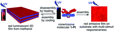
Rod-coil shaped amphiphilic bidentate dichloro(phenanthroline)platinum(II) complexes, 1-Pt and 2-Pt, which possess different hydrophilic lateral chains on one side of an identical aromatic rod core, are synthesized. 1-Pt first forms dimers then arranges into well-defined two-dimensional (2D) films consisting of highly ordered molecular arrays both from methanol and on substrate. 2-Pt, as a building-block, yields micrometer-sized rigid 2D sheets without formation of an initial dimer. Red luminescence of the film is induced by self-assembly of nonemissive 1-Pt molecules, whereas sheets based on nonemissive 2-Pt gives weak yellow emission. These results indicate that the coil-rod ratio plays an important role in the structure and optical properties of these self-assemblies. Moreover, the film on the substrate at the macroscopic scale, exhibits multi-stimuli responsiveness, which predicts its application in smart chemosensing devices and probes.


Introduction

The square-planar bidentate platinum(II) complexes have attracted much attention because of their particularly appealing photophysical and photochemical properties.1 These appealing properties are not observed in tetrahedral and octahedral metal complexes, since the forces, such as PtII⋯PtII and/or π–π between two molecules2 can be facilitated by cofacial molecular interactions in these platinum(II) complexes. The bidentate dichloro(diimine)platinum(II) complexes [Pt(diimine)Cl2], in which the chlorine atom as an auxiliary ligand, is one of a useful building blocks for constructing luminescent materials.3 However, Pt(diimine)Cl2 complexes are often found to be nonemissive in solution at room temperature because the molecules are extremely distorted at the low-lying d–d excited states in contrast to the ground state, which leads to nonradiative decay.1a To improve their emissive properties, either the cultivation of crystals, which induces the emission by enhancing PtII⋯PtII and/or π–π interactions between Pt(diimine)Cl2, or substitution of the auxiliary Cl with other groups, such as a strong σ-donor and a strong π-acceptor, is usually utilized.4 In this regard, we used another simple strategy, facile solution processable self-assembly approach, to increase PtII⋯PtII and/or π–π interactions between Pt(diimine)Cl2 to induce luminescence. In the self-assembling process, PtII⋯PtII and/or π-π interactions are convenient to be modulated by external influences, which is beneficial to generating the excellent luminescent materials. To date, there have been few examples of materials with induced luminescence by efficient self-assembly of bidentate Pt(diimine)Cl2.

On the other hand, luminescent organoplatinum(II) 2D materials, such as films, are promising candidates as active layers for chemosensing and organic electronic devices.5 Their structures and luminescence properties are governed by PtII⋯PtII and/or π–π interactions through control of the packing of molecules.6 Construction of such noncovalently linked quasi-2D structures are regarded as of interest because during self-assembly of small molecules into such structures at the nanoscopic or molecular levels, precise control of the bilateral intermolecular interactions is necessary but still remains a longstanding challenge.7 Without elaborate design of the building blocks, many defects are usually formed, leading to ill-defined and/or nonemissive films. Considering photoluminescence and application, the presence of highly ordered arrays by PtII⋯PtII and/or π–π interactions is essential to avoid unexpected agglomerates.8 Despite some luminescent nano-objects of organoplatinum(II) that have been constructed by crystallization, a facile solution-processable 2D material is more realistic for application. Up to now, reports on the preparation of highly ordered films based on Pt(diimine)Cl2 with induced luminescence are still limited.

Amphiphiles composed of rigid hydrophobic rod and hydrophilic coil segments are useful scaffolds for constructing switchable hierarchical structures responding to the selected conditions.9 The volume fraction of rod to coil segments, the cross-sectional area of the coil segment, or the shape of the rigid segment, greatly influence the molecules to form diversely structured nanomaterials.10 Amphiphiles consisting of rational coil and aromatic rod segments which could arrange side-by-side have the tendency to grow in two dimensions to form ordered self-assemblies.11 In the present study, we synthesize novel amphiphilic bidentate platinum(II) complexes 1-Pt and 2-Pt (Scheme 1). These complexes possess the same planar phenanthroline (Phen) aromatic rod but a different coil region. 1-Pt possesses four lateral triethylene glycol (OTEG) chains whereas 2-Pt contains two hydrophilic OTEG segments. 1-Pt molecules are successfully constructed into highly ordered microscopic and macroscopic 2D films from solution and on substrate with induced red luminescence by self-assembly. In the 2D films, the molecular motion which results in nonradiative decay inducing the luminescence quenching is achieved to be restrained. Compared with the weakly emissive sheets of 2-Pt, this work provides insight into the molecular structure–optical property relationships of organoplatinum(II) 2D self-assemblies at the molecule level.


image file: c6ra03553a-s1.tif
Scheme 1 Reaction scheme of the designed amphiphilic bidentate dichloro(phenanthroline)platinum(II) complexes, 1-Pt and 2-Pt.

Results and discussion

Synthesis of amphiphilic rod–coil Pt(Phen)Cl2 complexes

Target amphiphilic rod–coil 1-Pt and 2-Pt were synthesized following the synthetic route shown in Scheme 1. 1,10-Phenanthroline hydrate reacted with Br2 to prepare brominated phenanthroline 7. Compound 7 was oxidized to yield 3,8-dibromo-1,10-phenanthroline-5,6-dione 8. Compound 9 and 10 with different side chain was prepared by reaction of compound 8 with 2-bromomethyl-1,3-bis{2-[2-(2-methoxyethoxy)ethoxy]ethoxy}propane 5 and 1-(2-(2-methoxyethoxy)ethoxy)-2-bromoethane 6, respectively. Compound 9 reacted with biphenyl-4-ylboronic acid to afford 1. Amphiphilic rod–coil 1-Pt was obtained through reaction of 1 with K2PtCl4. 2-Pt was synthesized following the similar reaction route as 1-Pt from compound 10 by reacting with biphenyl-4-ylboronic acid and further with K2PtCl4.

2D self-assembly of Pt(Phen)Cl2 with induced luminescence from methanol solution

In CH2Cl2 and THF, the amphiphilic 1-Pt molecule, with four flexible side chains, formed a nonemissive transparent solution owing to their relatively high solubility in these solvents. Whereas, in water, 1-Pt did not exhibit a good solubility and formed an orange luminescent precipitate (Fig. S1). Interestingly, from methanol, 1-Pt formed a self-assembly with a red emission at high concentration (Fig. 1a inset).
image file: c6ra03553a-f1.tif
Fig. 1 (a) Emission spectra of 1-Pt with different concentrations of 1.0 × 10⁻⁷, 1.0 × 10⁻⁶, 5.0 × 10⁻⁶, 1.0 × 10⁻⁵, 5.0 × 10⁻⁵, 5.0 × 10⁻⁴, 6.0 × 10⁻⁴, 1.0 × 10⁻³, and 1.3 × 10⁻³ M, and (b) emission spectra of 2-Pt with different concentrations of 1.0 × 10⁻⁶, 5.0 × 10⁻⁶, 1.0 × 10⁻⁵, 1.0 × 10⁻⁴, 5.0 × 10⁻⁴, and 9.0 × 10⁻⁴ M in methanol, pass length = 1 cm, λex = 388 nm, slit width = 3. Inset (a) luminescent photo of 1-Pt complex and inset (b) photo of 2-Pt luminescence at concentration of 5.0 × 10⁻⁴ in methanol at room temperature irradiated by UV light at 365 nm.

Typically, the suspended methanol solution of 1-Pt was heated to 65 °C, resulting a transparent yellow solution without emission under irradiation with UV light at 365 nm. Upon cooling down to 25 °C, the yellow suspension emitting red emission was gradually formed within a few minutes. At a dilute concentration (1.0 × 10⁻⁶ M), self-assembly was not achieved and the solution was nonemissive (Fig. S2). In the electronic absorption spectrum (Fig. S3-a), the band at 376 nm and 419 nm originated from the singlet metal to ligand charge transfer (1MLCT) and the triplet metal to ligand or triplet metal to metal to ligand charge transfer (3MLCT/3MMLCT), respectively. These bands arose from the coordination of ligand 1 with PtII.12 A dimerization plot for the monomer–dimer equilibrium was performed, which yielded a linear plot with an R2 of 0.9988 (Fig. 2a and b). The results indicated the existence of dimer arising from metal–metal and/or π–π interactions.13 A weakly emissive band centered at 667 nm in the luminescence spectrum (Fig. S3-b) appeared, which was attributed to the presence of the dimer.14 In the concentration range from 1.0 × 10⁻⁷ to 5.0 × 10⁻⁵ M (Fig. 1a), two emission bands appeared at 438 nm from the monomer and 667 nm from the dimer respectively. With an increasing concentration, the intensity of the monomeric band initially increased and then decreased when the concentration reached to 5.0 × 10⁻⁵ M, which indicated the disappearance of monomer, while the band arising from the dimer15 exhibited an increased intensity and a gradual blue-shift. From 5.0 × 10⁻⁴ M to 1.3 × 10⁻³ M, the dimeric band centered at 667 nm blue-shifted to 614 nm,16 which were assignable to the formation of the self-assembly by further aggregation of dimers. In above aggregate, the free rotation of the molecules can be suppressed to reduce the inefficient radiation,17 in order to tune them from being quenched to emissive. Scanning electronic micrograph (SEM) of an air-dried suspension sample of 1-Pt from methanol (5.0 × 10⁻⁴ M) on a silicon wafer surface, showed a wrinkled 2D film with micrometer size (Fig. 3a). Transmission electronic microscopy (TEM) indicated the multilayer structure of the film (Fig. 3b). Interestingly, the magnified TEM micrograph showed that each layer was consisted of a highly ordered array of equidistant, parallel line-like architectures with spacing of 0.3–0.4 nm (Fig. 3c).


image file: c6ra03553a-f2.tif
Fig. 2 (a) UV-vis spectra of 1-Pt complex in methanol with the concentrations of 1.0 × 10⁻⁶, 2.5 × 10⁻⁶, 5.0 × 10⁻⁶, 1.0 × 10⁻⁵, 2.5 × 10⁻⁵, and 5.0 × 10⁻⁵ M, and (c) UV-vis spectra of 2-Pt complex in methanol at the concentrations of 1.0 × 10⁻⁶, 2.5 × 10⁻⁶, 5.0 × 10⁻⁶, 7.5 × 10⁻⁶, 1.0 × 10⁻⁵, 2.5 × 10⁻⁵, 5.0 × 10⁻⁵, and 7.5 × 10⁻⁵ M (arrow shows spectra with increasing concentration). (b) Dimerization plot for a monomer–dimer equilibrium of 1-Pt in methanol at 328 nm, R2 for the fitting of linear line is 0.9988. (d) Nonlinear dimerization plot for 2-Pt in methanol at 391 nm. Inset (a) and (c) are the corresponding zoomed-in UV-vis spectra.

image file: c6ra03553a-f3.tif
Fig. 3 (a) SEM, (b) TEM and (c) magnified TEM image of the 1-Pt film. (d) SEM, (e) TEM and (f) magnified TEM image of the sample of 2-Pt. SEM and TEM samples were prepared by drop-casting solution onto the silicon wafer and copper grid, respectively.

In contrast, 2-Pt, bearing two flexible hydrophilic side chains, easily precipitated from methanol, which was attributed to the reduced solubility and hydrophilicity. The yellow emissive precipitate of 2-Pt was obtained from a dilute methanol solution (1.0 × 10⁻⁶ M) by a similar heating and cooling process. For 2-Pt, no dimer was formed, which was indicated by the nonlinear dimerization plot (Fig. 2c and d). With increasing concentration, the corresponding dimeric band at 620–700 nm did not appeared in the concentration-dependent luminescence spectra (Fig. 1b), which indicated that the molecular arrangement of this yellow luminescent 2-Pt was different from that of red emissive 1-Pt. The SEM micrograph displayed that the sample consisted of large amounts of rigid 2D sheets in micrometer length and width (Fig. 3d). Differing from 1-Pt, the TEM image did not exhibit ordered arrays in the sheets (Fig. 3e and f). Combined with the former result of the nonlinear dimerization plot (Fig. 2d), it was concluded that the rigid 2D sheets arranged by 1-Pt without formation of initial dimer. This might arise from the quick precipitation of 2-Pt with relatively low solubility from methanol. These results demonstrated the critical role of the molecular structure, especially, the ratio of lateral coil to rod, both in the formation of highly ordered structure and corresponding optical properties.

X-ray diffraction (XRD) was used to study more details about the packing models and the luminescence mechanism. The XRD profile (Fig. 4b) of the 1-Pt film on a quartz surface showed that the d-spacings of 28.85, 16.16 and 9.50 Å (2θ < 10°) in the ratio of 1[thin space (1/6-em)]:[thin space (1/6-em)]2[thin space (1/6-em)]:[thin space (1/6-em)]3 indicated the formation of lamellar structure. From the density functional theory (DFT) calculation results of 1-Pt, the width of the 1-Pt head part was 6.91 Å (Fig. S4) along the b axis. A spacing of 9.50 Å, much bigger than the width of one molecular head and smaller than twice of them, indicated that two 1-Pt partially overlapped together. The peak with a d-spacing of 3.82 Å in the wide-angle X-ray diffraction (WAXD) pattern, corresponding to the Pt⋯Pt distance along c axis or π–π stacking, indicated Pt⋯Pt or π–π interactions between two overlapped 1-Pt molecules. These results demonstrated that two 1-Pt molecules stacked upon each other in a head-to-head manner with partial overlap, and the tails extended to the outer, to form a dimer, probably due to the steric hindrance of lateral coils. A spacing of 22.87 Å, smaller than the length of one dimer along the a axis, suggested that dimers might overlap side-by-side with a center-to-center (Pt-to-Pt) distance of 22.87 Å between two dimers to form 1D structure along the a axis (Fig. 4a).18 This configuration subsequently assembled into a 2D monolayer through weak π–π or Pt⋯Pt interactions (3.99 Å in WAXD pattern) along the c axis, which is in accordance with the spacing described in the magnified TEM image (Fig. 3c).18 The calculated thickness of the monolayer was about 44.52 Å. The atomic force microscopy (AFM) image of the film (Fig. S5), prepared by diluted solution (1.0 × 10⁻⁶ M), showed that the height of the layers was in 4.0–5.5 nm, in agreement with the calculated value, which confirmed the existence of monolayer structure. Such monolayer further cross-connected in a tail-to-tail method along the b axis to form lamellar structure, in which the center-to-center distance of the monolayers is 28.85 Å (Fig. 4a). These results suggested that the Pt⋯Pt and/or π–π interactions were enhanced and/or the motion of molecules were restrained by the dense packing of amphiphilic 1-Pt molecules in the 2D self-assembly to induce red luminescence. Whereas, for 2D sheets of 2-Pt with two hydrophilic tails, XRD patterns (Fig. S6) showed d-spacings of 17.52 Å and 8.77 Å (2θ < 10°) in the ratio of 2[thin space (1/6-em)]:[thin space (1/6-em)]1, which may suggest the formation of lamellar structure. But, in combination with the results of TEM micrograph, the 2D sheets were formed without highly ordered arrays. These results indicated the importance of the molecular structure in the packing model and luminescence properties of these 2D self-assemblies.


image file: c6ra03553a-f4.tif
Fig. 4 (a) Molecular packing model in 2D film of 1-Pt. (b) WAXD and SAXD profiles (inset) of 1-Pt 2D film which was prepared from methanol by drop-casting in heating and cooling process.

2D self-assembly of Pt(Phen)Cl2 on substrate

For practical application, the construction of luminescent organometallic films with highly ordered nanostructures on substrate is desired. 1-Pt is not only able to build 2D film from methanol solution but also on substrate. For example, a hot transparent methanol solution (65 °C) of amphiphilic 1-Pt, obtained by heating the suspension sample (5.0 × 10⁻⁴ M), was cast on a quartz surface and then cooled to 25 °C, upon spontaneous evaporation of methanol, to form yellow macroscopic film in a few minutes (Fig. 5a). Electronic absorption spectrum of the film (Fig. 5c) showed a band at 429 nm arising from 3MLCT/3MMLCT.3,12,13,19 The red-shifted band, compared with that at 419 nm of its hot transparent solution, implied stronger intermolecular interactions in the film than in solution state. The zoomed-in TEM image demonstrated that the film was composed of highly ordered arrays with width of 0.3–0.4 nm (Fig. 5d), which was identical to the film obtained from the methanol suspension by heating and cooling process. By drop-casting, 1-Pt easily constructed highly ordered 2D film with red luminescence on substrate (Fig. 5b). On the contrary, by drop-casting a hot (65 °C) methanol solution of 2-Pt (5.0 × 10⁻⁴ M) on quartz surface, a yellow bulk film (Fig. S7-a) exhibiting weak yellow-brown emission (Fig. S7-b) was obtained.
image file: c6ra03553a-f5.tif
Fig. 5 (a) Yellow-colored photograph and (b) red luminescence photograph (λex = 365 nm) of a 1-Pt film prepared by drop-casting the hot methanol solution (5.0 × 10⁻⁴ M) on a quartz substrate and then cooling it to room temperature (RT). (c) Electronic absorption spectra of hot methanol solution (5.0 × 10⁻⁴ M) of 1-Pt (dotted line) and film (solid line). (d) TEM micrograph of the film which was manufactured by drop-casting the heated methanol solution (5.0 × 10⁻⁴ M) on a copper grid and cooling to RT.

Multi-stimuli responsiveness of the 1-Pt film

Self-assembled square-planar platinum(II) complexes are sensitive to environmental factors such as temperature, pressure, pH and vapor, resulting in a change of the original color or luminescence, which are beneficial for use as sensor.20 Distinct from most single or dual stimuli-responsive organoplatinum materials, the red luminescent film of 1-Pt showed multi-stimuli-responsiveness to volatile organic compounds (Vocs), acids, and temperature (Fig. 6). When 1-Pt film was exposed to Vocs, its emissive color changed within a few seconds irreversibly (Fig. 6a). The luminescence color was converted differently which was due to different changes of intermolecular interactions induced by Vocs with variable polarity in the film.21 Furthermore, the film can respond to acidic stimuli in a few seconds. The vapor of strong acids, such as HCl, trifluoromethanesulfonic acid, and trifluoroacetic acid, quenched the luminescence unrecoverably (Fig. S8). Acetic acid can also quench the luminescence of the film in seconds. After removal of acetic acid by evaporation, the emission was switched on again (Fig. 6b), which may indicate the different responsive mechanism from the mentioned strong acids. Thermoresponsiveness was another property of this film. The emission was gradually quenched with increasing temperature from 25 to 110 °C, lower than the decomposition temperature of 1-Pt at 306 °C (obtained by TG analysis; Fig. S9), attributed to changes of the PtII⋯PtII and/or π–π interactions.22 The luminescence of the film was turned on again by cooling it to room temperature (Fig. 6c). The TEM image of the cooled film (Fig. S10) showed the similar ordered array as observed before heating up to 110 °C (Fig. 3c). These results demonstrate the potential for applications in chemical sensing and smart devices. Whereas, the film of 2-Pt only exhibited acid responsiveness and inconspicuous vapoluminescence (Fig. S11). In contrast to 1-Pt, the acidic quenched emission of the 2-Pt film was irreversible even after removal of acetic acid (Fig. S12).
image file: c6ra03553a-f6.tif
Fig. 6 (a) Diverse luminescence colors of the macroscopic film of 1-Pt on a quartz substrate after exposure to Vocs with different polarities. (b) Responsiveness of the film to acetic acid. (c) Luminescence color changes of the red emissive film triggered by temperature.

Conclusions

In summary, amphiphilic dichloro(phenanthroline)platinum(II) complexes, 1-Pt and 2-Pt, with different hydrophilic coil chains and the same lateral aromatic rod, are synthesized. 1-Pt successfully self-assembled in highly ordered 2D film from methanol solution. The close packing of molecules in this 2D film enhanced the PtII⋯PtII and π–π interactions leading to the induced red emission. Whereas, 2-Pt, with less hydrophilic coil, formed weak yellow luminescent 2D sheets. Moreover, the macroscopic film of 1-Pt can be efficiently gained by drop-casting the hot solution on the quartz substrate. By adjusting the lateral coil of platinum(II) complexes, the structure, optical property and multi-stimuli-responsiveness of these 2D self-assemblies can be modulated. Our work may offer intriguing potential for development of intelligent 2D chemosensing materials.

Experimental section

General

Unless otherwise noted, all commercial reagents were used as received without further purification. Methallyl dichloride (98%, GC) was purchased from TCI (Tokyo Chemical Industry) and methanol (99.9%, GC) was purchased from Adamas Reagent, Ltd. Tetrahydrofuran (THF) was refluxed over a mixture of sodium and benzophenone under argon and distilled just before use. Dichloromethane/CH2Cl2 (DCM) was dried over CaH2 under argon and freshly distilled prior to use. 1H NMR and 13C NMR spectra were recorded on a Bruker model Bruker AV-400 spectrometer, operating at 400 and 101 MHz, respectively. Matrix-assisted laser desorption ionization time-of-flight mass spectrometry (MALDI-TOF MS) was performed on an AB Sciex model AB Sciex 4800 Plus MALDI TOF/TOF analyzer, using dithranol as a matrix. Electronic absorption spectra were recorded on a PERSEE model TU-1901 spectrophotometer, using a quartz cell of 1 cm path length. Luminescent spectroscopy was conducted using a quartz cell of 1 cm path length on a HORIBA Scientific model Fluoromax-4 spectrophotometer. Scanning electron microscopy (SEM) was performed on a HITACHI model S-4800 FE-SEM electron microscope operating at 5 kV. Transmission electron micrographs (TEM) were recorded on a JEOL Model JEM-2100/2100 electron microscope operating at 200 kV. Atomic force microscopy (AFM) was performed on a Bruker model Dimension Icon microscope.

Synthesis of compounds

Synthesis of 3-{2-[2-(2-methoxyethoxy)ethoxy]ethoxy}-2-({2-[2-(2-methoxyethoxy)ethoxy]ethoxy}-methyl)propene (3). NaH (19.2 g, 0.8 mol) and methallyl dichloride (13.5 g, 0.11 mol) in freshly distilled dry THF (200 mL) were placed in a dry round-bottom flask under an argon atmosphere. Then triethylene glycol monomethylether (37.24 g, 0.23 mol) was added inside dropwise. The mixture was stirred for 15 min at room temperature and then heated up to 65 °C to react for 12 hours. After cooling to room temperature, the reaction mixture was quenched with water and extracted with DCM. The organic layer was then dried over anhydrous MgSO4. The solvent was then removed by Rotavapor. The residue was quickly and simply purified by column chromatography (SiO2, hexane/acetone/CH2Cl2/EtOH = 5/2/3/0.5, v/v) to give compound 3 as liquid. The compound 3 can be used in the next step without further purification.
Synthesis of 3-{2-[2-(2-methoxyethoxy)ethoxy]ethoxy}-2-({2-[2-(2-methoxyethoxy)ethoxy]ethoxy}-methyl)propanol (4). Compound 3 (3.3 g, 8.86 mmol) in freshly distilled dry THF (25 mL) was placed in a dry round-bottom flask under argon and cooled to 0 °C in an ice bath. BH3 (1 M, 16.5 mL) in THF (20 mL) was added slowly inside. The reaction mixture was stirred at 0 °C for 2 hours. Then it was quenched with NaOH aqueous solution (3 M, 16.5 mL) and stirred for 15 min, followed by adding 30% H2O2 aqueous solution (16.5 mL). The obtained mixture was stirred at room temperature for 30 min more and saturated with K2CO3, then extracted with DCM. The organic layer was dried over anhydrous MgSO4 and the solvent was removed by Rotavapor. Purification of the residue by silica gel flash column chromatography (SiO2, EA/CH3OH = 15/1, v/v) yielded 3.0 g of 4 (87%) as colorless liquid; 1H NMR (400 MHz, CDCl3): δ (ppm) 3.68 (t, J = 5.6 Hz, 1H, –OH), 3.60–3.47 (m, 30H), 3.32 (s, 6H), 2.10–2.04 (m, 1H). 13C NMR (101 MHz, CDCl3): δ (ppm) 72.1, 71.0, 70.8, 70.75, 70.7, 70.68, 70.6, 63.6, 59.2, 41.4. HRMS (FAB): calcd for C18H38O9 [M + H]+: m/z = 399.2594, found: 399.2601.
Synthesis of 2-bromomethyl-1,3-bis{2-[2-(2-methoxyethoxy)ethoxy]ethoxy}propane (5). The solution of CBr4 (3.28 g, 10 mmol) in dry DCM (5 mL) was slowly added into the solution of compound 4 (2.0 g, 5 mmol) in dry DCM (15 mL). The reaction mixture was cooled to 0 °C under argon atmosphere. Triphenyl phosphine (2.62 g, 10 mmol) in DCM (5 mL) was added dropwise into the mixture. Then it was stirred at 50 °C for 12 h. After cooling to room temperature, the solution was poured into water and extracted with DCM. The organic layer was dried over anhydrous MgSO4. The solvent was removed by Rotavapor, and the crude product was then purified by flash column chromatography (SiO2, PE/EA = 1/1, v/v), giving the product 5 as colorless liquid. Yield: 1.5 g (65%). 1H NMR (400 MHz, CDCl3): δ (ppm) 3.68–3.45 (m, 30H), 3.38 (s, 6H), 2.31–2.23 (m, 1H). 13C NMR (101 MHz, CDCl3): δ (ppm) 72.1, 70.8, 70.7, 70.6, 70.2, 59.2, 41.5, 33.6. HRMS (FAB): calcd for C18H37O8Br [M + H]+: m/z = 461.1750, found: 461.1744.
Synthesis of 1-(2-(2-methoxyethoxy)ethoxy)-2-bromoethane (6). The solution of CBr4 (6.56 g, 20 mmol) in dry DCM (5 mL) was slowly added into the solution of triethylene glycol monomethylether (1.64 g, 10 mmol) in dry DCM (15 mL). The reaction mixture was cooled to 0 °C under argon atmosphere. Triphenyl phosphine (5.24 g, 20 mmol) in DCM (5 mL) was added dropwise to the mixture. Then it was stirred at 50 °C for 12 h. After cooling to room temperature, the solution was poured into water and extracted with DCM. The organic layer was dried over anhydrous MgSO4. The solvent was removed by Rotavapor, and the crude product was then purified by flash column chromatography (SiO2, PE/EA = 5/1), giving the product 6 as colorless liquid. Yield: 1.6 g (70%). 1H NMR (400 MHz, CDCl3): δ (ppm) 3.81 (t, J = 6.3 Hz, 2H), 3.70–3.64 (m, 6H), 3.58–3.54 (m, 2H), 3.47 (t, J = 6.3 Hz, 2H), 3.39 (s, 3H). 13C NMR (101 MHz, CDCl3): δ (ppm) 30.3, 59.0, 70.5, 70.6, 71.2, 71.9. ESI mass: calcd for C7H15BrO3 [M + H]+: m/z = 227.03, found: 227.05. Anal. calcd for C7H15BrO3: C, 37.02; H, 6.66. Found: C, 36.92; H, 6.57.
Synthesis of 3,8-dibromo-1,10-phenanthroline (7). A solution of 1,10-phenanthroline hydrate (2 g, 10 mmol) in nitrobenzene (4 mL) was heated to 140 °C under argon atmosphere. Then bromine (0.77 mL, 15 mmol) in 2 mL nitrobenzene was added inside dropwise. The mixture was stirred at 140 °C for 48 hours. After adding the same equivalent of bromine, it continued to react for 48 hours. The reaction mixture was cooled to room temperature, and treated with aqueous ammonia solution, then extracted with DCM. The organic layer was dried over anhydrous MgSO4. The solvent was removed by Rotavapor, and the crude product was then purified by flash column chromatography (SiO2, CH2Cl2/EA = 10/15), giving the product 7 as white solid. Yield: 1.22 g (36%). 1H NMR (400 MHz, CDCl3): δ (ppm) 9.24 (s, 2H), 8.47 (s, 2H), 7.80 (s, 2H). 13C NMR (101 MHz, CDCl3): δ (ppm) 151.69, 144.15, 137.70, 129.70, 126.99, 120.35. HRMS (MALDI-TOF): calcd for C12H6Br2N2 [M + K]+: m/z = 374.8535, found: 374.6033.
Synthesis of 3,8-dibromo-1,10-phenanthroline-5,6-dione (8). Solid KBr (4.3 g, 36.3 mmol) was placed in a two-necked flask equipped with a condenser. The flask was placed in an ice bath, and then concentrated H2SO4 (98%, 6 mL) was added inside dropwise, followed by the addition of concentrated HNO3 (68%, 7.4 mL). 7 (1.22 g, 3.63 mmol) was dissolved in the concentrated H2SO4 (10 mL) and then added dropwise to the pre-cooled mixture. The resulting mixture was stirred for 20 min at room temperature and then for 3 h at 80 °C. After cooled down to room temperature, this mixture was poured into ice (100 g) and neutralized to pH ≈ 7 with aqueous ammonia solution (25%). The gained solution was then extracted with DCM. The organic layer was dried over anhydrous MgSO4. The solvent was removed by Rotavapor, and the crude product was then purified by flash column chromatography (SiO2, CH2Cl2), giving the product 8 as yellow solid. Yield: 1.02 g (77%). 1H NMR (400 MHz, CDCl3): δ (ppm) 9.14 (d, J = 2.3 Hz, 2H), 8.61 (d, J = 2.4 Hz, 2H). 13C NMR (101 MHz, CDCl3): δ (ppm) 177.13, 157.60, 150.47, 139.53, 128.44, 123.58. MALDI-TOF mass: calcd for C12H4Br2N2O2 [M + H]+: m/z = 366.87, found: 367.71. Anal. calcd for C12H4Br2N2O2: C, 39.17; H, 1.10; N, 7.61. Found: C, 39.21; H, 1.29; N, 7.52.
Synthesis of 5,6-bis[2-(2,5,8,11-tetraoxadodec-1-yl)-4,7,10,13-tetraoxatetradec-1-yl]oxy]-3,8-dibromo-1,10-phenanthroline (9) and 5,6-bis[2-[2-(2-methoxyethoxy)ethoxy]ethoxy]-3,8-dibromo-1,10-phenanthroline (10). Compound 8 (200 mg, 0.55 mmol), Bu4NBr (116 mg, 0.36 mmol), Na2S2O4 (575 mg, 3.3 mmol), and compound 5 (1.26 g, 2.73 mmol) or compound 6 (617 mg, 2.73 mmol) in the mixture solvent H2O/THF (4/8 mL) were stirred under argon atmosphere at room temperature. Then KOH aqueous solution (462 mg, 8.25 mmol) in 4 mL H2O was added inside. The mixture was stirred at 80 °C for 2 days. After cooling to room temperature, the solution was poured into water and extracted with DCM. The organic layer was dried over anhydrous MgSO4. The solvent was removed by Rotavapor, and the crude product was then purified by flash column chromatography (SiO2, CH2Cl2/acetone = 3/1 or EA), giving the product 9 or 10 as colorless liquid. Yield: 325 mg of 9 (53%) or 240 mg of 10 (67%) respectively. For 9, 1H NMR (400 MHz, CDCl3): δ (ppm) 9.06 (s, 2H), 8.71 (d, J = 1.7 Hz, 2H), 4.26 (d, J = 5.6 Hz, 4H), 3.71 (t, J = 5.4 Hz, 8H), 3.65–3.58 (m, 40H), 3.52–3.48 (m, 8H), 3.34 (s, 12H), 2.51 (dt, J = 11.6, 5.8 Hz, 2H). 13C NMR (101 MHz, CDCl3): δ (ppm) 150.07, 142.45, 141.09, 133.03, 127.49, 120.81, 72.43, 71.88, 70.64, 70.57, 70.48, 70.45, 69.26, 58.99, 40.70. HRMS (MALDI-TOF): calcd for C48H78Br2N2O18 [M + K]+: m/z = 1167.3253, found: 1166.8026; for 10, 1H NMR (400 MHz, CDCl3): δ (ppm) 9.09 (d, J = 1.5 Hz, 2H), 8.88 (d, J = 1.9 Hz, 2H), 4.46–4.42 (m, 4H), 3.84–3.80 (m, 4H), 3.70 (s, 8H), 3.66–3.63 (m, 4H), 3.54–3.51 (m, 4H), 3.35 (s, 6H). 13C NMR (101 MHz, CDCl3): δ (ppm) 150.35, 142.09, 141.97, 132.89, 127.52, 120.47, 72.87, 71.86, 70.66, 70.58, 70.11, 58.98. HRMS (MALDI-TOF): calcd for C26H34Br2N2O8 [M + K]+: m/z = 699.0319, found: 700.6271.
Synthesis of compound 1 and 2. [Pd(PPh3)4] (26 mg, 0.022 mmol, 10%) and 2 mol L−1 aqueous K2CO3 (3.6 mL) were added into the mixture solution of 9 (250 mg, 0.221 mmol) or 10 (200 mg, 0.302 mmol) and biphenyl-4-ylboronic acid (218 mg, 1.11 mmol or 299 mg, 1.51 mmol) in toluene (20 mL or 25 mL). The resulting mixture was stirred under argon atmosphere at 110 °C for 24 h. The solvent was removed in vacuo and the residue was dissolved in DCM. The organic layer was dried over MgSO4 and then concentrated by Rotavapor. The residue was purified by flash column chromatography (SiO2, CH2Cl2/acetone = 2/1 or CH2Cl2/CH3OH = 40/1), giving the product 1 as green solid or 2 as white solid. Yield: 120 mg of 1 (43%) or 138 mg of 2 (57%) respectively. For 1, 1H NMR (400 MHz, CDCl3): δ (ppm) 9.38 (s, 2H), 9.03 (s, 2H), 7.91 (d, J = 7.9 Hz, 4H), 7.85 (d, J = 8.0 Hz, 4H), 7.70 (d, J = 7.6 Hz, 4H), 7.52 (t, J = 7.4 Hz, 4H), 7.43 (t, J = 7.1 Hz, 2H), 4.40 (d, J = 5.2 Hz, 4H), 3.77 (d, J = 5.7 Hz, 8H), 3.62 (d, J = 4.5 Hz, 8H), 3.56 (s, 32H), 3.48 (d, J = 4.9 Hz, 8H), 3.32 (s, 12H), 2.60 (m, 2H). 13C NMR (101 MHz, CDCl3): δ (ppm) 147.32, 143.46, 142.35, 139.60, 138.32, 137.17, 134.11, 131.23, 129.03, 128.23, 128.05, 127.20, 127.10, 73.04, 71.87, 70.75, 70.56, 70.49, 70.44, 69.33, 59.05, 40.84. HRMS (MALDI-TOF): calcd for C72H96N2O18[M + H]+: m/z = 1277.6736, found: 1278.2584; for 2, 1H NMR (400 MHz, CDCl3): δ (ppm) 9.41 (s, 2H), 9.09 (s, 2H), 7.91 (d, J = 8.1 Hz, 4H), 7.82 (d, J = 8.1 Hz, 4H), 7.69 (d, J = 7.6 Hz, 4H), 7.51 (s, 4H), 7.42 (t, J = 7.4 Hz, 2H), 4.55 (s, 4H), 3.89 (s, 4H), 3.72–3.67 (m, 4H), 3.58 (t, J = 4.8 Hz, 4H), 3.52–3.48 (m, 4H), 3.45–3.40 (m, 4H), 3.29 (s, 6H). 13C NMR (101 MHz, CDCl3): δ (ppm) 147.26, 143.10, 141.85, 140.01, 136.90, 135.30, 130.12, 128.96, 128.06, 127.99, 127.85, 127.09, 126.95, 73.19, 71.82, 70.63, 70.57, 70.52, 70.23, 58.97. HRMS (MALDI-TOF): calcd for C50H52N2O8 [M + H]+: m/z = 809.3802, found: 808.9722.
Synthesis of compound 1-Pt and 2-Pt. A mixture of 1 (70 mg, 0.055 mmol) or 2 (44.49 mg, 0.055 mmol) and K2PtCl4 (25.03 mg, 0.060 mmol) in C2H5OH (35 mL) was stirred under reflux for 36 h at argon atmosphere. After cooling to room temperature, the reaction mixture was evaporated. It was dissolved into CH2Cl2 and reprecipitated by methanol. The gained product was further purified by thin layer chromatography (SiO2, CH2Cl2/methanol = 25/1 or CH2Cl2/methanol = 35/1). Yield: 50 mg of 1-Pt (60%) or 25 mg of 2-Pt (43%) respectively. For 1-Pt, 1H NMR (400 MHz, CDCl3): δ (ppm) 9.60 (s, 2H), 9.01 (s, 2H), 7.69 (dd, J = 17.2, 8.1 Hz, 8H), 7.53 (d, J = 7.3 Hz, 4H), 7.43–7.35 (m, 6H), 4.50 (d, J = 5.2 Hz, 4H), 3.79 (d, J = 5.7 Hz, 8H), 3.64 (d, J = 4.6 Hz, 8H), 3.57 (d, J = 4.7 Hz, 32H), 3.51–3.46 (m, 8H), 3.33 (s, 12H), 2.66–2.58 (m, 2H). 13C NMR (101 MHz, CDCl3): δ (ppm) 145.27, 144.70, 144.21, 141.90, 139.35, 137.03, 134.26, 130.88, 128.96, 128.62, 127.90, 127.89, 126.90, 73.21, 71.88, 70.65, 70.56, 70.47, 69.39, 58.99, 40.83. HRMS (MALDI-TOF): calcd for C72H96Cl2N2O18Pt [M + K]+: m/z = 1580.53200, found: 1581.51974; for 2-Pt, 1H NMR (400 MHz, CDCl3): δ (ppm) 9.30 (d, J = 1.6 Hz, 2H), 9.17 (d, J = 1.8 Hz, 2H), 7.60 (dd, J = 19.8, 8.4 Hz, 8H), 7.45 (dd, J = 6.5, 3.0 Hz, 4H), 7.38–7.34 (m, 6H), 4.74 (dd, J = 4.9, 2.9 Hz, 4H), 3.99–3.94 (m, 4H), 3.75–3.70 (m, 4H), 3.61–3.57 (m, 4H), 3.50 (dd, J = 5.8, 3.2 Hz, 4H), 3.42 (dd, J = 5.7, 3.2 Hz, 4H), 3.29 (s, 6H). 13C NMR (101 MHz, CDCl3): δ (ppm) 145.05, 144.69, 144.30, 141.85, 139.36, 136.77, 134.13, 131.34, 128.95, 128.86, 127.81, 126.88, 73.77, 71.86, 70.59, 70.53, 70.48, 58.99, 31.52, 31.45, 30.20, 29.70. HRMS (MALDI-TOF): calcd for C50H52Cl2N2O8Pt [M + Na]+: m/z = 1096.26462, found: 1097.58805.

Methods

The monomer–dimer equilibrium. KMM and Kππ for complex 1-Pt in methanol were determined in the concentration range of 1.0 × 10⁻⁶ to 5.0 × 10⁻⁵ M using the equation23 as shown in the following,
image file: c6ra03553a-t1.tif
where [C] is the concentration, A is the absorbance at a particular wavelength, εMM is the extinction coefficient (constant) of the MMLCT band. KMM and Kππ are the equilibrium constants of dimerization due to metal–metal and π–π interactions respectively by assuming that there are two dimerization processes as shown below:
image file: c6ra03553a-t2.tif

image file: c6ra03553a-t3.tif
DFT calculations. The geometry optimization of Pt(Phen)Cl2 was performed with Gaussian 09 (Rev. D. 01) by DFT method using B3LYP functional and the 6-311G* and LANL2DZ basis sets.24

Acknowledgements

This work was supported by National Foundation of Natural Science in China (No. 21172035 and No. 21302018), Research Fund for the Doctoral Program of Higher Education of China (No. 2012007511001 and No. 20120075120017), NRFM-K-12-01, The Science and Technology Commission of Shanghai Municipality (No. 12JC1400200) and Fundamental Research Funds for the Central University (15D210505 and 15D110566).

Notes and references

  1. (a) C. E. Buss, C. E. Anderson, M. K. Pomije, C. M. Lutz, D. Britton and K. R. Mann, J. Am. Chem. Soc., 1998, 120, 7783–7790 CrossRef CAS; (b) A. Amar, H. Meghezzi, J. Boixel, H. Le Bozec, V. Guerchais, D. Jacquemin and A. Boucekkine, J. Phys. Chem. A, 2014, 118, 6278–6286 CrossRef CAS PubMed; (c) V. N. Kozhevnikov, B. Donnio, B. Heinrich and D. W. Bruce, Chem. Commun., 2014, 50, 14191–14193 RSC; (d) S. W. Lai and C. M. Che, Top. Curr. Chem., 2004, 241, 27–63 CrossRef CAS; (e) T. K. Aldridge, E. M. Stacy and D. R. McMillin, Inorg. Chem., 1994, 33, 722–727 CrossRef CAS; (f) X. Yan, T. R. Cook, P. Wang, F. Huang and P. J. Stang, Nat. Chem., 2015, 7, 342–348 CrossRef CAS PubMed; (g) P. Wei, X. Yan and F. Huang, Chem. Soc. Rev., 2015, 44, 815–832 RSC.
  2. (a) Y. Chen, K. Li, W. Lu, S. S. Y. Chui, C. W. Ma and C. M. Che, Angew. Chem., Int. Ed., 2009, 48, 9909–9913 CrossRef CAS PubMed; (b) K. M. Wong and V. W. Yam, Acc. Chem. Res., 2011, 44, 424–434 CrossRef CAS PubMed.
  3. (a) C. Browning, V. N. Nesterov, X. P. Wang and M. A. Omary, J. Chem. Crystallogr., 2015, 45, 277–283 CrossRef CAS; (b) W. B. Connick, V. M. Miskowski, V. H. Houlding and H. B. Gray, Inorg. Chem., 2000, 39, 2585–2592 CrossRef CAS PubMed; (c) W. B. Connick, R. E. Marsh, W. P. Schaefer and H. B. Gray, Inorg. Chem., 1997, 36, 913–922 CrossRef CAS; (d) W. B. Connick, L. M. Henling, R. E. Marsh and H. B. Gray, Inorg. Chem., 1996, 35, 6261–6265 CrossRef CAS.
  4. (a) V. W. W. Yam, R. P. L. Tang, K. M. C. Wong and K. K. Cheung, Organometallics, 2001, 20, 4476–4482 CrossRef CAS; (b) K. M. C. Wong, W. S. Tang, X. X. Lu, N. Zhu and V. W. W. Yam, Inorg. Chem., 2005, 44, 1492–1498 CrossRef CAS PubMed.
  5. (a) V. W. W. Yam, V. K. M. Au and S. Y. L. Leung, Chem. Rev., 2015, 115, 7589–7728 CrossRef CAS PubMed; (b) O. S. Wenger, Chem. Rev., 2013, 113, 3686–3733 CrossRef CAS PubMed; (c) K. Li, Y. Chen, W. Lu, N. Zhu and C. M. Che, Chem.–Eur. J., 2011, 17, 4109–4112 CrossRef CAS PubMed.
  6. (a) S. M. Drew, L. I. Smith, K. A. McGee and K. R. Mann, Chem. Mater., 2009, 21, 3117–3124 CrossRef CAS; (b) W. R. Caseri, H. D. Chanzy, K. Feldman, M. Fontana, P. Smith, T. A. Tervoort, J. G. P. Goossens, E. W. Meijer, A. P. H. J. Schenning, I. P. Dolbnya, M. G. Debije, M. P. de Haas, J. M. Warman, A. M. van de Craats, R. H. Friend, H. Sirringhaus and N. Stutzmann, Adv. Mater., 2003, 15, 125–129 CrossRef CAS; (c) V. W. W. Yam, K. H. Y. Chan, K. M. C. Wong and N. Zhu, Chem.–Eur. J., 2005, 11, 4535–4543 CrossRef PubMed.
  7. (a) Z. Li, M. A. Hillmyer and T. P. Lodge, Nano Lett., 2006, 6, 1245–1249 CrossRef CAS PubMed; (b) J. K. Kim, E. Lee, Y. H. Jeong, J. K. Lee, W. C. Zin and M. Lee, J. Am. Chem. Soc., 2007, 129, 6082–6083 CrossRef CAS PubMed.
  8. (a) M. Krikorian, S. Liu and T. M. Swager, J. Am. Chem. Soc., 2014, 136, 2952–2955 CrossRef CAS PubMed; (b) Y. Li, L. Zhao, A. Y. Y. Tam, K. M. C. Wong, L. Wu and V. W. W. Yam, Chem.–Eur. J., 2013, 19, 14496–14505 CrossRef CAS PubMed.
  9. (a) J. H. Ryu, N. K. Oh, W. C. Zin and M. Lee, J. Am. Chem. Soc., 2004, 126, 3551–3558 CrossRef CAS PubMed; (b) M. Lee, B. K. Cho, Y. G. Jang and W. C. Zin, J. Am. Chem. Soc., 2000, 122, 7449–7455 CrossRef CAS; (c) Y. Yao, X. Chi, Y. Zhou and F. Huang, Chem. Sci., 2014, 5, 2778–2782 RSC; (d) X. Yan, S. Li, T. R. Cook, X. Ji, Y. Yao, J. B. Pollock, Y. Shi, G. Yu, J. Li, F. Huang and P. J. Stang, J. Am. Chem. Soc., 2013, 135, 14036–14039 CrossRef CAS PubMed; (e) G. Yu, Y. Ma, C. Han, Y. Yao, G. Tang, Z. Mao, C. Gao and F. Huang, J. Am. Chem. Soc., 2013, 135, 10310–10313 CrossRef CAS PubMed.
  10. (a) Z. Wang, K. Zhong, Y. Liang, T. Chen, B. Yin, M. Lee and L. Y. Jin, J. Polym. Sci., Part A: Polym. Chem., 2015, 53, 85–92 CrossRef CAS; (b) T. Fukino, H. Joo, Y. Hisada, M. Obana, H. Yamagishi, T. Hikima, M. Takata, N. Fujita and T. Aida, Science, 2014, 344, 499–504 CrossRef CAS PubMed.
  11. Y. Kim, S. Shin, T. Kim, D. Lee, C. Seok and M. Lee, Angew. Chem., Int. Ed., 2013, 52, 6426–6429 CrossRef CAS PubMed.
  12. (a) S. W. Lai, M. C. W. Chan, T. C. Cheung, S. M. Peng and C. M. Che, Inorg. Chem., 1999, 38, 4046–4055 CrossRef CAS; (b) J. A. G. Williams, A. Beeby, E. S. Davies, J. A. Weinstein and C. Wilson, Inorg. Chem., 2003, 42, 8609–8611 CrossRef CAS PubMed.
  13. C. Po, A. Y. Y. Tam, K. M. C. Wong and V. W. W. Yam, J. Am. Chem. Soc., 2011, 133, 12136–12143 CrossRef CAS PubMed.
  14. V. W. W. Yam, K. M. C. Wong and N. Zhu, J. Am. Chem. Soc., 2002, 124, 6506–6507 CrossRef CAS PubMed.
  15. J. Kalinowski, M. Cocchi, L. Murphy, J. A. G. Williams and V. Fattori, Chem. Phys., 2010, 378, 47–57 CrossRef CAS.
  16. J. B. Birks, D. J. Dyson and I. H. Munro, Proc. R. Soc. London, A, 1963, 275, 575–588 CrossRef CAS.
  17. Y. Hong, J. W. Lam and B. Z. Tang, Chem. Soc. Rev., 2011, 40, 5361–5388 RSC.
  18. D. J. Hong, E. Lee, J. K. Lee, W. C. Zin, M. Han, E. Sim and M. Lee, J. Am. Chem. Soc., 2008, 130, 14448–14449 CrossRef CAS PubMed.
  19. C. Yu, K. M. C. Wong, K. H. Y. Chan and V. W. W. Yam, Angew. Chem., Int. Ed., 2005, 44, 791–794 CrossRef CAS PubMed.
  20. J. Zhou, Q. Liu, W. Feng, Y. Sun and F. Li, Chem. Rev., 2015, 115, 395–465 CrossRef CAS PubMed.
  21. A. J. McConnell, C. S. Wood, P. P. Neelakandan and J. R. Nitschke, Chem. Rev., 2015, 115, 7729–7793 CrossRef CAS PubMed.
  22. (a) A. L. Balch, Angew. Chem., Int. Ed., 2009, 48, 2641–2644 CrossRef CAS PubMed; (b) M. Kato, S. Kishi, Y. Wakamatsu, Y. Sugi, Y. Osamura, T. Koshiyama and M. Hasegawa, Chem. Lett., 2005, 34, 1368–1369 CrossRef CAS; (c) X. Zhang, J. Y. Wang, J. Ni, L. Y. Zhang and Z. N. Chen, Inorg. Chem., 2012, 51, 5569–5579 CrossRef CAS PubMed; (d) P. Du, J. Schneider, W. W. Brennessel and R. Eisenberg, Inorg. Chem., 2008, 47, 69–77 CrossRef CAS PubMed.
  23. (a) K. W. Jennette, J. T. Gill, J. A. Sadownick and S. J. Lippard, J. Am. Chem. Soc., 1976, 98, 6159–6168 CrossRef CAS; (b) J. A. Bailey, M. G. Hill, R. E. Marsh, V. M. Miskowski, W. P. Schaefer and H. B. Gray, Inorg. Chem., 1995, 34, 4591–4599 CrossRef CAS; (c) K. M. C. Wong, N. Zhu and V. W. W. Yam, Chem. Commun., 2006, 3441–3443 RSC.
  24. M. J. Frisch, G. W. Trucks, H. B. Schlegel, G. E. Scuseria, M. A. Robb, J. R. Cheeseman, G. Scalmani, V. Barone, B. Mennucci, G. A. Petersson, H. Nakatsuji, M. Caricato, X. Li, H. P. Hratchian, A. F. Izmaylov, J. Bloino, G. Zheng, J. L. Sonnenberg, M. Hada, M. Ehara, K. Toyota, R. Fukuda, J. Hasegawa, M. Ishida, T. Nakajima, Y. Honda, O. Kitao, H. Nakai, T. Vreven, J. A. Montgomery Jr, J. E. Peralta, F. Ogliaro, M. Bearpark, J. J. Heyd, E. Brothers, K. N. Kudin, V. N. Staroverov, T. Keith, R. Kobayashi, J. Normand, K. Raghavachari, A. Rendell, J. C. Burant, S. S. Iyengar, J. Tomasi, M. Cossi, N. Rega, J. M. Millam, M. Klene, J. E. Knox, J. B. Cross, V. Bakken, C. Adamo, J. Jaramillo, R. Gomperts, R. E. Stratmann, O. Yazyev, A. J. Austin, R. Cammi, C. Pomelli, J. W. Ochterski, R. L. Martin, K. Morokuma, V. G. Zakrzewski, G. A. Voth, P. Salvador, J. J. Dannenberg, S. Dapprich, A. D. Daniels, O. Farkas, J. B. Foresman, J. V. Ortiz, J. Cioslowski and D. J. Fox, Gaussian, Inc., Wallingford CT, 2013.

Footnote

Electronic supplementary information (ESI) available. See DOI: 10.1039/c6ra03553a

This journal is © The Royal Society of Chemistry 2016