Design rules for the broad application of fast (<1 s) methylamine vapor based, hybrid perovskite post deposition treatments

Ting Zhao a, Spencer T. Williamsa, Chu-Chen Chueha, Dane W. deQuilettesb, Po-Wei Lianga, David S. Gingerb and Alex K.-Y. Jen*ab
aDepartment of Materials Science and Engineering, University of Washington, Seattle, WA 98195, USA. E-mail: ajen@u.washington.edu
bDepartment of Chemistry, University of Washington, Seattle, WA 98195, USA

Received 6th February 2016 , Accepted 8th March 2016

First published on 10th March 2016


Abstract

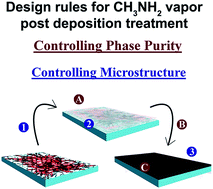
While organo-metal halide perovskite photovoltaics have seen rapid development, growth of high quality material remains a challenge. Herein, we report a facile post deposition treatment utilizing coordination between methylamine (CH3NH2) vapor and CH3NH3PbI3 perovskite that rapidly improves film quality, enhancing power conversion efficiency (PCE) by ∼9%. We further comprehensively analyze the physical impact of this process with regard to the material's optoelectronic properties and its detailed microstructural changes. Connecting this with an analysis of the source of organo-metal halide perovskite reactivity toward the vapor as well as phase behavior as a function of CH3NH2 vapor pressure and time, we provide design rules for the broad, rational extension of this process to new systems and scales.


Introduction

During the past few years, organic–inorganic hybrid perovskites have evolved from dye-sensitized solar cells (DSSCs)1,2 to solid state solar cells3 and have become a class of very promising materials for next-generation photovoltaic technologies. Very recently, a certified power conversion efficiency (PCE) over 20% has been accomplished.4 This class of material is collectively defined by the formula ABX3 with a lattice consisting of an interconnected network of BX6 octahedra. Exact crystal structure is determined both by the monovalent cation A (either inorganic (Cs+)) or organic (CH3NH3+ (MA+) or HC(NH2)2+ (FA+)), and the relative sizes of B and X, where B is a divalent metal cation (Pb2+ or Sn2+) and X is a halogen anion (Cl, Br, or I). These materials have excellent semiconducting properties including intense light absorption,5 small exciton binding energy (∼kT),6 ambipolar charge mobility,7 and long charge carrier diffusion lengths.8,9 More importantly, they possess low-cost solution processability, a factor that makes them serious rivals for existing photovoltaic materials.

This facile processing comes at the cost of complex and competing growth processes that frustrate control of film quality and coverage. Pin-hole formation, low crystallinity, and phase inhomogeneity are common obstacles to carrier transport and ultimate device performance. Compared to the mesoporous-structure based device configuration, morphological control is more challenging for the planar heterojunction (PHJ) structure because of complications in uniform perovskite nucleation caused by the lack of porosity.10,11 Nevertheless, the PHJ configuration still attracts significant research interests due to the versatility of emerging low-temperature fabrication techniques and materials.

Several methods have been developed to better control perovskite nucleation and growth. Sequential deposition from solution,12,13 dual-source evaporation,14 and the vapor-assisted solution process (VASP),15 the detailed processing methods of which have been introduced well in several review papers,16,17 all use a variety of phase transformations to circumvent direct perovskite growth from solution. For example, solution and vapor based two-step deposition methods both utilize a compact lead halide template to encourage uniform growth, eliminating the complexities inherent in direct MAPbI3 growth. To control this complexity rather than avoid it, techniques have focused on thermal processing18 and compositional tuning of the precursor solution through spectator ions,19,20 additives,21,22 and co-/anti-solvents,23 all of which provide improved film morphology through kinetic control of competing growth processes.17 Despite the significant improvements these strategies offer, the complexity of competing processes implicit in growth hinder reproducibility and mechanistic understanding. These challenges have motivated the development of many post deposition treatments that expand upon simple thermal annealing,24 such as solvent annealing,25 multi-cycled dimethylformamide (DMF) vapor treatment,26 and hot-pressing.27 While these post treatments markedly improve film morphology, they are relatively time-consuming and complex. Efficient, reproducible techniques are necessary to access the high throughput fabrication the material's solution processability enables.

In this work, we describe a fast (<1 s) and simple post deposition chemical treatment in which crystal reconstruction induced by a methylamine (MA0) vapor greatly improves perovskite film coverage, crystallinity, and device performance. We not only demonstrate the efficacy of this chemical treatment in improving the photovoltaic performance of a PHJ device, we also mechanistically study the process to reveal the role of hydrogen bonding between methylamine vapor and the perovskite's organic sublattice in mediating the material's reactivity toward the vapor through the exploration of a variety of hybrid perovskite systems. Furthermore, through detailed microscopy, we also demonstrate that the nature of MA0–perovskite coordination and its microstructural consequences are a function of MA0 vapor pressure, interaction time, and composition of the organic sublattice. Ultimately, we offer design rules for amine vapor based hybrid perovskite post chemical treatments.

Results and discussion

Vapor treatment and its physical consequences

Inspired by work reported by Zhu et al.28 in which a reversible perovskite phase transformation upon exposure to amine gas was revealed, we speculated that this kind of chemistry can be utilized to establish a facile post deposition treatment to improve perovskite crystallinity and quality through the recrystallization responsible for the regeneration of the perovskite phase from a similar transparent intermediate state. To explore this concept, we used methylamine rather than amine vapor because of its increased similarity to species already within the MAPbI3 lattice. To apply this in a device context, we first prepared a MAPbI3 film through simple one-step spin coating from DMF atop PEDOT:PSS, and then exposed this film to MA0 vapor (Fig. 1a). Immediate loss of pigment was observed in the perovskite upon introduction to a MA0-rich environment followed by rapid reversal back to perovskite upon removal of the film from the vapor. Herein, PEDOT:PSS serves as a hole-transporting layer (HTL) in our devices, but it also provides the necessary interfacial energy to prevent film dewetting during vapor treatment. This is an important component of this treatment's success in the PHJ architecture as compared to the findings of Cui et al. reported on a mesoporous growth substrate where capillary action helps direct material diffusion.29
image file: c6ra03485c-f1.tif
Fig. 1 Comparison of a MAPbI3 film before and after the vapor process in (a): (b) XRD, (c) and (d) SEM low magnification, and (e) and (f) SEM high magnification.

This technique potentially enables the use of one-step solution processing without the meticulous control typically required for high quality material growth. To apply this methodology broadly at many scales with many materials, we need a rigorous physical and chemical understanding of the process as a whole. Macroscopically, XRD suggests that perovskite crystallinity markedly increases upon vapor exposure and subsequent annealing at 100 °C for 15 min (Fig. 1b). SEM of the film before and after vapor treatment (Fig. 1c–f) reveals massive material diffusion resulting in almost complete film coverage, a huge improvement over the very poor coverage generated by unmodified 1-step perovskite growth from solution. It should be noted that ethanol itself does not induce any color or morphology change of the prepared MAPbI3 thin film. While key to the utility of the process, these two insights leave a significant hole in our understanding: What within the material is changing and how is it affecting performance and photophysical properties?

To address this, we first generated PHJ devices with the configuration in Fig. 2a. Control devices fabricated with unmodified 1-step perovskite growth from DMF showed a low PCE of ∼3%, but after the simple vapor treatment devices reached 12% PCE through marked increases in all photovoltaic parameters (Fig. 2b, Table S1). More detailed device characterizations including hysteresis, stabilized power output and EQE measurement are in the ESI (Fig. S1 and 2). From XRD and SEM (Fig. 1), this is likely due to a combination of shunt pathway elimination through improved film coverage and possible material enhancement.


image file: c6ra03485c-f2.tif
Fig. 2 The JV curve of the planar heterojunction photovoltaic device architecture in (a) with and without MA0 vapor treatment is in (b). MAPbI3's photophysical quality as an absorber before and after vapor treatment is analyzed with (c) transient and (d) steady state PL spectroscopy. With transient PL spectroscopy, we examine both the material on glass and on C60. (e), (f) and (g), (h) are bright and dark field TEM images, respectively, of an identical region before and after vapor exposure.

To better understand how MA0 vapor treatment affects the photophysical properties of MAPbI3, we studied the photoluminescence (PL) decay kinetics from perovskite films on glass. To maintain similar film quality of perovskite on glass compared to on PEDOT:PSS, we used slightly different vapor treatment process (see Methods for details) and verified the expected MA0-induced changes in crystallinity with XRD (Fig. S4). Fig. 2c shows representative PL decays of the perovskite before and after MA0 vapor exposure on glass measured in air.12 After MA0 treatment, the PL decay is faster and the PL intensity is slightly decreased (Fig. 2d, see more data in Fig. S5a and c). Since we do not see an increase in the PL lifetime or integrated intensity indicative of defect annihilation or surface passivation22 we conclude that while MA0 vapor treatment may improve device performance by removing macroscopic film defects (e.g. pinholes) as observed in SEM (Fig. 1), it does not effectively eliminate the structural defects responsible for non-radiative recombination losses.

In the steady state PL spectrum in Fig. 2d, MA0 treated films show a slight reduction in the PL intensity, consistent with the lifetime measurements. We also observe a ∼7 nm blue-shift upon vapor treatment, and also a slight reduction in the full width at half maximum (∼10%) (see Fig. S6 for other examples). We observed similar trends for films encapsulated with poly(methyl methacrylate) (PMMA) in a nitrogen atmosphere (Fig. S5g and h), suggesting that the reduction in PL is intrinsic to the MA0/perovskite interaction and not a result of improved diffusion to (and quenching at) the perovskite/air interface. The correlation with improved crystallinity in the films studied and the PL shift may suggest a link between disorder23,24 and emissive states within the band gap, but the shift is small enough to have multiple possible origins.30

While intrinsic material properties are important, comparison of PL quenching before and after interfacing the material with a charge extraction layer is more immediately relevant to device performance. We constructed a perovskite/C60 bilayer film and found that vapor treated films quenched photogenerated charges more efficiently (∼90% quenching) than untreated films (∼75% quenching), which is evidenced by the faster PL decay (Fig. 2c). In the treated films, these data suggest the charge transfer process to C60 is more efficient in treated films and likely outcompetes any additional non-radiative pathways introduced after MA0 vapor treatment. We attribute faster PL decay to the increased interfacial quality between perovskite and C60 in vapor treated films. In contrast, Cui et al.29 observed an increase in photoluminescence intensity after vapor treatment for films on compact TiO2, which is opposite to what one would expect for more efficient charger transfer in MA0 treated films.

These data illustrate an important device relevant consequence of MA0 vapor treatment, but it leaves significant questions about the microstructural consequences of this process. Thus, we fabricated perovskite thin-films for transmission electron microscopy (TEM) via a methodology identical to that used for device fabrication with the exception of using a diluted perovskite solution and a PEDOT:PSS coated TEM grid substrate.

Although it may be tempting to conclude from SEM that grain size doesn't change greatly upon MA0 vapor treatment (Fig. 1e and f), bright and dark field TEM (Fig. 2e, f and g, h respectively) reveal that vapor treatment induces gain size reduction rather than growth by more than an order of magnitude. Final microstructure after MA0 vapor treatment can be best described as a densely packed distribution of nanoscale crystallites. This microstructural change correlated with a decrease in PL intensity (Fig. 2c) and points to increased grain boundary area as a limitation of this process.31 Grain size reduction initially seems contradictory to the increase in crystallinity (Fig. 1b) and the suggestion of less disorder in the steady state PL of the vapor treated film (Fig. 2d).

A closer analysis of perovskite grain structure before vapor exposure with dark field TEM reveals a variety of morphological features that are persistently unapparent both in SEM and often even in bright field TEM (Fig. S7). Generally, we find (1) buried polycrystallinity, (2) highly strained and defective single crystals, and (3) densely packed, highly oriented grain boundaries throughout films grown from unmodified 1-step deposition. This is in addition to the predominantly disordered regions that remain undetectable and “dark” in dark field imaging. This level of microstructural complexity is unsurprising because of the existence of both MAPbI3·DMF32 and inherently disordered33,34 phases prominent in spin-cast film growth, compounded with the transformation from cubic to tetragonal MAPbI3 in the temperature window typical annealing traverses that is known to induce grain boundary formation in similar materials through processes like twinning in previously single crystalline domains.35 This complexity makes it challenging to define an exact grain size, as there is a continuum of disorder and a variety of grain and grain boundary sizes and aspect ratios. Ultimately, the shift from this microstructure to the nanocrystalline microstructure shown in Fig. 2f & h is beneficial for device function, but this analysis reveals the important diminishing returns implicit in the application of this process. There is a short discussion of the interpretation of these dark field data.

Chemical sources of reactivity toward MA0 vapor

To broaden the application of this process to the expanding family of hybrid organic–inorganic perovskites, factors determining lattice reactivity toward MA0 vapor must be established. The coordination chemistry between MAPbI3 and neutral molecules such as DMF,36 DMSO,4 and H2O (ref. 37) has garnered a great deal of interest because of its importance in growth and degradation. Amine gases like NH3,28 MA0, and larger analogues29 demonstrate the ability to disrupt the MAPbI3 lattice with differing degrees of readiness and reversibility. Van der Waals interaction, hydrogen bonding, water bridging, ion-dipole coordination, and electron transfer have all been proposed to govern host–guest interactions during the intercalation of neutral species in a solid.38 The diverse coordination chemistry of lead halides and their hybrid analogues in both solution39,40 and the solid state28,29,32,37,41 makes the mechanisms that govern MA0 vapor's interaction with the lattice somewhat difficult to pinpoint. It has been suggested that Lewis bases, specifically pyridine and thiophene, preferentially coordinate Pb dangling bonds42 which points to the metal ion's potential reactivity. The MAPbI3·DMF crystal structure32 suggests that DMF, also a Lewis base, preferentially hydrogen bonds with MA+ which points to the potential reactivity of the organic sublattice. While this at first seems to simplify the matter, the halogen ion's reactivity toward the vapor cannot be neglected by virtue of the key role bonding between the halide and the organic cation plays in stabilizing the hybrid perovskite's 3D inorganic sublattice over the 2D lattice of PbI2.43

To build an understanding of how these competing factors determining hybrid perovskite reactivity toward MA0 vapor, we systematically varied each component of the lattice and examined the response to vapor exposure. First, and most importantly, exposure of PbI2 to MA0 vapor causes a transition to a transparent state followed by the brief formation of brown perovskite before most of the film reverts to yellow PbI2 which is accompanied by a drastic change in microstructure (Fig. S8a and b). This is unsurprising as the PbI2·xMA0 system is essentially analogous to MAPbI3·xMA0 with an iodide deficiency. The fact that MA0 vapor does not possess the formal positive charge of methylammonium while demonstrating a similar capacity for transformation and even perovskite formation is an excellent illustration of the importance of hydrogen bonding in the formation and cohesion of MAPbI3 itself. This is a simple but poignant demonstration of the core importance of the nature of the inorganic halide polyhedra in determining reactivity, and the rapid generation of the 3D perovskite analogue from the 2D PbI2 lattice shows that interaction between MA0 and the lead halide framework is not limited to intercalation.

Bonding within each hybrid perovskite lattice is unique, and thus the intrinsic reactivity of the metal halide framework toward MA0 vapor is likely altered by changes in components of the crystal. We first addressed the influence of halogen and metal ions by fabricating MAPbBr3 and MASnI3 films respectively. Upon vapor exposure, we found the same rapid, reversible transformation to the transparent intermediate state and subsequent rapid reversion to the original perovskite that is characteristic of MAPbI3, and again this transformation is accompanied by dramatic microstructural changes (Fig. S8c–f). As such, we more closely investigated the role of the organic cation by fabricating FAPbI3 and CsPbI3 films. As both the capacity to hydrogen bond and the cohesivity offered to the lattice as a whole vary as a function of the composition of the organic sublattice, we expected significant differences between each system's response to MA0 vapor exposure. In keeping with the other systems, FAPbI3 rapidly becomes transparent upon vapor exposure, but reverts back to its initial state from this transparent intermediate state much less readily, even after annealing at 100 °C for 15 min. Even though the intermediate phase is far more stable in the case of FAPbI3 exposed to MA0 vapor, there are still huge microstructural changes that occur (Fig. S8g and h). Finally, we explore the more extreme case of CsPbI3 in which we find no reactivity toward the vapor by virtue of no change in the film's pigment. SEM reveals no significant microstructural changes upon vapor exposure which, when combined with the behavior of FAPbI3, indicates that the nature of bonding within the organic sublattice strongly mediates the reactivity toward MA0 vapor that is initially imparted to a hybrid perovskite lattice by the nature of the inorganic polyhedra contained within. This means that Cui et al.'s assumption that the source of reactivity is the presence of lead halide polyhedra is reasonable, and the organic sublattice determines the final nature of this reactivity after a hybrid lattice has been established.

By virtue of the fact that in MAPbI3 the conduction and valence band edges are composed primarily of I 5p and Pb 6s orbitals,44 the transparency of the intermediate state in these various systems likely results from a complete loss of structure within the inorganic sublattice. This loss of pigment upon vapor exposure can point both to a solvation event29 or a dramatic but coherent change in structure via the formation of a crystalline intermediate. Although Cui et al. did an excellent job of showing the accessibility of the solvated state and its prevalence under a high concentration of MA0 vapor, they also demonstrated that although a transparent state can be established under NH3 vapor,28 complete solvation is not feasible. This illustrates the potential existence of multiple intermediate states within a single system, the nature of which likely playing a decisive role in determining the morphological consequences of the process.

Mapping vapor induced transformations

It has been demonstrated that in solution the nature of perovskite formation depends upon methylammonium concentration.45 Specifically, low concentrations allow topotactic transformation preserving structural elements of PbI2, while at high concentrations dissolution occurs resulting in structure determined by the precipitation event rather than the nature of the starting material. Because of the dramatic but unique responses of MAPbI3 and FAPbI3 to the vapor, we compared their transformations under high and low methylamine vapor pressure to more completely map MA0 induced hybrid perovskite transformations. High vapor pressure exposure is identical to the vapor treatment process discussed above, and enabled by the high volatility of MA0 in ethanol at room temperature. Low vapor pressure exposure entails placing MAPbI3 and FAPbI3 films in chambers with a slow leak of MA0 vapor for three hours. Although direct analysis of intermediate states during vapor exposure is complicated by the incredible rapidity of transformation under high vapor pressure and the necessity of a closed system under low vapor pressure, we conducted XRD analysis of each perovskite film before vapor exposure, after exposure, and after annealing at 100 °C for 15 min. For MAPbI3 exposed to a high concentration of MA0 vapor (Fig. 3a), the intermediate state is too transient to be observed after vapor removal, but based on the work of Cui et al. it is clear that solvation occurs under these conditions. Perovskite crystallinity dramatically increases immediately after vapor exposure, and increases relatively little upon subsequent annealing. Exposure of MAPbI3 to a low concentration of MA0 vapor results in a relatively long lived, structurally coherent transparent state characterized by reflections marked as I1 in (Fig. 3b). The solitary strong reflection at low 2θ of the intermediate's primary peak (∼7.2°) suggests a swelling of the lattice consistent with the perturbation of the inorganic sublattice implicated by the intermediate's transparency, but we lack sufficient detail to solve the structure. Annealing almost completely regenerates MAPbI3, but with less crystallinity than in the case of high concentration MA0 vapor exposure. For continued reference to the vapor treatments with high and low MA0 vapor concentration, we will used the terms high [MA0] treatment and low [MA0] treatment.
image file: c6ra03485c-f3.tif
Fig. 3 Phase evolution through XRD analysis of MAPbI3 under (a) high [MA0] and (b) low [MA0] treatments, as well as FAPbI3 under (c) high [MA0] and (d) low [MA0] treatments. Although detail is limited, the phases indicated are listed in the legend located in the center right of the figure. FAPbI3 (y) and FAPbI3 (b) indicate the yellow and black FAPbI3 polymorphs respectively.

Intermediate states formed by FAPbI3 are distinct between high and low [MA0] treatments, and one is long lived even after annealing. Because of the well-known instability of the black polymorph at room temperature.46 The FAPbI3 film contains both yellow and black polymorphs before vapor exposure. Upon high [MA0] treatment (Fig. 3c), the black polymorph disappears and is replaced by the yellow polymorph and an intermediate phase distinct from I1 with a primary peak at ∼11° 2θ which we have marked as I2. Annealing, causes little change in this phase distribution. Low [MA0] treatment (Fig. 3d), leads primarily to a greater fraction of stable I2 and an phase bearing the same solitary strong peak at ∼7.2° 2θ as I1. This intermediate was labeled as I1.

As a function of time (Fig. S9), low [MA0] treatment of FAPbI3 initially causes I2 formation followed by I1 formation over time. This indicates that while I2 forms more rapidly under MA0 vapor, the state analogous to MAPbI3's preferred state under low concentration MA0 is the same eventual fate of FAPbI3. Annealing primarily causes elimination of I1 and the generation of only a small amount of MAPbI3 while the fractions of the yellow FAPbI3 polymorph and I2 remain relatively unchanged. This suggests that while the primary product of I1 is MAPbI3, the readiness with which the transformation proceeds is significantly reduced compared to I1, which is part of the reason we draw a distinction between them. Although detail in Fig. 3 is limited, the structural similarities between I1 and I1 suggest that atomic spacing may primarily be determined by the neutral MA0 molecule's interaction with the lattice. MAPbI3 formation within FAPbI3 is a demonstration of the proton transfer between cations in the lattice and MA0. The stark kinetic differences in the transformation of each intermediate discussed above shows that bonding within the hybrid perovskite organic sublattice plays a significant role in determining the readiness with which vapor induced intermediate states regenerate the desired perovskite, and in the extreme case of CsPbI3 prevents reaction entirely.

While the phase evolution presented in Fig. 3 illustrates the role of vapor concentration and organic sublattice composition in mediating available transformations, we cannot directly discern whether these intermediate phases persist exclusively during exposure or they are the result of equilibration after vapor removal. That said, through the microstructural consequences of each transformation just discussed, we can gain increased mechanistic insight as well as improved intuition in the rational design of vapor based post deposition treatments. To this end we correlated SEM and detailed TEM analysis at low magnification (Fig. 4 and 5) and high magnification (Fig. S10 and S11) of regions in each system before vapor exposure, after vapor exposure, and after subsequent annealing. Because films for SEM analysis are fabricated under conditions identical to device fabrication, we can directly see the macroscopic consequences of the vapor treatments in a device context. As TEM samples are fabricated via an identical method with the only exception of a diluted perovskite precursor solution, we have analogous growth conditions with reduced coverage and film density enabling us to more easily define microscopic differences in material diffusion and grain reconstruction inherent to each process.


image file: c6ra03485c-f4.tif
Fig. 4 SEM of (a–c) MAPbI3 and (d–f) FAPbI3 before and after MA0 vapor exposure for both high and low [MA] treatments.

image file: c6ra03485c-f5.tif
Fig. 5 Each pane compared an identical region before and after MA0 vapor exposure with bright field imaging (top), select area electron diffraction (inset), and dark field imaging (bottom) with transmission electron microscopy of (a, b) MAPbI3 and (c, d) FAPbI3 at both (a & c) high and (b & d) low [MA0] treatments.

SEM characterization of each transformation shows a great degree of diffusion in all cases. Both high [MA0] cases (Fig. 4a, b and d, e) result in excellent film coverage. Both low [MA0] cases (Fig. 4a & c, d & f) result in coarsening which generated thick and barren regions. Even though plentiful pin-holes are in the initial films (Fig. 4a and d), the density of material is great enough so that a reasonably large amount of material surrounds any given point in the film. This is not the case in the TEM samples in which coverage was sparse enough to isolate individual features and observe their microscopic transformation upon vapor exposure, a key distinguishing feature between the data sets in Fig. 4 and 5. Each pane of Fig. 5 correlates bright field TEM images (top), select area electron diffraction (SAED) patterns (inset), and one representative dark filed image (bottom) of a region before and after vapor treatment. The dark field images in Fig. 5, however, just show one representative dark field image, and if the beam is rotated we can detect some kind of diffracted signal from majority of apparent grains present in the sample. In general, SAED patterns show little contribution from intermediate phases which is unsurprising in such a high vacuum environment. This makes Fig. 3 a more meaningful analysis of phase evolution. Throughout the different treatments, TEM SAED shows no clear change in collective crystal orientation which supports the interpretation of increased crystallinity from the XRD. In all cases the diffraction pattern becomes more ring-like and closer to a true powder after vapor treatment, a consequence of the reduction in grain size.

For MAPbI3 high [MA0] exposure, the phase purity confirmed by SAED (Fig. 5a inset) and the dramatic reduction in grain size apparent in the dark field images (Fig. 5a, bottom) were already discussed in detail at the beginning of the manuscript. The bright field images (Fig. 5a, top) show a great deal of diffusion unconstrained by the initial physical bounds of the material consistent with the liquid intermediate discussed by Cui et al. In contrast, diffusion in MAPbI3 during low [MA0] exposure (Fig. 5b, top) remained constrained to the physical extent of the material before vapor exposure. Although coarsening still drives large scale diffusion which thickens some regions and depletes others, we can more clearly see the way diffusion is facilitated by the preexisting solid framework, an issue distinguishing low [MA0] treatment from the high [MA0] case. The dark field images (Fig. 5b, bottom, and the higher magnification analogues in Fig. S11) show that although grain size is still reduced compared to the initial state, final average grain size is slightly larger than in the high [MA0] treatment. This is likely due both to grain growth within the solid intermediate and the reduction of perovskite nucleation rate caused by the more gradual change in MA vapor pressure upon process completion inherent to the low [MA0] treatment.

Even though transformation back from the intermediate state is incomplete in high [MA0] treated FAPbI3, grain size revealed by dark field imaging (Fig. 5c) is quite comparable to that of high [MA0] treated MAPbI3 indicating that dramatic disruption to the original perovskite grain structure occurs upon initial intermediate formation, an issue related to the swelling of the lattice. That said, diffusion is once again constrained by material distribution before exposure, implicating the prevalence of a solid intermediate and suggesting that pervasive lattice disruption occurs regardless of solvation.

Low [MA0] treatment of FAPbI3 leads to diffusion that is again facilitated by the solid framework existing before exposure (Fig. 5d). Coarsening facilitated by large scale diffusion similar to MAPbI3 under low [MA0] vapor and consistent with Fig. 4d can be observed. Also in keeping with MAPbI3, average grain size is smaller than it was initially but larger than the high [MA0] analogue. Coupled with the distinctly different phase content of each system (Fig. 3), this points to MA0 vapor pressure as a function of time as a dominant determinant of final grain size even though the nature of accessible phases and their transformations are largely determined by the composition of the organic sublattice. This slight increase in grain size comes at the cost of coverage and overall perovskite crystallinity in the low [MA0] treatment of both systems.

The dramatic changes in morphology evident in Fig. 4 show that regardless of intermediate state, MA0 vapor facilitates massively enhanced diffusion. The more subtle distinctions apparent in Fig. 5 suggest that MAPbI3 can readily reach an essentially solvated state under high MA0 vapor pressure, but both reducing the vapor pressure and altering composition of the organic sublattice (i.e. FAPbI3 and CsPbI3) impedes the formation of this truly solvated state. Combining this with the sharp kinetic distinction between the intermediates and the complete transformation back to perovskite from this solvated state versus the slow and incomplete transformation after structurally coherent intermediate stabilization under low [MA0] vapor, it becomes apparent that circumventing the formation of metastable states during vapor based post deposition treatment may be an important component of gaining the full value the process offers in increased film quality. The solvation event thus have the dual purpose of facilitating diffusion and kinetically excluding metastable, crystalline intermediates that frustrate transformation.

Fig. 6 summarizes this and the other observations above graphically to generate a summary of the transformations at play. Background colors indicate preferred state as a function of MA0 vapor pressure and time, and the colors of the paths superimposed on this represent the actual state of a system being taken through the conditions indicated. The important aspects to notice are the differences in the readiness of MAPbI3 and FAPbI3 solvation, prevention of I1 formation during high [MA0] treatment because of solvation, and differing degrees of metastability of I1, I1, and I2. As we lack quantitative vapor pressures, these are only schematic. It's important to note, that in generating this graphical summary we had to neglect some of the complexity inherent to the FAPbI3 system, specifically with regard to the multiple accessible FAPbI3 polymorphs as well as the MAPbI3 generation upon long vapor exposure.


image file: c6ra03485c-f6.tif
Fig. 6 Schematic representations of (a) MAPbI3 and (b) FAPbI3 phase evolution during and after MA0 vapor exposure as a function of MA0 vapor pressure and time. The background shows the preferred phase under the indicated conditions as a function of MA0 vapor pressure and time, and the paths emulate the high and low [MA0] treatments discussed in the text. The color of the each path represents the unique phase evolution during that process.

Conclusion

From our physical and chemical analysis of hybrid perovskite MA0 vapor post deposition treatment, we offer the design rules summarized in Fig. 7 for the broad application of this process in contexts involving different materials and scales. In general, obliteration of original perovskite grain structure occurs regardless of solvation. The cost of massively improved film coverage is greatly reduced grain size regardless of the nature of the intermediate formed during vapor exposure, and promoting grain growth is a challenge because of a dependence on vapor removal and coarsening processes. Our PL results show the utility this process offers in establishing a perovskite interface with more intimate contact for charge extraction, as well as the diminishing returns inherent in the process. Taken together, this work demonstrates the successful implementation of this process in PHJ architecture. Using the mechanistic insight generated here, it is clear that these microstructural trends and physical insights have very little dependence upon initial perovskite growth route and thus starting morphology. While it is clear that a variety of amine vapors can induce massive material changes, choice of vapor determines both accessible phases and how the system can move through them, as is visualized in Fig. 6 for MAPbI3 and FAPbI3.
image file: c6ra03485c-f7.tif
Fig. 7 Design rules for MA0 vapor based post deposition treatments for organo-metal halide perovskites in general.

The methodology of this facile and high throughput post deposition treatment is potentially useful for translation to industry, and sensitivity to the issues summarized in Fig. 7 may minimize the detriments and maximize the benefits it entails. Ultimately, this comes down to controlling the microstructural consequences of the process and ensuring phase purity upon completion. These issues quickly become complex when large scale systems with implicit inhomogeneities and non-ideal time scales are implemented, especially with materials with more complex phase behavior like FAPbI3. We hope to extend the understanding here to such processes.

Experimental

Materials

All materials were purchased from Sigma-Aldrich without further purification unless stated specifically. CH3NH3I was synthesized by reacting 33 wt% CH3NH2 in ethanol with 57% HI in water solution at a 2[thin space (1/6-em)]:[thin space (1/6-em)]1 molar ratio of CH3NH2 to HI and 0 °C for 2 h.3 The product was collected by removing solvent through rotary evaporation, followed by diethyl ether washing until colorless and recrystallized twice in methanol. The crystalline white powder was finally dried in a vacuum oven at 60 °C for 24 h.

Substrate cleaning, CH3NH3PbI3 film fabrication, and vapor treatment process

ITO (15 ohm sq−1) glass substrates were cleaned sequentially with detergent, deionized water, acetone, and isopropanol under sonication for 10 min each. After drying under a N2 stream, substrates were further cleaned by a UV ozone treatment for 10 min.

PEDOT:PSS (Baytron P VP Al 4083, filtered through a 0.45 μm nylon filter) was first spin-coated onto the substrates at 5k rpm for 30 s and annealed at 150 °C for 10 min in air. To avoid any possible influence from oxygen and moisture, the substrates were transferred into a N2-filled glovebox. The perovskite precursor solution was made by dissolving an equimolar ratio of PbI2 (1 M) and CH3NH3I (1 M) in DMF at 60 °C and filtering through 0.45 μm PTFE filter. Perovskite layers were formed by spin-coating the precursor solution at 6k rpm for 45 s and then annealing at 100 °C for 15 min.

After annealing, MA0 vapor treatment was conducted by holding the perovskite film upside down right above a 20 mL open vial (28 mm outer diameter × 61 mm height and 22 mm aperture diameter) with 6 mL of 33 wt% CH3NH2 solution in ethanol at room temperature. Upon turning clear (less than 1 s), the film was immediately removed vertically. Rapid vertical removal of the film from the MA0 vapor source is important to prevent inhomogeneities that develop when the film is moved laterally through a MA0 concentration gradient. The film was annealed again at 100 °C for 15 min to further increase thin-film crystallinity (as shown in Fig. 3a and video in SI).

The perovskite films of differing compositions fabricated for comparison of reactivity were fabricated using an analogous method as that just described, but complete details are presented in the ESI. The low [MA0] vapor treatment was conducted by sealing perovskite films in a container with a source of MA0 vapor for 3 hours. The detailed experimental set up is discussed in the ESI and presented in Fig. S12.

Device fabrication and characterization

ITO glass cleaning, PEDOT:PSS deposition, and CH3NH3PbI3 film growth are discussed above in the vapor treatment section. Atop the CH3NH3PbI3 film, C60 (15 mg mL−1 in ortho-dichlorobenzene (DCB)) and C60-bis surfactant (2 mg mL−1 in isopropyl alcohol) were sequentially deposited by spin coating at 1k rpm for 60 s and 3k rpm for 60 s, respectively. Silver electrodes with a thickness of 120 nm were finally evaporated under vacuum (<2 × 10⁻⁶ Torr) through a shadow mask. The device area was defined as 3.14 mm2. All JV curves were recorded using a Keithley 2400 source meter unit. The device photocurrent was measured upon illumination from a 450 W thermal Oriel solar simulator (AM 1.5G) calibrated with a standard Si photodiode detector equipped with a KG-5 filter, which can be traced back to the standard cell of the National Renewable Energy Laboratory (NREL).

Characterization

X-ray diffraction (XRD) experiments were performed using a Bruker F8 Focus Powder XRD operating at 40 kV and 40 mA with a Cu K-alpha (1.54 A) X-ray source. Secondary electron images were taken with a FEI Sirion scanning electron microscope at 5 kV.

Time-resolved PL decay traces were acquired using a PicoHarp 300 time-correlated single photon counter (TCSPC). A 470 nm pulsed diode laser (PDL-800 LDH-P-C-470B, 300 ps pulse width) was used for excitation at a fluence of 10 nJ cm−2 (n0 ∼ 1015 cm−3) with a repetition rate of 500 kHz for time resolved measurements and a continuous-wave 532 nm (CrystaLaser, GCL532-005-L) at a power density of 90 mW cm−2 (∼1.5 sun) was used for steady state measurements. Samples were excited face-on (not through the substrate) and the emission was filtered through a 700–850 nm bandpass filter (700 LP and 850 SP). Photoluminescence from the sample was directed to a Micro Photon Devices (MPD) PDM Series single photon avalanche photodiode with a 50 μm active area for TRPL measurements or a portable charge coupled device spectrometer (USB2000, Ocean Optics) for steady state PL measurements. Perovskite films for PL studies were prepared on glass to highlight the intrinsic PL behavior before and after MA0 treatment. In order to make high-quality films on glass, we increased the MA0 vapor pressure within the head space of the vial by increasing the MA0 solution volume from 6 to 10 mL (Fig. S4). To evaluate the quenching efficiency, C60 (15 mg mL−1 in DCB) was spin-coated onto the original neat perovskite films. As these photoluminescence measurements were conducted in air, we also studied MAPbI3 films encapsulated with PMMA (30 mg mL−1 in chlorobenzene). We find that the MAPbI3/air interfacial chemistry may affect carrier lifetime during measurement (Fig. S2a–f), and the vapor treatment greatly alters the geometry of this interface. In addition, we observe slightly different ratios between the integrated PL intensity observed in the time-resolved PL versus the steady state PL spectra in the samples measured in air, we attribute this to sample degradation between the two separate measurements. Importantly, we observed the same trends in PL intensity before and after treatment regardless of the measurement conditions.

TEM samples were prepared by first adhering a finder TEM grid with a Carbon B coating to a cleaned ITO glass substrate (specifically EF400-Ni from Electron Microscopy Services). Before mounting, grids were glow discharge treated for 30 seconds with an oxygen/hydrogen plasma with a Solarus 950 Gatan Advanced Plasma System. The grid is mounted in slight tension on a glass slide with scotch tape to ensure intimate thermal contact. The same deposition and heat treatment procedure for first PEDOT:PSS then the desired perovskite are followed as above with the exception of the use of a 0.4 M perovskite precursor solution. Vapor exposure was also identical except for the use of a MA0 vapor source with a smaller aperture (4 mL vial with a 10 mm diameter neck rather than 20 mL with a 22 mm diameter neck) for high vapor pressure exposure. When the grid is removed from the glass slide for characterization, the points of contact between the tape and the grid are carefully cleaned with IPA to prevent contamination of the high vacuum environment of the TEM. A Tecnai G2 F20 transmission electron microscope was used at 200 kV for all TEM measurements.

Conflict of interest

The authors declare no competing financial interest.

Acknowledgements

This work is supported by the Office of Naval Research (N00014-14-1-0170), the Department of Energy SunShot (DE-EE0006710), and the Asian Office of Aerospace R&D (FA2386-11-1-4072). A. K.-Y. Jen thanks the Boeing-Johnson Foundation for financial support. S. T. Williams and D. W. deQuilettes acknowledges support from a National Science Foundation Graduate Research Fellowship (DGE-0718124 and DGE-1256082). This work was made possible by the Molecular Analysis Facility at the University of Washington.

Notes and references

  1. J.-H. Im, C.-R. Lee, J.-W. Lee, S.-W. Park and N.-G. Park, Nanoscale, 2011, 3, 4088 RSC.
  2. A. Kojima, K. Teshima, Y. Shirai and T. Miyasaka, J. Am. Chem. Soc., 2009, 131, 6050–6051 CrossRef CAS PubMed.
  3. H.-S. Kim, C.-R. Lee, J.-H. Im, K.-B. Lee, T. Moehl, A. Marchioro, S.-J. Moon, R. Humphry-Baker, J.-H. Yum, J. E. Moser, M. Grätzel and N.-G. Park, Sci. Rep., 2012, 2, 591 Search PubMed.
  4. W. S. Yang, J. H. Noh, N. J. Jeon, Y. C. Kim, S. Ryu, J. Seo and S. Il Seok, Science, 2015, 348, 1234–1237 CrossRef CAS PubMed.
  5. M. A. Green, A. Ho-Baillie and H. J. Snaith, Nat. Photonics, 2014, 8, 506–514 CrossRef CAS.
  6. A. Miyata, A. Mitioglu, P. Plochocka, O. Portugall, J. T.-W. Wang, S. D. Stranks, H. J. Snaith and R. J. Nicholas, Nat. Phys., 2015, 11, 582–587 CrossRef CAS.
  7. J. H. Heo, S. H. Im, J. H. Noh, T. N. Mandal, C.-S. Lim, J. A. Chang, Y. H. Lee, H. Kim, A. Sarkar, M. K. Nazeeruddin, M. Grätzel and S. Il Seok, Nat. Photonics, 2013, 7, 486–491 CrossRef CAS.
  8. G. Xing, N. Mathews, S. Sun, S. S. Lim, Y. M. Lam, M. Grätzel, S. Mhaisalkar and T. C. Sum, Science, 2013, 342, 344–347 CrossRef CAS PubMed.
  9. S. D. Stranks, G. E. Eperon, G. Grancini, C. Menelaou, M. J. P. Alcocer, T. Leijtens, L. M. Herz, A. Petrozza and H. J. Snaith, Science, 2013, 342, 341–344 CrossRef CAS PubMed.
  10. T. Salim, S. Sun, Y. Abe, A. Krishna, A. C. Grimsdale and Y. M. Lam, J. Mater. Chem. A, 2014, 1–27 Search PubMed.
  11. C. Zuo, H. J. Bolink, H. Han, J. Huang, D. Cahen and L. Ding, Adv. Sci., 2016 DOI:10.1002/anie.201504379.
  12. J. Burschka, N. Pellet, S.-J. Moon, R. Humphry-Baker, P. Gao, M. K. Nazeeruddin and M. Grätzel, Nature, 2013, 499, 316–319 CrossRef CAS PubMed.
  13. Z. Xiao, C. Bi, Y. Shao, Q. Dong, Q. Wang, Y. Yuan, C. Wang, Y. Gao and J. Huang, Energy Environ. Sci., 2014, 7, 2619–2623 CAS.
  14. M. Liu, M. B. Johnston and H. J. Snaith, Nature, 2013, 501, 395–398 CrossRef CAS PubMed.
  15. Q. Chen, H. Zhou, Z. Hong, S. Luo, H.-S. S. Duan, H.-H. H. Wang, Y. Liu, G. Li and Y. Yang, J. Am. Chem. Soc., 2014, 136, 622–625 CrossRef CAS PubMed.
  16. S. D. Stranks, P. K. Nayak, W. Zhang, T. Stergiopoulos and H. J. Snaith, Angew. Chem.,Int. Ed., 2015, 54, 3240–3248 CrossRef CAS PubMed.
  17. S. T. Williams, C.-C. Chueh and A. K.-Y. Jen, Small, 2015, 11, 3087 CrossRef.
  18. W. Nie, H. Tsai, R. Asadpour, J.-C. Blancon, A. J. Neukirch, G. Gupta, J. J. Crochet, M. Chhowalla, S. Tretiak, M. A. Alam, H.-L. Wang and A. D. Mohite, Science, 2015, 347, 522–525 CrossRef CAS PubMed.
  19. M. M. Lee, J. Teuscher, T. Miyasaka, T. N. Murakami and H. J. Snaith, Science, 2012, 338, 643–647 CrossRef CAS PubMed.
  20. W. Zhang, M. Saliba, D. T. Moore, S. K. Pathak, M. T. Hörantner, T. Stergiopoulos, S. D. Stranks, G. E. Eperon, J. A. Alexander-Webber, A. Abate, A. Sadhanala, S. Yao, Y. Chen, R. H. Friend, L. A. Estroff, U. Wiesner and H. J. Snaith, Nat. Commun., 2015, 6, 6142 CrossRef CAS PubMed.
  21. P.-W. Liang, C.-Y. Liao, C.-C. Chueh, F. Zuo, S. T. Williams, X.-K. Xin, J. Lin and A. K.-Y. Jen, Adv. Mater., 2014, 26, 3748–3754 CrossRef CAS PubMed.
  22. C. Zuo and L. Ding, Nanoscale, 2014, 6, 9935–9938 RSC.
  23. N. J. Jeon, J. H. Noh, Y. C. Kim, W. S. Yang, S. Ryu and S. Il Seok, Nat. Mater., 2014, 1, 1–7 Search PubMed.
  24. A. Dualeh, N. Tétreault, T. Moehl, P. Gao, M. K. Nazeeruddin and M. Grätzel, Adv. Funct. Mater., 2014, 24, 3250–3258 CrossRef CAS.
  25. Z. Xiao, Q. Dong, C. Bi, Y. Shao, Y. Yuan and J. Huang, Adv. Mater., 2014, 26, 6503–6509 CrossRef CAS PubMed.
  26. W. Zhu, T. Yu, F. Li, C. Bao, H. Gao, Y. Yi, J. Yang, G. Fu, X. Zhou and Z. Zou, Nanoscale, 2015, 7, 5427–5434 RSC.
  27. J. Xiao, Y. Yang, X. Xu, J. Shi, L. Zhu, S. Lv, H. Wu, Y. Luo, D. Li and Q. Meng, J. Mater. Chem. A, 2015, 3, 5289–5293 CAS.
  28. Y. Zhao and K. Zhu, Chem. Commun., 2014, 50, 1605–1607 RSC.
  29. Z. Zhou, Z. Wang, Y. Zhou, S. Pang, D. Wang, H. Xu, Z. Liu, N. P. Padture and G. Cui, Angew. Chem., Int. Ed., 2015, 9705–9709 CrossRef CAS PubMed.
  30. V. D'Innocenzo, A. R. S. Kandada, M. De Bastiani, M. Gandini, A. Petrozza, A. R. Srimath Kandada, M. De Bastiani, M. Gandini and A. Petrozza, J. Am. Chem. Soc., 2014, 136, 17730–17733 CrossRef PubMed.
  31. D. W. de Quilettes, S. M. Vorpahl, S. D. Stranks, H. Nagaoka, G. E. Eperon, M. E. Ziffer, H. J. Snaith and D. S. Ginger, Science, 2015, 348, 683–686 CrossRef CAS PubMed.
  32. F. Hao, C. C. Stoumpos, Z. Liu, R. P. H. Chang and M. G. Kanatzidis, J. Am. Chem. Soc., 2014, 136, 16411–16419 CrossRef CAS PubMed.
  33. B. Park, B. Philippe, T. Gustafsson, K. Sveinbjörnsson, A. Hagfeldt, E. M. J. Johansson and G. Boschloo, Chem. Mater., 2014, 26, 4466–4471 CrossRef CAS.
  34. J. J. Choi, X. Yang, Z. M. Norman, S. J. L. Billinge and J. S. Owen, Nano Lett., 2014, 14, 127–133 CrossRef CAS PubMed.
  35. D. Damjanovic, Rep. Prog. Phys., 1999, 61, 1267 CrossRef.
  36. W. Yongzhen, A. Islam, X. Yang, C. Qin, J. Liu, K. Zhang, W. Peng, L. Han, Y. Wu, A. Islam, X. Yang, C. Qin, J. Liu, K. Zhang, W. Peng, L. Han, W. Yongzhen, A. Islam, X. Yang, C. Qin, J. Liu, K. Zhang, W. Peng and L. Han, Energy Environ. Sci., 2014, 7, 2934 Search PubMed.
  37. A. M. A. Leguy, Y. Hu, M. Campoy-Quiles, M. I. Alonso, O. J. Weber, P. Azarhoosh, M. van Schilfgaarde, M. T. Weller, T. Bein, J. Nelson, P. Docampo and P. R. F. Barnes, Chem. Mater., 2015, 27, 3397–3407 CrossRef CAS.
  38. G. Pedro and C. Sanchez, Functional Hybrid Materials, John Wiley & Sons, 2006 Search PubMed.
  39. Y. Wu, A. Islam, X. Yang, C. Qin, J. Liu, K. Zhang, W. Peng and L. Han, Energy Environ. Sci., 2014, 7, 2934 CAS.
  40. K. Yan, M. Long, T. Zhang, Z. Wei, H. Chen, S. Yang and J. Xu, J. Am. Chem. Soc., 2015, 137, 4460–4468 CrossRef CAS PubMed.
  41. N. Ahn, D.-Y. Son, I.-H. Jang, S. M. Kang, M. Choi and N.-G. Park, J. Am. Chem. Soc., 2015, 137, 8696–8699 CrossRef CAS PubMed.
  42. N. K. Noel, A. Abate, S. D. Stranks, E. S. Parrott, V. M. Burlakov, A. Goriely and H. J. Snaith, ACS Nano, 2014, 8, 9815–9821 CrossRef CAS PubMed.
  43. D. B. Mitzi, Prog. Inorg. Chem., 1999, 1–121 CrossRef CAS.
  44. J. Kim, S.-H. H. Lee, J. H. Lee and K.-H. H. Hong, J. Phys. Chem. Lett., 2014, 5, 1312–1317 CrossRef CAS PubMed.
  45. S. Yang, Y. C. Zheng, Y. Hou, X. Chen, Y. Chen, Y. Wang, H. Zhao and H. G. Yang, Chem. Mater., 2014, 26, 6705–6710 CrossRef CAS.
  46. T. M. Koh, K. Fu, Y. Fang, S. Chen, T. C. Sum, N. Mathews, S. G. Mhaisalkar, P. P. Boix and T. Baikie, J. Phys. Chem. C, 2014, 118, 16458–16462 CAS.

Footnotes

Electronic supplementary information (ESI) available. See DOI: 10.1039/c6ra03485c
Ting Zhao and Spencer T. Williams contributed equally to this work.

This journal is © The Royal Society of Chemistry 2016