DOI:
10.1039/C6RA03434A
(Paper)
RSC Adv., 2016,
6, 38052-38063
Study on the cross-linking effect of a natural derived oxidized chitosan oligosaccharide on the porcine acellular dermal matrix
Received
5th February 2016
, Accepted 24th March 2016
First published on 24th March 2016
Abstract
The purpose of this study is to investigate the cross-linking interaction between a natural derived oxidized chitosan oligosaccharide (OCOS) and the porcine acellular dermal matrix (pADM), and further evaluate the varying properties of the pADM after cross-linked. The shrinkage temperature (Ts) and cross-link density study indicate that the amino groups of lysine or hydroxylysine side groups of collagen could predominantly react with the available aldehyde group of OCOS to form a more stable Schiff's base leading to the improved hydrothermal stability of pADM. Fourier transform infrared (FTIR) spectroscopy, X-ray diffraction (XRD), scanning electronic microscopy (SEM) and atomic force microscopy (AFM) analysis indicate that the structural integrity of collagen (i.e. D-periodicity structure and triple helix) is still maintained after the OCOS treatment. Meanwhile, the mechanical properties, hydrophilicity, collagenase degradation and cytotoxicity of pADM after cross-linking have been all promoted obviously. Above all, the cytocompatibility analysis implies that when the dosage of OCOS is less than 8%, introducing OCOS into pADM might be favorable for cell adhesion, growth and proliferation. Taken as a whole, the present study demonstrates that OCOS could serve as an ideal cross-linker for the chemical fixation of pADM, which are compatible with its potential applications in biomaterials.
1 Introduction
The porcine acellular dermal matrix (pADM) has been extensively utilized as tissue scaffold on account of its specific three-dimensional (3D) architecture that mimics the biological characteristics of the native extracellular matrix (ECM). The basic component of pADM is collagen molecules, which are the most abundant and wildly distributed protein, comprising the major protein family in the extracellular matrix and is found in skin, bone, tendons, visceral intercellular substances, ligaments, vessels, sclera and other animal parts.1,2 Collagen has multiple advantages, such as low immunogenicity, low irritation, low cytotoxicity, good biocompatibility, good ability of promoting the cell growth, etc., which make it increasingly widespread in the biomedical field.3–5 Collagen-based biomaterials for different applications have various requirements, including mechanical property and degradation rat.5,6 Chemical cross-linking is always the most common method for improving mechanical property, stability, reducing antigenicity and adjusting appropriate degradability of collagen.7,8 Because of the high cytotoxicity and easy calcification, classical glutaraldehyde cross-linking already tends to be eliminated.9 Carbodiimide was once deemed as a perfect zero length cross-linker, owing to its special reaction mechanism which would not introduce itself into matrix,10 but it is limited by low degree of cross-linking and byproducts. Recently, genipin is believed as a better agent as its high degree of cross-linking and low cytotoxicity.11 However, genipin is not only expensive but also appends the darker color to collagen. Therefore, it is still not an ideal cross-linker.
Natural carbohydrates exist extensively in the nature, possessing many properties such as nontoxicity, degradation in vivo, high bioactivity, etc. Although blending collagen with carbohydrates could improve the natural physicochemical performances to some degree, the phase separation of the mixture is likely to happen in its applications, which affects the performances of the product. In order to strengthen the chemical combination between collagen and carbohydrates, some studies reported that carbodiimide can be the medium for the cross-linking between them.12,13 Nevertheless, because of the huge effects of steric hindrance caused by big molecular weight of carbohydrates and the limitation of reaction activity of groups, it is hard for carbodiimide to attack with the effective groups in collagen, resulting in low degree of cross-linking, which cannot satisfy the requirements. In recent years, alginate dialdehyde was used as a cross-linker for chemical modification of collagen, which stabilized collagen to some extent and preserved the triple helical structure.14 Besides, cellulose dialdehyde and chitosan dialdehyde were also applied for cross-linking collagen.15,16 Although, some weaknesses (like large molecular weight, big steric hindrance, poor solubility in water, and poor permeability) still unavoidably existed, especially for some matrix with dense surface, which would easily cause surface cross-linking and less effective in cross-linking.
Chitosan oligosaccharide (COS) is the degradation product of chitosan, consisting of 2–10 glucosamine units, which is a unique oligosaccharide among natural carbohydrates. The advantages of COS, such as good solubility in water, little steric hindrance, and well absorbability, make it well used in nutrients, food additives, plant growth regulators, beverage additives, etc.17–19 Oxidized chitosan oligosaccharide (OCOS) could be prepared by selectively oxidation of COS to generate aldehyde groups with high chemical reactivity. Thus, these active groups could react with the free amino groups to form Schiff's base in the same way as glutaraldehyde, which could possess incomparable superiority beyond mixture. Compared with oxidized polysaccharide, OCOS has low molecular weight, good solubility, small steric hindrance, good permeability and high reactivity. Since there are a large amount of free amino groups within collagen molecules, it can be presumed that pADM could be cross-linked by OCOS to some extent. Hence, glycosylated collagen could be prepared by means of cross-linking with OCOS by covalent bond, which may endow collagen matrix with stable structure. Meanwhile, the mechanism of interactions and structure–property relationship between pADM and OCOS remains largely unknown. Thus, it is meaningful to further investigate the effects on OCOS cross-linking with pADM, and the interaction between OCOS and pADM.
In this context, pADM was used as a model of collagen, the effects of the structure of pADM in the presence of OCOS were investigate by Fourier transform infrared (FTIR) spectroscopy, X-ray diffraction analysis (XRD), scanning electron micrograph (SEM), atomic force microscopy (AFM). Meanwhile, the mechanical properties, hydrophilicity, collagenase degradation have been studied. Furthermore, the cytocompatibility of pADM after cross-linking by OCOS has been also detected by using methyltetrazolium (MTT) in accordance with international standards (ISO10993-5). And the morphologies of cells seeded on the pADM before and after cross-linking are obtained by SEM. Our aim is to further explore the interaction between collagen and OCOS, and understand the properties of the OCOS modified pADM.
2 Experimental
2.1 Materials
pADM was prepared strictly according to our previous report6 and patent ZL200410022506.9 and the chitooligosaccharides dialdehyde (OCOS, the value of oxidation degree was about 50%) was self-prepared according to selective oxidation by sodium periodate. Unless noted otherwise, all chemicals and reagents were purchased from Sigma-Aldrich (St. Louis, MO, USA).
2.2 pADM treatment with OCOS
To elucidate the cross-linking effects of OCOS on the natural characteristics of pADM, different dosages of OCOS (1, 2, 4 or 8%) were dissolved in 50 mL sodium carbonate buffer solution (pH 9.4), and then 1 g of pADM was soaked in the above OCOS solutions. The reaction mixture was continuously magnetic stirred at 37 °C. After the reaction, 0.1 M NH4Cl solution was added to terminate the reaction. Each sample was washed thoroughly and further lyophilized before testing.
The shrinkage temperature (Ts) of the cross-linked specimens (OCOS–pADM) was tested by a shrinkage temperature tester (SW-II, Changchun Hardware Tools Factory, China). Each test was conducted on five samples and the Ts values were calculated from five replicates.
2.3 Cross-link density
The cross-link density was characterized by free amino content analysis.6 The amount of free amino groups, after heating with ninhydrin, is proportional to the optical absorbance of the solution. A ninhydrin reaction was performed to determine the free amino content of each sample. And the cross-linking degree was calculated by following equation:
where NH2,before is the amount of free amino groups in the pADM before cross-linking and NH2,after is the amount of free amino groups in the samples after cross-linking.
2.4 FTIR spectra measurements
Fourier transform infrared (FTIR) spectra were obtained from tablets containing of samples and KBr (mKBr/msample ≈ 100) with a FTIR spectrophotometer (Nicolet iS10, Thermo Scientific CO., America). The FTIR spectra were recorded in the range of 4000–400 cm−1, and the spectra plots represented the average of 32 scans. All measurements were carried out at room temperature and with humidity around 65%.
2.5 X-ray diffraction analysis
Crystalline structure analysis was conducted using an X'Pert X-ray diffractometer (Philips, Holland) with Cu Kα-radiation.
2.6 Scanning electronic microscopy (SEM) and energy dispersive X-ray spectrometer (EDS)
The morphology of pADM and OCOS–pADM was recorded by SEM (JSM-7500F, JEOL, Japan), all the specimens were sputter coated with aurum and imaged at accelerating voltage of 5 kV. Under the 100 times magnification, used the voltage of 15 kV, and selected the area of about 1000 μm × 1000 μm to carry out the energy spectrum scanning. 3 areas in each sample were selected and the average was calculated.
2.7 AFM observations
The atomic force microscopy (AFM) was used to evaluate the topographic change of collagen fibrils by OCOS. pADM and OCOS–pADM were sectioned with a thickness of 9 μm by frozen sectioning (CM1950, Leica, Germany), then the slices were adhered to a glass sheet by protein glue. After that, the samples was observed by atomic force microscope (AFM, SHIMADZU, SPM-9600, Japan) at room temperature in tapping mode at a fixed scanning rate of 1 Hz.
2.8 Mechanical properties
Samples were cut into dog bone specimens with a gauge length and width of 20 mm and 4 mm, respectively. Mechanical testing of the samples was carried out using a universal testing machine (AI-7000S, Gotech, China) with a strain rate of 5 mm min−1. Each test was conducted on five samples and the average was calculated. From the mean and standard deviation (STD), error bars were determined.
2.9 Hydrophilicity test
The hydrophilicity test was carried out by measuring the moisture rate, the water absorption rate, the capillary water absorption rate and the water contact angle of the samples. Briefly, the samples were cut into squares with a length of 50 mm and placed at room temperature and with humidity around 65% for 24 h. Then after measuring the dry weight (W0), the samples were soaked in distilled water for 24 h. Subsequently, samples were pressed by a clean tweezers and hung in the air for 30 seconds, after that, the wet weight (W1) of samples were measured. At last, the samples were weighted (W3) after centrifuging at a speed of 10![[thin space (1/6-em)]](https://www.rsc.org/images/entities/char_2009.gif) 000 rpm min−1 for 15 minutes. The moisture rate, the water absorption rate and the capillary water absorption rate was calculated by following equations:
000 rpm min−1 for 15 minutes. The moisture rate, the water absorption rate and the capillary water absorption rate was calculated by following equations:| |
 | (1) |
| |
 | (2) |
| |
 | (3) |
Each test was conducted on five samples and the average was calculated.
The surface water contact angles (WCA) of the samples were measured with a goniometer (Dataphysics OCA-H200, Germany) at room temperature.14 Briefly, 5 μL per drop of distilled water was carefully deposited onto the surface of the samples, angles were measured on five different regions of each surface and averaged.
2.10 Degradation properties
The resistance of pADM and OCOS–pADM to degradation was determined by exposing the samples to bacterial type I collagenase (Sigma-Aldrich, America). In brief, dry samples were cut into 10 mm diameter circles (n = 5 per group) and weighted (W1), then incubated at 37 °C for 1, 2, 4, 7 days with the medium of collagenase (1 U mL−1, 3 mL per mg of sample). Samples were removed from the medium at each time point, and were washed five times with distilled water. Subsequently, the samples were lyophilized and weighed again (W2). The degradation rates samples were calculated by the following equation:
2.11 Cell culture and analysis
The cytocompatibility of samples were assessed in vitro using L929 fibroblasts. In brief, OCOS–pADM was cut into 40 mm × 70 mm samples and sealed before 60Co irradiation sterilization. The samples were soaked in 9.3 mL serum-free culture medium supplemented with 1% (v/v) antibiotic solution and incubated for 24 hours at 37 °C. L929 fibroblasts were seeded in 96 well plastic tissue culture plates with the concentration of 1 × 10⁴ cells per well in RPMI medium 1640 medium with 1% (v/v) antibiotic solution and 10% calf serum at 37 °C with 5% CO2 for 1 day, then the medium were carefully replaced with 200 μL extract liquid and 10% (v/v) calf serum, then the 96 well plates were incubated for 1, 3 and 5 days at 37 °C with 5% CO2, respectively. At each time point, 20 μL 3-(4,5-dimethylthiazol-2-yl)-2,5-diphenyltetrazoliumbromide (MTT) was added into the culture plate and cultured at 37 °C for 4 h to form formazan crystals, subsequently, DMSO (200 μL per well) was added to dissolve the formazan crystals, and optical density values (OD) were measured at 492 nm with a microplate reader (Model 550, Bio Rad Corp., USA) to evaluate the grade of cytotoxicity. In addition, OCOS–pADM samples were cut into small pieces (10 mm × 10 mm) and placed into 24 well culture plate after sterilization, then the samples were cultured with the L929 fibroblasts at a density of 1 × 10⁴ cells per well in 1640 medium with 1% (v/v) antibiotic solution and 10% calf serum at 37 °C with 5% CO2 for 3 days, the medium was changed every 2 days. Then the samples were washed slightly with phosphate buffer solution (PBS, pH 7.4) and fixed by 4% paraformaldehyde in PBS for 30 min at 4 °C. Subsequently, samples seeded with L929 fibroblasts were dehydrated through a degraded ethanol series (30%, 50%, 75%, 80%, 90%, 95% and 100%) and critical point drying. Dried samples were coated with aurum and examined using a scanning electron microscopy (SEM, JSM-7500F, JEOL, Japan).
3 Results and discussion
3.1 Effects of dosages of OCOS on cross-linking characteristics
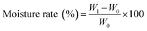
pADM is constructed of three-dimensional collagen type I fibers, the thermal stability of cross-linked collagen is an important characteristic of the effectiveness of cross-linking. In gradually heated water, the temperature at which pADM shrinks is defined as the hydrothermal shrinkage temperature (Ts). That happens due to the thermally process can decrease the forces (hydrogen bonds, cross-link, etc.) between and within collagen fibers, which would cause the destabilization of the structure of collagen. The basic forces (covalent bonds, hydrogen bonds, van der Waals force) which are responsible for the high thermostability of collagen, could be attributed to long range interactions.15 OCOS contains amounts of aldehyde groups, which can react with the amino groups of collagen to form Schiff's base between OCOS and collagen. These covalent bonds within collagen can stable the structure of collagen, which leads to improving the hydrothermal stability of collagen. Increase in resistance against hydrothermal stress is one of the important aspects in the stabilization of collagen matrix, therefore, Ts is an indicator of the cross-linking degree of collagen. The higher Ts usually indicates higher cross-link density and therefore a better biostability and durability of the fixed collagen.11,15
Presuming that, pADM is constructed of pure collagen fibers after removing the non-collagen compositions, and the amino groups only exist in Lys and Hly, in that way, the content of free amino groups in collagen is 0.37 mmol g−1 theoretically. In fact, the amount of free amino groups is lower than that value, due to the self-cross-linking within collagen which took place by amino groups and other reactive groups in collagen, such as Schiff's base cross-linking reaction, β-aldol cross-linking reaction, etc.20 In this study, the density of the free amino groups is about 0.207 mmol g−1 according to the ninhydrin reaction. The free amino content in pADM can be effectively used to characterize the reacting degree at the site of the amino group, which was marked as the cross-link density. A higher cross-link density often implies a lower level of free amino groups left in the fixed tissue. When chemical reaction takes place, the increase in the dosage of reactant can lead to the increase in the amount of resultant. The amount of the amino groups on collagen is certain. In order to make a positive effect on the chemical reaction equilibrium, enlarging the dosage of OCOS is necessary. Therefore, the cross-linking degree can be controlled by changing the dosage of OCOS. Every OCOS molecule has more than two aldehyde groups, if two of these aldehyde groups react with the amino groups in collagen, a bridge within the collagen can be formed, which is the so called cross-linking. In addition, if only one aldehyde group reacts with an amino group, it leads only to capping of the reacted amino group but could not form a cross-link, which is called masking.21 The increase of Ts is thought to result primarily from cross-linking, rather than masking, of the amino groups within the collagen. With the increase of the dosage of OCOS, Ts of OCOS–pADM changed due to the new chemical bonds formed between inter- and intra-collagen molecules, compared with the blank group Ts of 63 °C, the highest Ts around of 78 °C appeared in the 4% and 8% group, as displayed in Fig. 1. Meanwhile, the cross-link density continually increased along with the increase of OCOS, which is in line with the mechanism of the reaction. In the first stage, enlarging the dosage of OCOS, more aldehyde groups were consumed to form cross-link bonds and to make a contribution to higher Ts, the cross-linking reaction played a major role. In particular, Ts of OCOS–pADM stayed steady when the dosage of OCOS is greater than 4%, while the cross-link density grew continuously. That happened because of the masking reaction took place except for some cross-linking reaction which could consume the free amino groups but could not make an evident contribution to the increase in Ts. Moreover, it is known that reduction of free amino groups in the biological tissue diminishes its antigenicity.21
 |
| | Fig. 1 The effect of dosage to the Ts and cross-linking density of OCOS–pADM. | |
3.2 FTIR analysis
Fourier transform infrared (FTIR) spectra were carried out to investigate the variation of the structure of collagen in the presence of OCOS. Collagen has a special triple helix conformation which can be directly shown by the characteristic infrared absorption peak, as exhibited in Fig. 2. The characteristic absorption bands at 1650 cm−1 represent the amide I, which is produced by proteinic amide C![[double bond, length as m-dash]](https://www.rsc.org/images/entities/char_e001.gif) O stretching vibrations. The amide II bands at 1545 cm−1 is caused by bending vibration of N–H and stretching vibration of C–N. More, the amide III at 1235 cm−1 is contributed by the stretching vibration of C–N and bending vibration of N–H in plane and wagging vibration from CH2 groups in the glycine backbone and proline side-chains. Meanwhile, the characteristic absorption bands at 3410 cm−1 and 3082 cm−1 represent the amide A and amide B, and are caused by the stretching vibration of N–H. It is known that the amide I and amide II are directly related to the α-helix, β-sheet, angle, and curly stack with no rules of the secondary structural components of collagen.16,22 In addition, the bands at 1733 cm−1 is assigned to the symmetric vibration of CO in aldehyde groups, while the bands at 1080 cm−1 is caused by the stretching vibration of C–O, C–O–C and the frame vibration of rings in oligosaccharides. Fig. 6 suggests that all characteristic absorption bands in all samples were maintained, with only small changes which demonstrated that the backbone of collagen in the presence of OCOS did not change along with the increasing of OCOS content, that is to say, adding OCOS did not damage the natural structure of collagen, which is important to a cross-linker and the biological activity of collagen. Comparisons between the FIIR spectra of pADM and OCOS–pADM revealed that the peak at 1080 cm−1 had obvious enhancement after cross-linking which might indicate that the structural units of OCOS was introduced into collagen, and that peak became stronger along with the increase of dosage of OCOS. Nevertheless, the amide A reduced and moved toward lower wavenumbers, the amide I reduced as well. The possible reason is that OCOS can react with collagen to form stable Schiff's base which might destroy the hydrogen bands between the peptide chains of collagen.16
O stretching vibrations. The amide II bands at 1545 cm−1 is caused by bending vibration of N–H and stretching vibration of C–N. More, the amide III at 1235 cm−1 is contributed by the stretching vibration of C–N and bending vibration of N–H in plane and wagging vibration from CH2 groups in the glycine backbone and proline side-chains. Meanwhile, the characteristic absorption bands at 3410 cm−1 and 3082 cm−1 represent the amide A and amide B, and are caused by the stretching vibration of N–H. It is known that the amide I and amide II are directly related to the α-helix, β-sheet, angle, and curly stack with no rules of the secondary structural components of collagen.16,22 In addition, the bands at 1733 cm−1 is assigned to the symmetric vibration of CO in aldehyde groups, while the bands at 1080 cm−1 is caused by the stretching vibration of C–O, C–O–C and the frame vibration of rings in oligosaccharides. Fig. 6 suggests that all characteristic absorption bands in all samples were maintained, with only small changes which demonstrated that the backbone of collagen in the presence of OCOS did not change along with the increasing of OCOS content, that is to say, adding OCOS did not damage the natural structure of collagen, which is important to a cross-linker and the biological activity of collagen. Comparisons between the FIIR spectra of pADM and OCOS–pADM revealed that the peak at 1080 cm−1 had obvious enhancement after cross-linking which might indicate that the structural units of OCOS was introduced into collagen, and that peak became stronger along with the increase of dosage of OCOS. Nevertheless, the amide A reduced and moved toward lower wavenumbers, the amide I reduced as well. The possible reason is that OCOS can react with collagen to form stable Schiff's base which might destroy the hydrogen bands between the peptide chains of collagen.16
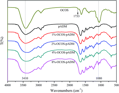 |
| | Fig. 2 FTIR spectra of OCOS–pADM with different dosage of OCOS. | |
3.3 X-ray diffraction analysis
X-ray diffraction is a physical method to determine the spatial structure of the collagen and to understand the triple helix of the collagen molecule and the axial arrangement of the collagen fibrils. Normally, the X-ray diffraction pattern of collagen mainly has three diffraction peaks. The first one is 2θ = 7–8° (about 1.2 nm), as the crystal diffraction peak, the Bragg spacing is composed of collagen fibers in the molecular chain between horizontal distance. The second one is at about 22°, the emergence of a very wide peak for collagen fiber structure which is caused by diffuse scattering. The third one is 2θ = 31–32° (about 0.28 nm), there is a small broad peak reflecting the collagen helix rotation within the height corresponding to the axial period, that is a typical symbol of the triple helix structure.22 It can be seen in Fig. 3 that the X-ray diffraction spectrum diagram of all samples shaped similarly especially the peaks around 31–32° which indicates that pADM maintained the native conformation of collagen fibers and there was no appreciable damage to the triple helix after cross-linking. In addition, the diffraction peaks at 2θ = 7–8° moved a little bit towards the high degrees of 2θ, showing that the space between collagen molecular chains became smaller.
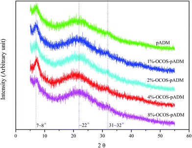 |
| | Fig. 3 XRD diagrams of pADM treated with different dosages of OCOS. | |
In order to understand the changes, the distances were calculated from 2θ at peak 1 (d1) and peak 3 (d2) by Bragg equation 2dsinθ = nλ (λ = 0.154 nm), and the results are listed in Table 1. From Table 1, the d1 of 4%-OCOS–pADM decreased from 1.221 nm to the minimum 1.187 nm, other groups decreased slightly compared with pADM, while the values of d2 didn't change much. The results illustrated that cross-linking by OCOS would not have an evil influence on the native structure of collagen, which is important for the cross-linked matrix to maintain the biological properties of collagen.
Table 1 The distance between collagen chains and the axial repeat per residue
| Group |
d1/(nm) |
d2/(nm) |
| pADM |
1.221 |
0.282 |
| 1%-OCOS–pADM |
1.219 |
0.282 |
| 2%-OCOS–pADM |
1.211 |
0.282 |
| 4%-OCOS–pADM |
1.187 |
0.281 |
| 8%-OCOS–pADM |
1.190 |
0.280 |
3.4 SEM and EDS analysis
PADM is weaved by collagen fibers randomly, which could be fixed and maintained after cross-linking with OCOS. As illustrated in Fig. 4, the microfiber morphology and the 3D architecture of pADM before and after cross-linking stayed almost the same, the collagen fibers had a layer by layer structure with suitable porosity, and the pore size is about 50 μm to 200 μm. Pore size and porosity of collagenous tissue are important to cell adhesion and proliferation. Normally, the range of suitable pore size is from 20 μm to 200 μm,23 the pore size of OCOS–pADM is within this range which demonstrates that OCOS–pADM has potentials in the use of scaffoldings for cell growth. In addition, the appearance of color of OCOS–pADM was changed from white to yellow through a visual observation showed in (F) of Fig. 4, the more dosage of OCOS, the darker of the color, which is closely related to the formation of the Schiff's base.
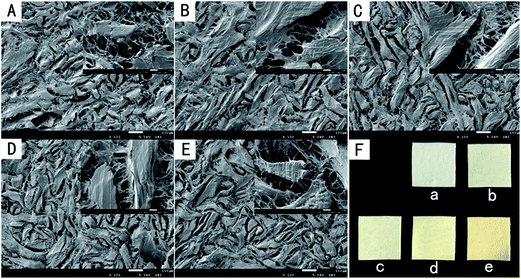 |
| | Fig. 4 SEM micrographs of pADM treated with different dosage of OCOS under different magnifications (100×, 1000×), (A) pADM, (B) 1%-OCOS–pADM, (C) 2%-OCOS–pADM, (D) 4%-OCOS–pADM, (E) 8%-OCOS–pADM. And photographs of pADM treated with different dosage of OCOS (F), (a) pADM, (b) 1%-OCOS–pADM, (c) 2%-OCOS–pADM, (d) 4%-OCOS–pADM, (e) 8%-OCOS–pADM. | |
OCOS, made of COS, has the primary structure of the oligosaccharide. It is clear that the ratio of elements in OCOS differs widely with pADM. Therefore, energy dispersive X-ray spectrometer (EDS) was executed to analyze the elements content on the surface of pADM before and after cross-linking. Fig. 5 shows that the atomic ratio of oxygen element was about 24.93% in pADM, which was rose up to 25.38%, 26.10%, 26.47%, 26.65% after cross-linking with OCOS (1%, 2%, 4%, 8%) respectively. The oxygen element content increased due to OCOS is abundant of oxygen element (the atomic ratio of O is about 41% by theoretical calculation) which could be introduced into pADM by cross-linking. The result also evinced that with the increase in dosage of OCOS, the cross-link density increased as well as the ratio of oxygen, which is agreeable with the result of cross-linking characteristics.
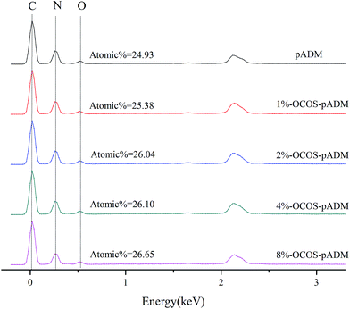 |
| | Fig. 5 The elements contents on the surface of pADM treated with different dosage of OCOS. | |
3.5 AFM analysis
Collagen fibril is an aggregate of collagen molecules, which did not advance side by side, but mutually staggered around a certain distance. The adjacent tropocollagen molecules staggered axially by a distance, d, which is the sum of gap and overlap regions. The most characteristic feature of collagen fibrils is that there are D-periodicity with a length of 67 nm2. It is significative to observe the D-periodicity of cross-linked collagen fibrils since the presence of D-periodicity is believed to be important for the mechanical and biological functions of collagenous matrix.24 According to Fig. 6, the D-periodicity in cross-linked matrix was clearly visible, indicating that the micro structure of all samples was maintained, which was beneficial to cross-linked matrix to keep the antigenicity and biological compatibility of native collagen.
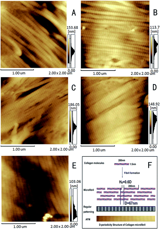 |
| | Fig. 6 AFM images of pADM treated with different dosage of OCOS under different magnifications. (A) pADM, (B) 1%-OCOS–pADM, (C) 2%-OCOS–pADM, (D) 4%-OCOS–pADM, (E) 8%-OCOS–pADM. (F) The diagram of D-periodicity of collagen. | |
3.6 Mechanical properties
Tensile strength and elongation at break testing were carried out to evaluate the effects of different dosages of OCOS cross-linking on the mechanical properties of pADM. Good mechanical strength can guarantee the ability of collagenous tissue to avoid deformation due to the extrusion and stretch in the use of biomaterials. Tensile strength, an important indicator of mechanical properties of collagen, characterizes the ability of collagen to resist the breakage causing by external forces. Fig. 7 reveals that the tensile strength of OCOS–pADM was superior to pADM, besides, the 4% dosage of OCOS ranked first, while the 8% group was lower than 4% group. The result was consistent with the hydrothermal stability of the samples. Normally, the ability to resist external forces mainly benefited from the chemical bonds, van der Waals forces and hydrogen bonds within collagen, which indicated that cross-links were formed within collagen. Meanwhile, the higher dosage of OCOS did not lead to the expected higher elongation at break. The decrease of elongation at break showed that the brittleness of matrix increased, and this kind of phenomenon often occurs in the process of cross-linking of collagenous matrix.25,26 This might be due to the conformation of the intramolecular cross-linking, intermolecular cross-linking and cross-linking between microfibrils, which can promote collagen fibers to form a relatively stable rigid structure. In the process of stretch, fibers were directional aggregation to some extent and were difficult to be deformed but easy to cause stress concentration, leading to the occurrence of brittleness, thus reducing the corresponding elongation at break.
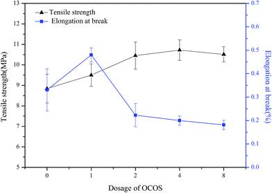 |
| | Fig. 7 Tensile strength and elongation at break of pADM treated with different dosage of OCOS. | |
3.7 Hydrophilicity analysis
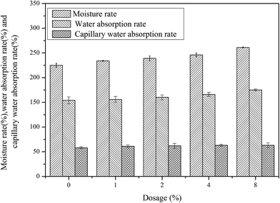
Water contact angle, the moisture rate, the water absorption rate and the capillary water absorption rate were tested to explore the correlation between the microstructure and surface property of OCOS–pADM. As is illustrated by Fig. 8, with the increase in dosage of OCOS, the water contact angle was reduced gradually. It is known that the lower the water contact angle, the better hydrophilicity. In addition, the moisture rate and the water absorption rate increased along with the increase in OCOS content as shown in Fig. 9, which comports with the result of water contact angle. That is because of cross-linking can introduce the polar groups (hydroxyl groups and glycosidic bonds) of OCOS to collagen which was demonstrated by the EDS analysis, and these polar groups can provide good hydrophilicity. Moreover, from Fig. 9, the capillary water absorption rate was increased slightly after cross-linking after the high speed centrifugal process. The possible reason is that the distance between the collagen fibers changed a little, so as well as the capillary effect. During the use of biomaterials, the forces like extrusion, pull, etc. may squeeze the adsorbed water out. The result of the capillary water absorption rate showed that the performance of OCOS–pADM would not be affected by the external forces. As biomaterials, the surface properties are very important. A well hydrophilic property is helpful to the adhesion and proliferation of cells, which is beneficial to wound healing.27
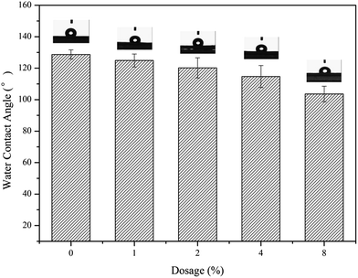 |
| | Fig. 8 Contact angle of pADM treated with different dosage of OCOS. | |
 |
| | Fig. 9 Moisture rate, water absorption rate and capillary water absorption rate of pADM treated with different dosage of OCOS. | |
3.8 Degradation properties
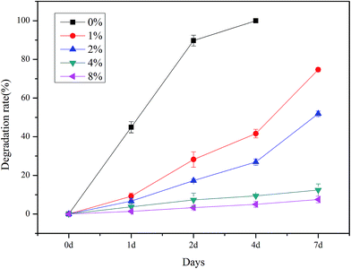
To regulate the biodegradation rate of collagen scaffolds, chemical cross-linking is a necessary method. Before cross-linking, it is difficult for pADM scaffolds to resist degradation in the presence of collagenase. Fig. 10 indicates that all groups presented signs of degradation to some extent after incubating with bacterial collagenase type I. pADM group, showed the worst resistance to degradation in all groups, was almost degraded completely after 2 days, while cross-linking groups showed good performance. After 7 days exposing to collagenase, 1%, 2%, 4%, 8% OCOS groups were degraded ∼74.7%, ∼51.9%, ∼12.4%, ∼7.5%, respectively, the increased cross-linking degree of pADM resulted in the promoted biostability. It is known that, the bacterial collagenase can catalyze hydrolysis and cut off the peptide bonds in nonpolar region of collagen, in a single α chain segment, or in the cross-section of triple helix. Cross-linking can promote the ability of collagen to resist to the action of cutting off α chain by collagenase. Therefore, it can be speculated that, the cross-linking may occur in a single chain segment, between chains, molecules, or microfibrils of collagen.
 |
| | Fig. 10 Resistance to degradation of pADM treated with different dosages of OCOS. | |
3.9 Cytocompatibility analysis
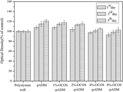
To determine whether different dosages of OCOS treatments could induce any extra cytotoxicity of pADM, cytocompatibility study was conducted by direct contact and indirect extraction. As shown in Fig. 11, the optical density of all groups increased with the increase of culture time. At each time point, the optical density values showed that, with the increase of OCOS content, the relative proliferation rate of the cultured cells was decreased, even though, cross-linked pADM showed a significant promotion on the proliferation of L929 fibroblasts except for the 8% group, while the lowest relative proliferation rate of 93% appeared in the 8% OCOS group. The main component of pADM is type I collagen, which is favorable to fibroblasts due to its specific biological properties and it can be the attachment and support for L929 fibroblasts to promote adhesion, growth and proliferation, that is why the pure pADM group showed higher optical density than control group. The results showed that adding in a small amount of OCOS could promote the growth and proliferation of cells, and it also showed that the cross-linked pADM had little cytotoxicity when the dosage of OCOS reached to 8%. The reason may be that most of the aldehyde groups reacted with reactive groups in collagen in excess of OCOS, so there was still a small amount of excess aldehyde groups which may react with the protein and polysaccharide in fibroblasts.16 In general, when the dosage of OCOS is less than 8%, introducing OCOS into pADM might be favorable for the cell's proliferation which was in accordance with the requirements of the surface use of biomaterials.
 |
| | Fig. 11 Proliferation of L929 fibroblasts cultured in the extraction liquid of pADM treated with different dosages of OCOS. | |
Fig. 12 shows the SEM image of L929 fibroblasts adhesive on OCOS–pADM. After 3 days culture, the cells on OCOS–pADM showed almost similar morphologies to those on pADM, except for the 8% OCOS group. Fibroblasts cells showed typical spindle shape of L929 fibroblasts and spread well within collagen fibers, and there were large amount of extracellular secretions on the surface of cells. In addition, the cell could not only firmly adhere on collagen fibers, but also go deep into the pores among collagen fibers. What's more, the pseudopodia of cells fixed on the different collagen fibers respectively so that cells could grow across the gap of collagen fibers. In the 8% OCOS group (E group in Fig. 12), condition of cells was inferior to other groups, however, this result is consistent with the MTT assay.
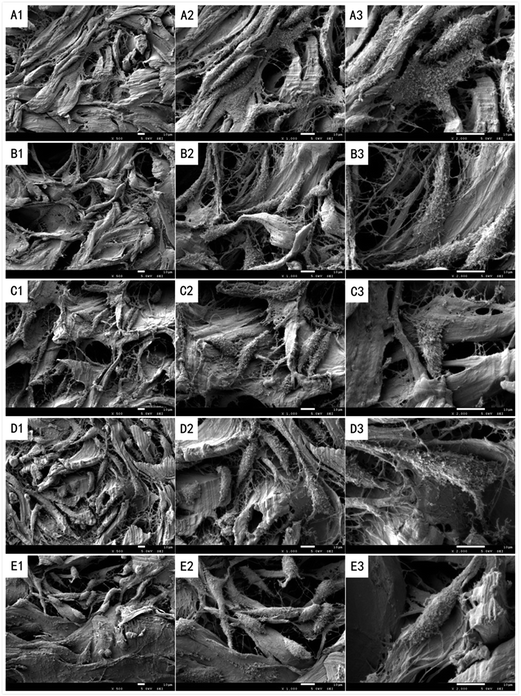 |
| | Fig. 12 SEM photographs of L929 fibroblasts cultured for 3 days on pADM treated with different dosages of OCOS under different magnifications (200×, 500× and 2000×). (A) pADM, (B) 1%-OCOS–pADM, (C) 2%-OCOS–pADM, (D) 4%-OCOS–pADM, (E) 8%-OCOS–pADM. | |
3.10 The reaction between collagen and OCOS
Porcine acellular dermal matrix, as a kind of extracellular matrix, is porous enough for mass transfer and growth of cells. Cross-linking enabled the formation of new chemical bonds within pADM, thus making the structure more stable, and improving the ability of resistance to degradation. The specific structure of native collagen leads to the distance between the active groups on collagen molecule, therefore the cross-linker must have proper length or size to attach the active groups. Carbodiimide can only cause intramolecular cross-linking of carboxyl and amino groups on collagen without introducing itself into the collagen, which is known as the “zero length cross-linking” agent. So it has an inherent defect that it is powerless for the far spaced active groups. The “size-cross-linking” agents can overcome the shortcomings of “zero length cross-linking” agent, help to the formation of three kinds of cross-linking. One is intramolecular cross-linking (longer than “zero length cross-linking”), which can affect the denaturation and tensile strength of materials. Another is intermolecular cross-linking, which can make effects on swelling capacity and surface extensibility of materials. The third is the cross-linking between collagen microfibrils (Fig. 13). These three ways of cross-linking improve stability of collagen from a variety of spatial cross-linking. Glutaraldehyde, a wild used cross-linker, has a length of five-carbon chain containing two aldehyde groups on each side, which can react with amino groups on collagen to form Schiff's base to improve stability of collagen greatly. However, the use of glutaraldehyde is limited by the high toxicity and the calcification of the cross-linked tissue. The double functionalized cross-linker (like glutaraldehyde) is often considered to form a linear cross-linking, and the multi-functionalized cross-linker is often considered to be more complex (3D cross-linking) in the conformation of cross-linking structure, which can make greater contribution to the stability of collagen. Polyalcohol glycidyl ether contains two or more epoxy groups, the length and structure can be designed to meet requires, the cross-linking effect is good.28 But, due to the use of organic chemical synthesis method, it is inevitable that the by-products are recommended, if left in the collagen, will have a negative impact on biocompatibility. The more epoxy groups, the stronger the cross-link performance, but the worse the water solubility. Collagen is a hydrophilic substance, which is difficult to be compatible with oil soluble epoxy compounds. The reaction system has poor performance on mass transfer. If the organic solvent is added, it may lead to residual solvent and cause the limit of the application of the epoxy compound. Genipin (226 Da) is a kind of iridoid extracted from plants, can form a dialdehyde structure under alkaline condition, so it has good cross-linking behavior.11 As a natural product, it can overcome the toxicity of the synthetic chemicals, and the biological compatibility of the cross-linked product is perfect. But genipin is expensive and hard to industrialize. OCOS, a series of homologues contains 2–10 monosaccharide units, with the molecular weight distribution from 320 to 1600 Da. It has a variety of different molecular scale to meet the needs of various chain lengths, so it can match the different distance between free amino groups of collagen. Each OCOS molecule contains two or more aldehyde groups, which can form firmer Schiff's base bonds with amino groups. Therefore, the three dimensional net structure is formed by the intramolecular cross-linking, intermolecular cross-linking and cross-linking between microfibrils. After cross-linking by OCOS, the natural D-periodicity microstructure (Fig. 6) and the porous structure (Fig. 4) of matrix were maintained, which is beneficial for maintain the original biological activity of collagen. The “rigid structure”, including covalent cross-linking bond, polycyclic of OCOS and the polycyclic formed by cross-linking, improved the stability of collagen greatly, so OCOS–pADM can resist the change of the external environment. The increase of the Ts (Fig. 1) is just benefited from this “rigid structure”, so do the tensile strength (Fig. 7) and the ability of resistance to degradation (Fig. 10). Moreover, as derivative of the chitosan degradation products, OCOS is an animal-derived carbohydrate, that is to say it has a better homology than plant-derived genipin. The chitosan oligosaccharide chain was introduced into collagen by cross-linking, which helped to endows the function of COS to collagen matrix (such as hydrophilicity, Fig. 8 and 9). Besides, the COS modified collagen is more similar to natural collagen glycoprotein. Due to the fact that the COS itself has good biocompatibility and the cross-linked pADM kept the good natural microstructure (triple helix, D-periodicity and porous structure), the COS modified collagen matrix (COS-Col) had a good biocompatibility (Fig. 12). The degradation products would be monosaccharide units, collagen degradation products, as well as small molecules of Cos-Col, which is expected to ensure the safety if implant in vivo. In addition, to meet the requirements of different biological materials, the cytotoxicity and the ability of resistance to degradation can be regulated by controlling the degree of oxidation of OCOS or/and the dosage of OCOS added. COS is abundant, widely available, and inexpensive. The preparation method of OCOS is simple, and OCOS has the advantages of a variety of cross-linkers, which is expected to be widely used in the field of biological materials.
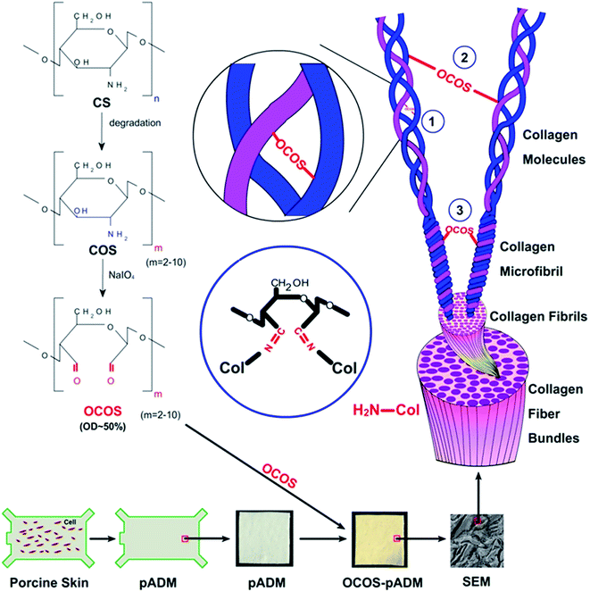 |
| | Fig. 13 The schematic diagram of cross-linking reaction between pADM and OCOS, ① intramolecular cross-linking, ② intermolecular cross-linking, ③ cross-linking between collagen microfibrils. | |
4 Conclusions
OCOS, an animal-derived cross-linking agent, prepared from the natural products COS. It not only inherits the excellent biocompatibility of the chitosan oligosaccharide, but also has excellent cross-linking performances. It is believed that, OCOS has the smaller molecular weight, the better solubility in water when compared with polysaccharide derivatives. Hence, it is especially suitable for cross-linking with the compact structure of biological materials, such as pADM, because it can penetrate deeply into the structure of biological materials, and produce more stable cross-linked structure. Using OCOS to modify pADM can improve the structural stability, hydrothermal stability, resistance to degradation, mechanical properties and hydrophilicity significatively. Monosaccharide units were successfully introduced into the matrix after cross-linked, which have no undue adverse impact on the triple helix and the D-periodicity of pADM. Therefore, the cross-linked matrix has good biological compatibility. As a cross-linker, OCOS can balance chemical activity and biocompatibility, which is a new generation of cross-linker for collagen matrix.
Acknowledgements
This work is supported by National Natural Science Foundation of China (contract grant number 51473001) and Fundamental Research Funds for the Central Universities of China (SCU2014d005).
References
- K. Gelse, E. Poschl and T. Aigner, Adv. Drug Delivery Rev., 2003, 55, 1531–1546 CrossRef CAS PubMed.
- M. D. Shoulders and R. T. Raines, Annu. Rev. Biochem., 2009, 78, 929–958 CrossRef CAS PubMed.
- Y. Hu, L. Liu, W. H. Dan, N. H. Dan and Z. P. Gu, J. Appl. Polym. Sci., 2013, 130, 2245–2256 CrossRef CAS.
- V. Ottania, M. Raspantib and A. Ruggeri, Micron, 2011, 32, 251–260 CrossRef.
- S. Chattopadhyay and R. T. Raines, Biopolymers, 2014, 101, 821–833 CrossRef CAS PubMed.
- Y. Hu, L. Liu, W. H. Dan, N. H. Dan, Z. P. Gu and X. X. Yu, Int. J. Biol. Macromol., 2013, 55, 221–230 CrossRef CAS PubMed.
- Y. Di and R. J. Heath, Polym. Degrad. Stab., 2009, 94, 1684–1692 CrossRef CAS.
- S. C. Baker, G. Rohman, J. Southgate and N. R. Cameron, Biomaterials, 2009, 30, 1321–1328 CrossRef CAS PubMed.
- M. Sato, Y. Hiramatsu, S. Matsushita, S. Sato, Y. Watanabe and Y. Sakakibara, J. Artif. Organs, 2014, 17, 265–271 CrossRef CAS PubMed.
- N. Nakajima and Y. Ikada, Bioconjugate Chem., 1995, 6, 123–130 CrossRef CAS PubMed.
- N. H. Dan, S. W. Xiao, Y. Hu, Y. N. Chen and W. H. Dan, J. Soc. Leather Technol. Chem., 2015, 99, 176–182 CAS.
- X. M. Zhu, X. Y. Zhou, J. Y. Yi, J. Tong, H. Wu and L. H. Fan, Int. J. Biol. Macromol., 2014, 70, 300–305 CrossRef CAS PubMed.
- L. H. Fan, M. Cao, S. Gao, T. Wang, H. Wu, M. Peng, X. Y. Zhou and M. Nie, Carbohydr. Polym., 2013, 93, 380–385 CrossRef CAS PubMed.
- Y. Hu, L. Liu, Z. P. Gu, W. H. Dan, N. H. Dan and X. X. Yu, Carbohydr. Polym., 2014, 102, 324–332 CrossRef CAS PubMed.
- S. V. Kanth, A. Ramaraj, J. R. Rao and B. U. Nair, Process Biochem., 2009, 44, 869–874 CrossRef CAS.
- X. H. Liu, N. H. Dan, W. H. Dan and J. X. Gong, Int. J. Biol. Macromol., 2016, 82, 989–997 CrossRef CAS PubMed.
- S. Y. Chae, M. K. Jang and J. W. Nah, J. Controlled Release, 2005, 102, 383–394 CrossRef CAS PubMed.
- Y. J. Jeon and S. K. Kim, Carbohydr. Polym., 2000, 41, 133–141 CrossRef CAS.
- F. Q. Hu, M. D. Zhao, H. Yuan, J. You, Y. Z. Du and S. Zeng, Int. J. Pharm., 2006, 315, 158–166 CrossRef CAS PubMed.
- J. M. Pachence, J. Biomed. Mater. Res., 1996, 33, 35–40 CrossRef CAS PubMed.
- H. W. Sung, C. S. Hsu, Y. S. Lee and D. S. Lin, J. Biomed. Mater. Res., 1996, 31, 511–518 CrossRef CAS PubMed.
- S. W. Xiao, W. H. Dan and N. H. Dan, RSC Adv., 2015, 5, 88324–88330 RSC.
- H. M. Powell and S. T. Boyce, Biomaterials, 2006, 27, 5821–5827 CrossRef CAS PubMed.
- Y. P. Li, A. Asadi, M. R. Monroe and E. P. Douglas, Mater. Sci. Eng., C, 2009, 29, 1643–1649 CrossRef CAS.
- B. Balakrishnan, M. Mohanty, P. R. Umashankar and A. Jayakrishnan, Biomaterials, 2005, 26, 6335–6342 CrossRef CAS PubMed.
- B. Balakrishnan and A. Jayakrishnan, Biomaterials, 2005, 26, 3941–3951 CrossRef CAS PubMed.
- G. D. Winter, Nature, 1995, 4, 293–2944 Search PubMed.
- H. W. Sung, W. H. Cheng, I. S. Chiu, H. L. Hsu and S. A. Liu, J. Biomed. Mater. Res., 1996, 33, 177–186 CrossRef CAS PubMed.
|
| This journal is © The Royal Society of Chemistry 2016 |
Click here to see how this site uses Cookies. View our privacy policy here. 


