Conductive and SERS-active colloidal gold films spontaneously formed at a liquid/liquid interface†
Xiuxiu Yina,
Yossef Peretza,
Pola G. Oppenheimerb,
Leila Zeiric,
Alexandra Masarwaa,
Natalya Frouminc and
Raz Jelinek*ac
aDepartment of Chemistry, Ben Gurion University of the Negev, Beer Sheva 84105, Israel. E-mail: razj@bgu.ac.il
bSchool of Chemical Engineering, University of Birmingham, Edgbaston, Birmingham B15 2TT, UK
cIlse Katz Institute for Nanotechnology, Ben Gurion University of the Negev, Beer Sheva 84105,
First published on 29th March 2016
Abstract
We show that water-soluble gold thiocyanate salt [KAu(SCN)4] spontaneously formed electrically-conductive and surface enhanced Raman scattering (SERS)-active films at the interface between water and pentane. Microscopy and spectroscopic analyses reveal that the films comprised of condensed Au nanoparticles, assembled via crystallization of KAu(SCN)4 and self-reduction of the Au3+ ions by the thiocyanate ligands. Importantly, the pentane phase was crucial in promoting the crystallization/reduction reactions. The Au films exhibited useful applications, including excellent conductivity and SERS sensing. We also show that the new film self-assembly process could be readily harnessed for producing macro-scale Au patterns.
Introduction
Construction of nanostructured metallic films through “bottom-up” (e.g. self-assembly) chemical schemes has attracted significant interest in recent years as a viable alternative to “top down” lithography methods which are generally expensive, require sophisticated instrumentation, and often utilize hazardous reagents.1–3 Self-assembled nanostructured gold films have been fabricated through diverse strategies, such as Langmuir–Blodgett techniques,4 and layer-by-layer deposition.5 Self-assembly at the air/water and liquid/solid interfaces has been widely employed for Au film fabrication, since these two-dimensional interfaces provide intrinsic templates for Au nanostructures.6–9 Most interface-based Au film formation strategies, however, require initial synthesis of Au nanoparticles (NPs) and/or coating NPs with specific functional units, overall making such schemes rather complex.10The liquid–liquid interface offers a useful platform for creating organized NP assemblies, as self-assembly processes in such interfaces are primarily dictated by minimization of the interfacial energy and surface tension.11 Electrochemical deposition of metal NPs at organic solvent/water interfaces has been also reported.12,13 However, similar to the scenarios described above, production of organized assemblies in liquid/liquid interfaces also required pre-synthesis of the NPs and careful adjustment of experimental parameters, including types and compositions of liquids utilized, NP concentrations and sizes, and co-addition of residues used for preventing NP aggregation.14–16
Here, we demonstrate that Au nano-crystalline films were spontaneously formed at the interface between water and pentane through incubation of gold thiocyanate [Au(SCN)4−] in the aqueous phase, without co-addition of reducing nor stabilization agents. The self-assembled films comprised of a dense layer of Au nanoparticles, were remarkably robust and could be easily transferred onto solid substrates without further purification steps. Creation of patterned films could be readily accomplished through placement of “stamps” exhibiting desired shapes at the liquid–liquid interface. Crucially, different from previous studies of NP assemblies at liquid/liquid interfaces, the one-step process described here involves simultaneous formation of the Au NPs and their assembly at the water/pentane interface. Notably, the oil layer was intimately involved both in Au NP formation and their assembly at the interface. The Au nanocrystal films exhibited high electrical conductivity and also functioned as effective SERS-active substrates, underscoring the versatility and potential practical uses of this simple bottom-up process.
Results and discussion
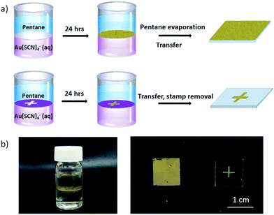
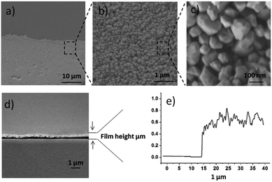
The preparation procedure of the nanostructured Au film is depicted in Fig. 1. Freshly-prepared KAu(SCN)4 aqueous solution was placed in a glass vial and a pentane layer was deposited above the water phase. Following 24 hour incubation a uniform thin film was formed at the water/pentane interface (Fig. 1a). After evaporation of pentane, the Au sheath could be transferred onto solid substrates through bottom horizontal lifting. Fig. 1a also outlines the formation of a patterned film by placing a “stamp” floating upon the water sub-phase underneath the pentane layer. Fig. 1b presents photographs illustrating film formation. Specifically, the picture in Fig. 1b (left) shows the Au film appearing at the pentane/water interface following 24 hour incubation. The photographs in Fig. 1b (right) underscore the uniform appearance of the Au film following transfer onto a solid surface, and a “plus” pattern produced according to the procedure in Fig. 1a.Fig. 2 presents microscopic characterization of the films, specifically their morphology and thickness. The scanning electron microscopy (SEM) images of a film extracted from the water/pentane interface after 24 hour (Fig. 2a–c) reveal a dense layer of irregularly-shaped colloidal nanoparticles, exhibiting diameters of between 50 and 100 nm. The cross section SEM image in Fig. 2d points to a relatively uniform film thickness of approximately 0.6 μm. This measurement was corroborated by the atomic force microscopy (AFM) height profile (Fig. 2e). The surface roughness apparent in the AFM height profile in Fig. 2e reflects the colloidal morphology of the film.
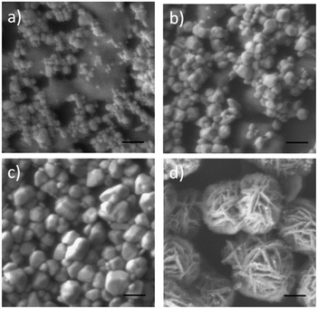
The evolution of film structure and specific contribution of the water/pentane interface to film features are highlighted in Fig. 3. Fig. 3a–c depicts SEM images of films extracted from the water surface (after evaporation of the organic solvent) at different incubation times of the gold thiocyanate complex in the water sub-phase. The SEM experiment shows that after two-hour incubation the film comprised of regions containing nanoparticles and abundant amorphous surfaces among them (Fig. 3a). After longer incubation time (8 h, Fig. 3b), shrinkage of the spread-out domains between the nanoparticles was apparent, and the nanoparticles themselves appeared larger. After 24 hours, a dense film of irregularly-shaped NPs exhibiting diameters of between 50–100 nm was recorded (Fig. 3c).
An important question we addressed in the SEM experiment in Fig. 3d concerns the contribution of the pentane layer towards film assembly and its morphology. The SEM image in Fig. 3d shows sizeable spherical aggregates formed after incubating KAu(SCN)4 for 24 hours in water, without the presence of a pentane layer. The aggregates exhibit significantly different morphology as compared to the film formed at the water/pentane interface (e.g. Fig. 3a–c). Specifically, the large aggregates display elongated crisscrossing nanowires (Fig. 3d), likely originating from aurophilic interactions between KAu(SCN)2 crystallites, formed through partial reduction of KAu(SCN)4.17 Notably, the aggregates depicted in Fig. 3d slowly precipitated and did not form films upon the water surface, unlike the situation in the air/pentane system.
Notably, organic solvents other than pentane that were placed upon the water subphase did not generate uniform Au NP films. For example, a hexane/water interface yielded incomplete and non-uniform distribution of Au NPs (Fig. S1†). The SEM experiments in Fig. 3 and Fig. S1† underscore the central role of the pentane/water interface in promoting assembly of the nanoparticle film. While the precise mechanism accounting for film growth has not been deciphered, the significance of distinct molecular interactions involving the organic solvent molecules (pentane, hexane, etc.) are expected.
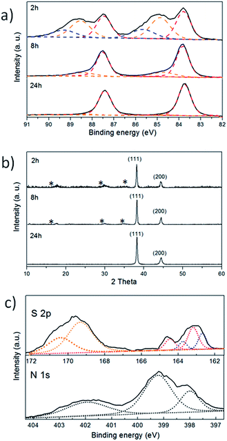
Fig. 4 presents X-ray photoelectron spectroscopy (XPS) and X-ray diffraction (XRD) data, which illuminate the crystalline properties and reaction pathways responsible for film assembly. The Au 4f spectra in Fig. 4a highlight the evolution of gold species during the film assembly process. Specifically, Fig. 4a shows that the percentage of metallic gold (Au0), corresponding to the peaks at 83.8 eV and 87.5 eV, gradually increased from approximately 40% after two hours, to ∼80% after 8 hours, and to almost 100% after 24 hours. In parallel, the intensities of the Au 4f peaks ascribed to AuIII and AuI were reduced, reflecting gradual reduction of the Au ions.
The XRD spectra in Fig. 4b underscore the evolution of the crystalline properties of the Au film assembled at the water/pentane interface. Consistent with the XPS data in Fig. 4a, the XRD patterns indicate that crystalline Au0 became the most prominent film feature as incubation progressed. Specifically, Fig. 4b demonstrates that the peaks corresponding to the (111) and (200) Au0 crystal planes were dominant after two-hour incubation, becoming the sole diffraction peaks after 24 hours. It should be noted that XPS experiments generally illuminate localized areas close to the film surface, rather than the entire film volume contributing to XRD results.18 The XRD peaks highlighted by stars (*) in Fig. 4b, recorded in films extracted after 2 h and 8 h, respectively, are ascribed to intermediate gold/organic hybrid structures. In particular, the peaks at around 30° and 35° (reflecting 3.0 Å and 2.6 Å crystal planes, respectively) correspond to crystalline KAu(SCN)2 assembled through aurophilic interactions,17 while the peak at 20° is likely associated with other crystalline KAu(SCN)4 species. Overall, the XRD patterns in Fig. 4b corroborate the XPS results, confirming the gradual spontaneous self-reduction of the gold ions in KAu(SCN)4, ultimately generating metallic crystalline Au NPs.
Self-reduction of KAu(SCN)4 to yield crystalline metallic Au0 likely occurred through the following reaction:
Reaction 1:
| 4H2O + 2Au(III)(SCN)4− → 2Au(0) + SO42− + CN− + 8H+ + 7SCN− |
The sulfur and nitrogen XPS spectra in Fig. 4c provide experimental evidence for the occurrence of Reaction 1, and aid identification of the reaction products associated with the films. Specifically, the de-convoluted S 2p peaks at 162.2 eV and 163.7 eV in Fig. 4c are attributed to SCN− residues bound to the Au NPs.19,20 Similarly, the XPS peaks at 163.2 and 164.5 eV are assigned to thiocyanate physically-adsorbed to the film surface.21,22 Importantly, the intense S 2p signals at 169.3 eV and 170.4 eV (Fig. 3c) correspond to oxidized sulfur in SO4− residues,23 confirming oxidation of SCN− following transfer of the electrons to the Au ions in KAu(SCN)4. The N 1s spectra in Fig. 3c corroborate the above interpretations. The de-convoluted peak at 399.2 is attributed to the un-reacted thiocyanate units,24 while the XPS signal at 398 eV corresponds to CN− residues adsorbed on the gold surface following thiocyanate-induced reduction of the gold ions.22 Zincon colorimetric method also revealed the presence of CN− ions in the aqueous phase (data not shown). The N 1s signal at 402 eV is assigned to N–O surface contamination.18 C 1s XPS data further confirm the presence of SCN− and CN− adsorbed onto the film surface (Fig. S2†).
The microscopy and spectroscopy data in Fig. 3 and 4 illuminate the reaction mechanism and the central role of the water/pentane interface in promoting film formation. A mechanism pertinent with the experimental results is depicted in Fig. 5. According to the proposed model, KAu(SCN)4 and partially-reduced KAu(SCN)2 crystallization nuclei which are sparingly soluble in the aqueous environment migrate to the pentane/water. Accumulation of the crystallites at the pentane/water interface is also energetically-favored as film formation between the two liquids reduces the surface energy through assembly of a more stable organic/solid/water interface.12 Further growth of the potassium gold thiocyanate crystallites at the water/pentane interface and simultaneous reduction of the AuIII ions generates the colloidal Au nanoparticle film. The prominent role of surface energy in film formation might also account for the specific contribution of pentane to the phenomenon. Indeed, interactions of the nucleating seeds formed at the liquid/liquid interface with the pentane molecules are different than the corresponding interactions with hexane (or other organic solvents), dictating distinct film formation profiles for each solvent.
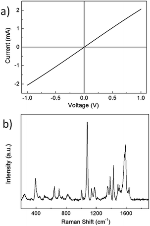
The nanocrystalline Au film exhibited practical functionalities (Fig. 6). Fig. 6a depicts the current/voltage curve recorded between two metal electrodes placed upon the film at a spacing of 1 cm. The ohmic behavior reflected in the linear I/V graph and relatively high currents (corresponding to resistivity of ∼5 Ω sq−1) underscore excellent electronic transport. Notably, the conductivity was measured for the as-assembled film, without additional treatments such as washing, sintering, or gold-coating enhancement. Indeed, annealing of the Au film (through 3 hour heating at 300 °C) yielded only marginally higher conductivity (Fig. S3†).
The nanostructured Au film assembled at the water/pentane interface was further examined as a substrate for surface-enhanced Raman scattering (SERS, Fig. 6b). SERS analysis was carried out by placing varying concentrations of the SERS-active probe p-aminothiophenol (PATP) upon the Au films. Fig. 6b, for example, depicts the spectrum of 10−8 M PATP upon the Au film, showing strong Raman signals at 818, 1008, 1077, 1145, 1187, 1392, 1440 and 1579 cm−1 that are consistent with reported values.25 Notably, typical SERS spectra of PATP were obtained down to 10−10 M concentrations, demonstrating remarkable sensitivity of the Au film (Fig. S4†). The pronounced PATP signals indicate that the nanocrystalline Au film presents abundant Raman “hot-spots”.
Experimental
Materials
HAuCl4·3H2O KSCN and p-aminothiophenol were purchased from Sigma-Aldrich and used as received. n-Pentane (98%) was purchased from Alfa Aesar. Water used in the experiments was doubly purified by a Barnstead D7382 water purification system (Barnstead Thermolyne, Dubuque, IA), at 18.3 mΩ cm resistivity.Synthesis of the KAu(SCN)4 complex
1 mL of an aqueous solution of HAuCl4·3H2O (24 mg mL−1) was added to 1 mL of a solution of KSCN in water (60 mg mL−1). The precipitate formed was separated by centrifugation at 4000g for 10 min. The supernatant was decanted, and the residue was dried at room temperature.Gold film growth at the interface
5 mL of freshly prepared KAu(SCN)4 aqueous solution (3 g L−1) was taken in a vial along with 5 mL of pentane resulting in a biphasic mixture with the pentane on top and aqueous solution below. The vial is then incubated at 25 °C for 24 h. A golden film is formed slowly at the liquid/liquid interface. After evaporation of pentane, the interfacial gold films were lifted onto silicon wafers or glass substrates for further analysis. Patterned gold films were prepared by placing a Kapton mask, which has a hollow-out plus sign with a thickness of 200 μm, at the interface before incubation.SERS measurements
The Raman system comprised a Jobin-Yvon LabRam HR 800 micro-Raman system, equipped with a liquid-N2-cooled detector. The excitation sources were a He–Ne laser (633 nm) with a power of 5 mW on the sample. The laser was focused with a 100 long-focal-length objective to a spot of about 2 mm. Most measurements were taken with the 600 g mm1 grating and a confocal microscope with a 100 mm hole with a typical exposure time of 1 min. PATP was dissolved in ethanol at concentrations of 10−8, 10−9 and 10−10 M. The gold film (previously transferred on to silicon wafer see above) was dipped in the analyte solution and dried.Conductivity measurements
Electrical measurements were performed on a Keithley 2400 sourcemeter in a two-probe configuration. For the sample preparation of electrical measurements, the substrate containing the gold film was mounted in a thermal evaporator with a stainless steel shadow mask attached. Cr (10 nm) and Au (90 nm) were evaporated onto the substrates to obtain the electrode patches at a predefined spacing.Instruments and characterization
Scanning electron microscopy (SEM) was conducted with a JEOL (Tokyo, Japan) model JSM-7400F scanning electron microscope equipped with EDS (Thermo Scientific). Powder X-ray diffraction (XRD) measurements were carried out on a Panalytical Empyrean powder diffractometer equipped with a parabolic mirror on the incident beam providing quasi-monochromatic Cu Kα radiation (λ = 1.54059 Å) and an X'Celerator linear detector. X-ray photoelectron spectroscopy (XPS) analysis was conducted using a Thermo Fisher ESCALAB 250 instrument with a basic pressure of 2 × 10⁻⁹ mbar. The samples were irradiated in two different areas using monochromatic Al Kα, 1486.6 eV X-rays, using a beam size of 500 μm. Atomic force microscopy (AFM) analysis was conducted with a Dimension 3100 SPM instrument from Digital Instruments (Veeco, NY).Conclusions
In conclusion, we demonstrate assembly of colloidal nanocrystalline Au films at the interface between two immiscible solvents, water and pentane. The process is based upon crystallization/reduction of KAu(SCN)4 initially dissolved in the water phase, resulting in formation of dense colloidal nanoparticle Au film at the water/pentane interface. Different than almost all reported schemes for construction of nanoparticle-based films at liquid/liquid interfaces in which nanoparticles have been prepared prior to film deposition, the process shown here involves in situ crystallization and reduction of the gold complex yielding metallic Au0 nanoparticles. Indeed, Au nanoparticle assembly occurred predominantly at the water/oil interface. Microscopic and spectroscopic analyses illuminate the mechanistic aspects of the self-assembly process, in particular the reaction pathway in which the thiocyanate ligands contribute the reducing electrons for the gold ions, and the prominent role of the pentane phase in affecting film assembly.The nanocrystalline Au films are amenable to practical applications. We demonstrated formation of macroscopically-patterned structures through placing a stamp with the desired shape at the water/oil interface. The Au films exhibited excellent electrical conductivities and strong SERS enhancement capabilities, pointing to possible sensing applications. Overall, the new self-assembly phenomenon based upon spontaneous crystallization and reduction of gold thiocyanate at the water/pentane interface constitutes a versatile means for producing functional gold films.
Acknowledgements
We thank Dimitry Mogiliansky for XRD measurements, and Jurgen Jopp for AFM analysis.Notes and references
- M. Kwiat, S. Cohen, A. Pevzner and F. Patolsky, Nano Today, 2013, 8, 677 CrossRef CAS.
- N. A. Melosh, A. Boukai, F. Diana, B. Gerardot, A. Badolato, P. M. Petroff and J. R. Heath, Science, 2003, 300, 112 CrossRef CAS PubMed.
- D. Wang, B. Sheriff, M. McAlpine and J. Heath, Nano Research, 2008, 1, 9 CrossRef CAS.
- K. Mayya, V. Patil and M. Sastry, Langmuir, 1997, 13, 2575 CrossRef CAS.
- Y. Liu, Y. Wang and R. O. Claus, Chem. Phys. Lett., 1998, 298, 315 CrossRef CAS.
- T. Hassenkam, K. Nørgaard, L. Iversen, C. J. Kiely, M. Brust and T. Bjørnholm, Adv. Mater., 2002, 14, 1126 CrossRef CAS.
- K. Ohno, K. Koh, Y. Tsujii and T. Fukuda, Angew. Chem., Int. Ed., 2003, 42, 2751 CrossRef CAS PubMed.
- H. Ma and J. Hao, Chem.–Eur. J., 2010, 16, 655 CrossRef CAS PubMed.
- T. Vinod and R. Jelinek, ACS Appl. Mater. Interfaces, 2014, 6, 3341 CAS.
- K. Kim, H. S. Han, I. Choi, C. Lee, S. Hong, S.-H. Suh, L. P. Lee and T. Kang, Nat. Commun., 2013, 4, 2182 Search PubMed.
- W. H. Binder, Angew. Chem., Int. Ed., 2005, 44, 5172 CrossRef CAS PubMed.
- S. G. Booth, A. Uehara, S. Y. Chang, J. F. W. Mosselmans, S. L. Schroeder and R. A. Dryfe, J. Phys. Chem. C, 2015, 119, 16785 CAS.
- Y. Grunder, Q. M. Ramasse and R. A. W. Dryfe, Phys. Chem. Chem. Phys., 2015, 17, 5565 RSC.
- P.-P. Fang, S. Chen, H. Deng, M. D. Scanlon, F. Gumy, H. J. Lee, D. Momotenko, V. Amstutz, F. Cortés-Salazar, C. M. Pereira, Z. Yang and H. H. Girault, ACS Nano, 2013, 7, 9241 CrossRef CAS PubMed.
- J. Wang, D. Wang, N. S. Sobal, M. Giersig, M. Jiang and H. Möhwald, Angew. Chem., Int. Ed., 2006, 45, 7963 CrossRef CAS PubMed.
- B. Su, N. Eugster and H. H. Girault, J. Am. Chem. Soc., 2005, 127, 10760 CrossRef CAS PubMed.
- A. Morag, N. Froumin, D. Mogiliansky, V. Ezersky, E. Beilis, S. Richter and R. Jelinek, Adv. Funct. Mater., 2013, 23, 5663 CrossRef CAS.
- J. F. Moulder, J. Chastain and R. C. King, Handbook of X-ray photoelectron spectroscopy: a reference book of standard spectra for identification and interpretation of XPS data, Perkin-Elmer Eden Prairie, MN, 1992 Search PubMed.
- K. G. Thomas, J. Zajicek and P. V. Kamat, Langmuir, 2002, 18, 3722 CrossRef CAS.
- Z. Zhang, J. Zhang, C. Qu, D. Pan, Z. Chen and L. Chen, Analyst, 2012, 137, 2682 RSC.
- C. Shen, M. Buck, J. D. Wilton-Ely, T. Weidner and M. Zharnikov, Langmuir, 2008, 24, 6609 CrossRef CAS PubMed.
- J. W. Ciszek and J. M. Tour, Chem. Mater., 2005, 17, 5684 CrossRef CAS.
- G. Rousseau, C. Lavenn, L. Cardenas, S. Loridant, Y. Wang, U. Hahn, J.-F. Nierengarten and A. Demessence, Chem. Commun., 2015, 51, 6730 RSC.
- J. W. Ciszek, M. P. Stewart and J. M. Tour, J. Am. Chem. Soc., 2004, 126, 13172 CrossRef CAS PubMed.
- T. P. Vinod, S. Zarzhitsky, A. Morag, L. Zeiri, Y. Levi-Kalisman, H. Rapaport and R. Jelinek, Nanoscale, 2013, 5, 10487 RSC.
Footnote |
| † Electronic supplementary information (ESI) available: Additional SERS spectra, I–V curve and XPS spectrum of C 1s. See DOI: 10.1039/c6ra03403a |
| This journal is © The Royal Society of Chemistry 2016 |

