
Electrodeposition of Au nanoparticles on poly(diallyldimethylammonium chloride) functionalized reduced graphene oxide sheets for voltammetric determination of nicotine in tobacco products and anti-smoking pharmaceuticals†
Yanqiu Jinga,
Erge Lina,
Xinhong Sub,
Yingjie Liuc,
Huaiqi Lid,
Xiuxiu Yuana,
Lu Pingd and
Yikuan Fan*b
aCollege of Tobacco Science, Henan Agricultural University, Zhengzhou, 450000, China
bResearch Institute of Henan Tobacco Corporation, Zhengzhou, 450000, China. E-mail: yikuanfan@126.com
cZhengzhou Branch of Henan Tobacco Corporation, Zhengzhou, 450000, China
dTechnology Center of Chinese Tobacco Industrial Company of Henan, 450000, Zhengzhou, China
First published on 2nd March 2016
Abstract
The principal objective of this study was to develop a sensitive and selective electrochemical sensor for nicotine detection based on a novel poly(diallyldimethylammonium chloride) functionalized reduced graphene oxide–gold nanoparticle (PDDA-RGO/Au) nanocomposite. The PDDA-RGO/Au nanocomposite was synthesized by a wet chemical approach followed by an electrodeposition process. The synthesized nanocomposite was characterized by FTIR, Raman spectroscopy, SEM and XRD. The sensor showed electrocatalytic activity in both aqueous and micellar media toward the oxidation of nicotine in Britton–Robinson buffer solution using cyclic voltammetry and electrochemical impedance spectroscopy (EIS) techniques. The results showed that the PDDA-RGO/Au nanocomposite could greatly enhance the electrochemical oxidation of nicotine. The linear response range of the sensor was between 0.5 μM and 300 μM with a detection limit of 0.12 μM. Excellent results were achieved for the determination of nicotine in tobacco products and anti-smoking pharmaceuticals.
1. Introduction
Nicotine, 3-(1-methyl-2-pyrrolidinyl)pyridine, is the main alkaloid in tobacco leaves (Nicotiana tabacum L., Solanaceae). Nicotiana tabacum is cultivated throughout the world for the preparation of cigars, cigarettes, and pipe and chewing tobacco. Although there are more than 4000 compounds in cigarette smoke, nicotine alone is primarily considered to be the addictive and reinforcing agent responsible for continued smoking behaviour.1 Pharmacologically, nicotine is a compound which acts on the central nervous system in the form of mood elevations, a sense of euphoria and revitalizing energy.2 However, nicotine has been suspected to causes cancer of various organs involving lung, nose, oral cavity, kidney, stomach, bladder and colon.3 Reduction in the nicotine content of cigarettes can result in an abstinence syndrome. The quantity of NIC considered as the lethal dose for children is 10 mg; the level for adults is 40–60 mg. Moreover, in order to support people who want to quit smoking, various pharmaceutical companies have come up with popular anti-smoking products which support the user to refrain from smoking. Therefore, the determination of nicotine is important in the fields of chemistry, toxicology, pharmacology, environmental and clinical areas.To date, the analytical methods reported for determination of nicotine include gas chromatography,4,5 high performance liquid chromatography,6,7 capillary electrophoresis,8,9 flow injection analysis,10,11 spectrophotometry,12 radiation immunity test13 and electroanalysis.14 Each of these methods has its own advantages as well as its limitations. For example, high performance liquid chromatography operation requires highly trained technical staff. The instrument of the flow injection analysis is expensive. In contrast, electrochemical approach is an alternative way for nicotine detection due to its simplicity, low-cost, high accuracy and low detection limit.15–24 Suffredini and co-workers demonstrated the electro-oxidation process of nicotine at a boron-doped diamond electrode, including formation of methanol and substitution of CH3 to OH in the tertiary nitrogen of pyrrolidine ring with two-electron transition.25 However, direct electrochemical determination of nicotine is hindered by slow electrode kinetics and extremely positive or negative redox potentials which are out of the potential window of conventional electrodes. In order to overcome this problem, chemically modified electrodes are common in electrochemical determination due to higher rates of electrochemical reaction, higher sensitivity and selectivity they offer than bare electrodes. So for, several materials such as cerium nanoparticles, poly(4-amino-3-hydroxynaphthalene sulfonic acid),26 nano-TiO2 (ref. 27–30) and poly-o-phenylenediamine31 were used for electrochemical determination of nicotine. Accordingly, this area remains a large challenge to construct a simple, low-cost, high sensitivity electrochemical sensor for nicotine determination in biological fluids.
Graphene has attracted a great deal of interest since it has been discovered in 2004 due to its extraordinary properties, such as excellent electronic conductivity, large specific surface area and enhanced electrocatalytic activity.32 Therefore, graphene is considered as an excellent candidate for electrode surface modification for specific target molecule detection. However, most of the graphene used in the modification process is in the reduced form of graphene oxide (GO). The reduced graphene oxide (RGO) is prone to irreversible spontaneous agglomeration and lowers the performance of modified electrode. In order to overcome this problem, poly(diallyldimethylammonium chloride) (PDDA) has been studied for functionalizing GO to a solution-processable RGO.33 On the other hand, gold nanoparticles (Au NPs) are widely used nanomaterials for the fabrication of electrochemical biosensors because they can provide suitable microenvironments for immobilization of biomolecules with excellent electrocatalytic property.34–38
In this work, we prepared a PDDA functionalized RGO (PDDA-RGO) using a simple and facial wet chemical method using urea as reducing agent. The PDDA-RGO/Au nanocomposite was then formed by electrodeposition of Au NPs at PDDA-RGO surface. To our knowledge, there is no work had been made on graphene–Au based nanocomposite sensor for the determination of nicotine. Several factors affecting the electrocatalytic performances of the proposed nicotine sensor were also further optimized. We also studied the analytical applications of the sensor for determining nicotine in tobacco products and anti-smoking pharmaceuticals.
2. Experimental
2.1 Chemicals and materials
Synthetic graphite (average particle diameter <20 μm), poly(diallyldimethylammonium chloride) (20 wt% in water) (PDDA), N2H4 and HAuCl4 were purchased from Sigma-Aldrich. Nicotine standard samples (99%) were provided by Chinese tobacco industrial company of Henan. Britton–Robinson (BR) (0.1 M) supporting electrolyte buffer solutions of pH range (2.0–8.0) (CH3COOH + H3BO3 + H3PO4) were used for preparing the standard solutions of nicotine. Other chemicals were of analytical reagent grade and used without further purification. The used water was doubly distilled. Modified Hummers method was used for GO preparation by strong oxidize graphite.39 In a typical process, 4 g graphite powder was put into 60 mL of concentrated H2SO4 and then add 1.25 g NaNO3. After gentle mixing, 15 g of KMnO4 was then added followed by mixing. In order to increase the reaction temperature, the entire process was conducted in an ice bath. Then, the mixture was stirred at 40 °C for 1 h. After that, 200 mL water was added and the temperature of the solution was increased to almost boiling. This condition was kept for half hour. Then, the reaction was terminated by adding 500 mL of water with certain of H2O2 solution. Centrifugation was used for separating solid sample from the solution. Wash and re-disperse process were repeated until all impurities were removed. The solid sample was then dried in a vacuum oven.2.2 Synthesis of PDDA-RGO/Au nanocomposite
PDDA-RGO composite was prepared by a simple wet chemical method according to the literatures with some modifications.33,40 Briefly, 5 mg GO was dispersed in 10 mL water and then 2 mL PDDA (0.5 wt%) was added. The aggradation of dispersion was resolved by 10 min sonication. Then 0.1 mL of N2H4 was added into dispersion, and the mixture was heated to 90 °C for 30 min. After cool down, the mixture was centrifuged three times followed by water washing process for removing excess PDDA and N2H4. Finally, the PDDA-RGO was re-dispersed into 10 mL water to obtain 0.5 mg mL−1 PDDA-RGO. RGO without PDDA functionalization was also prepared using a similar method expect adding PDDA.For glassy carbon electrode modification, 3 μL of PDDA-RGO dispersion (1 mg mL−1) was dropped onto the GCE surface and dried at room temperature. The electrochemical deposition of Au NPs was performed in 1 M H2SO4 solution containing 1% HAuCl4 using chronoamperometry at an applied potential of −0.2 V for 120 s. A CH Instruments 660A electrochemical workstation was used for all electrochemical experiments. Three electrode system was used for the experiment. An Ag/AgCl (3 M KCl) as the reference electrode and a platinum electrode was used as the auxiliary electrode.
For electrochemical determination of nicotine, bare GCE (without any modification) was used as the control group. The CV measurements were carried out at scan range between 0.1–1.5 V with scan rate of 50 mV s−1. The EIS measurements were carried out in 0.1 M PBS (pH = 8.0) containing 5 mM Fe(CN)63−/4− (1![[thin space (1/6-em)]](https://www.rsc.org/images/entities/char_2009.gif) :1) with 0.1 M KCl at scan rate of 100 mV s−1. The DPV measurements were carried out at scan range between 0.1–1.5 V. The modulation time was set as 0.05 s with a time interval of 0.2 s and a step potential of 0.2 mV s−1.
:1) with 0.1 M KCl at scan rate of 100 mV s−1. The DPV measurements were carried out at scan range between 0.1–1.5 V. The modulation time was set as 0.05 s with a time interval of 0.2 s and a step potential of 0.2 mV s−1.
2.3 Characterization
The morphology and structure of the prepared samples were characterized using a field emission scanning electron microscope (FeSEM, ZEISS SUPRA 40VP, Germany) and an X-ray diffractometer (D8-Advance XRD, Bruker, Germany) with Cu Kα radiation, respectively. Raman spectroscopy was performed at room temperature using a Raman Microprobe (Renishaw RM1000) with 514 nm laser light. Fourier transform infrared spectroscopy (FTIR, Nicolet iS5, Thermo Scientific, USA) was used for analysing surface functional groups of sample.2.4 Sample preparation
Commercial cigarettes were purchased in a supermarket. The cigarettes were freed from the rolling paper and the filter. Tobacco from ten cigarettes of each brand was mixed and dried in an oven for 1 h at 50 °C. Subsequently, the tobacco powder (1 g) was placed into a 50 mL beaker, then 20 mL of deionized water was added and the receptacle was capped. The mixture was sonicated for 30 min in an ultrasonic water bath at room temperature, and then the slurry was filtered. The clear filtrate was collected as sample. Anti-smoking pharmaceuticals were purchased in a local pharmacy. The chewing gum (5 mg nicotine per gum) was cut into small pieces and placed in a separatory funnel. The gum pieces were dissolved in 20 mL of hexane and extracted with 20 mL of the supporting electrolyte. The residues were rinsed again with 20 mL of BR puffer solution and the combined extract was filtered and made up to 100 mL with supporting electrolyte in a volumetric flask.3. Results and discussion
3.1 Characterization of PDDA-RGO/Au nanocomposite
The surface morphology of the RGO, PDDA-RGO and PDDA-RGO/Au nanocomposite were observed using SEM. As shown in the Fig. 1, the RGO displayed a stack layered morphology due to the van der Waals interactions. In contrast, PDDA-RGO nanocomposite showed a well dispersed morphology with some wrinkles. Therefore, the surface functionalization of RGO with PDDA is an effective route for preventing agglomeration and could retain a large surface area of RGO. After chronoamperometric deposition of Au nanoparticles (Fig. 1C), the surface of PDDA-RGO sheets have shown the decoration of many spherical Au nanoparticles with an average diameter of 18 nm (based on more than 300 Au nanoparticles). Elemental mapping (Fig. 1D) confirmed the uniform distribution of the Au nanoparticles on the PDDA-RGO sheets. TEM analysis (Fig. 1E) further confirmed the Au nanoparticles randomly attached on the PDDA-RGO sheets with high density.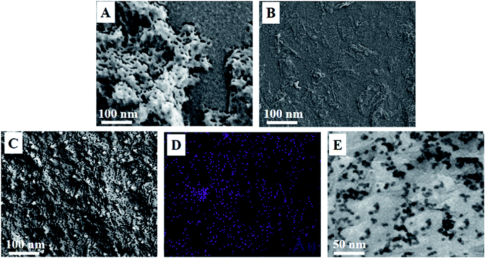 | ||
| Fig. 1 SEM images of (A) RGO, (B) PDDA-RGO and (C) PDDA-RGO/Au nanocomposite. (D) Elemental mapping of the PDDA-RGO/Au nanocomposite. (E) HRTEM image of the PDDA-RGO/Au nanocomposite. | ||
The reduction and PDDA surface functionalization processes were firstly confirmed by FTIR study. Fig. 2A showed the FTIR spectra of GO, RGO and PDDA-RGO nanocomposite. The spectrum of GO showed peaks at 1718 cm−1, 1589 cm−1, 1410 cm−1 and 1023 cm−1, which are assigned to the C![[double bond, length as m-dash]](https://www.rsc.org/images/entities/char_e001.gif) O stretching of COOH groups, CC vibrations, C–OH stretching vibrations and C–O vibrations from alkoxy groups, respectively.41–43 After hydrazine reduction process, the intensity of these characteristic peaks significantly decreased or even vanished in the both RGO and PDDA-RGO nanocomposite, which indicates that GO has been successfully reduced. Meanwhile, two extra peaks are observed in the spectrum of PDDA-RGO nanocomposite at 842 and 1501 cm−1, which are attributable to the N–C bond of PDDA, indicating the successful surface functionalization process.44,45 The reduction and PDDA surface functionalization processes were also confirmed by Raman spectra analysis. Fig. 2B displayed the Raman spectra of GO, RGO and PDDA-RGO. Three samples all displayed two characteristic bands at ∼1570 and ∼1340 cm−1, corresponding to the graphite (G band, first-order scattering of E2g phonons by sp2 carbon atoms) and diamondoid (D band, breathing mode of k-point photons of A1g symmetry) bands, respectively.46–49 The intensity ratio between D band and G band (ID/IG) increased clearly from GO to RGO, indicating the occurrence of the reduction process due to the formation of defect-rich graphite domains, which further confirmed the reduction process by the reduction.50 The deposition of the Au nanoparticles on the PDDA-RGO sheets did not change the ID/IG ratio, indicating the carbon state of the RGO remains same. Furthermore, the G band of PDDA-RGO nanocomposite showed a small shift from 1570 to 1586 cm−1. This small shift can be ascribe to the electron transfer from RGO to the adsorbed PDDA.44,51–53
O stretching of COOH groups, CC vibrations, C–OH stretching vibrations and C–O vibrations from alkoxy groups, respectively.41–43 After hydrazine reduction process, the intensity of these characteristic peaks significantly decreased or even vanished in the both RGO and PDDA-RGO nanocomposite, which indicates that GO has been successfully reduced. Meanwhile, two extra peaks are observed in the spectrum of PDDA-RGO nanocomposite at 842 and 1501 cm−1, which are attributable to the N–C bond of PDDA, indicating the successful surface functionalization process.44,45 The reduction and PDDA surface functionalization processes were also confirmed by Raman spectra analysis. Fig. 2B displayed the Raman spectra of GO, RGO and PDDA-RGO. Three samples all displayed two characteristic bands at ∼1570 and ∼1340 cm−1, corresponding to the graphite (G band, first-order scattering of E2g phonons by sp2 carbon atoms) and diamondoid (D band, breathing mode of k-point photons of A1g symmetry) bands, respectively.46–49 The intensity ratio between D band and G band (ID/IG) increased clearly from GO to RGO, indicating the occurrence of the reduction process due to the formation of defect-rich graphite domains, which further confirmed the reduction process by the reduction.50 The deposition of the Au nanoparticles on the PDDA-RGO sheets did not change the ID/IG ratio, indicating the carbon state of the RGO remains same. Furthermore, the G band of PDDA-RGO nanocomposite showed a small shift from 1570 to 1586 cm−1. This small shift can be ascribe to the electron transfer from RGO to the adsorbed PDDA.44,51–53
 | ||
| Fig. 2 (A) FTIR and (B) Raman spectra of (a) GO, (b) RGO and (c) PDDA-RGO nanocomposite. | ||
The interlayer changes and crystalline structure of GO, RGO and PDDA-RGO/Au nanocomposite were analyzed by XRD and depicted in Fig. 3. As expected, the XRD pattern of GO shows a characteristic (001) peak at 11.1° with a d-spacing value of 0.71 nm.54 However, the RGO displays an almost featureless XRD pattern, suggesting the occurrence of reduction of GO. A small peak centred at 22.8° is due to the presence of stacked graphene layers of RGO.55 The XRD spectrum of the RGO/Au nanocomposite shows diffraction peaks located at 39.7°, 460°, 67.2° and 81.3°, which are assigned to (111), (200), (220) and (311) planes of face-centered-cubic (fcc) crystallographic structure of Au (JCPDS 4-0783), respectively, confirming the successful electrodeposition of Au nanoparticles.
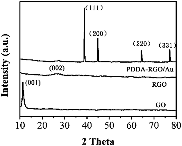 | ||
| Fig. 3 XRD patterns of GO, RGO and PDDA-RGO/Au nanocomposite. | ||
3.2 Electrochemical determination of nicotine
The electrochemical properties of the bare GCE (curve a), PDDA-RGO/GCE (curve b) and PDDA-RGO/Au/GCE (curve c) were examined by CV (Fig. 4A) and electrochemical impedance spectroscopy (EIS) (Fig. 4B) using Fe(CN)63−/4− redox probe. As shown in the Fig. 4A, a pair of well-defined redox peaks at 0.15 and 0.22 V was observed at bare GCE. At PDDA-RGO/GCE, the peak intensities were slightly increased, suggesting that the electron conductivity of PDDA-RGO modified GCE better than that of the bare GCE, and thus that the PDDA-RGO modification enhanced the electron transfer between the electrode and solution to some extent. In comparison with the bare GCE and the PDDA-RGO/GCE, the PDDA-RGO/Au/GCE showed notably further larger capacitive currents indicating the enhanced electroactive surface area as a result of the high electron-conductivity and the large specific area of PDDA-RGO/Au/GCE. Fig. 4B shows the Nyquist plots of the bare GCE, PDDA-RGO/GCE and PDDA-RGO/Au/GCE. Charge transfer resistance (Rct) is regarded as an effective parameter of electrode reaction, and can be measured by the semicircle diameter of Nyquist plots obtained from EIS. Randles equivalent circuit model was used for analysing the performance of each electrodes, where the Ret, Rs, Cdl and Zw represents the electron transfer resistance, solution transfer resistance, double layer capacitance and Warburg impedance, respectively. The semicircular part diameter is equivalent to the electron transfer resistance. The linear part at lower frequencies corresponds to the diffusion process. A semicircle portion results from the parallel combination of electron transfer resistance (Ret) and Cdl resulting from electrode impedance.56 As shown in the figure, the bare GCE shows the largest circuit diagram, indicating the lowest electroactivity of the Fe(CN)63−/4− redox probe on the electrode surface. In contrast, both PDDA-RGO/GCE and PDDA-RGO/Au/GCE had smaller circuit diagram, suggesting the enhanced electroactivity. The rapid electron transfer at the nanocomposite can be ascribed to the excellent conductivity. The obtained Rct values have confirmed the electroactivity sequence of the results obtained from the CV experiments. Therefore, based on above characterizations, PDDA-RGO/Au/GCE was expected had best performance towards electrochemical based application. | ||
| Fig. 4 (A) Cyclic voltammograms (scan rate for CV: 50 mV s−1) and (B) Nyquist diagrams of (a) GCE, (b) PDDA-RGO/GCE and (c) PDDA-RGO/Au/GCE in 2 mM K4[Fe(CN)6] + 0.1 M KCl. Inset is the Randles equivalent circuit. | ||
The electrochemical performance of the PDDA-RGO/Au nanocomposite towards detecting nicotine was investigated. Fig. 5 displays the CVs of bare, PDDA-RGO/GCE and PDDA-RGO/Au/GCE in pH 7.0 BR with absence and presence of nicotine. It can be seen that no electrode response was observed for PDDA-RGO/Au/GCE with the absence of nicotine. However, in the presence of 50 μM nicotine, a small oxidation peak was observed on the bare GCE. In contrast, well-defined oxidation peaks were observed on the PDDA-RGO/GCE and PDDA-RGO/Au/GCE with reduction potential at 1.23 and 1.07 V, respectively. Moreover, it illustrated a significant current enhancement on the PDDA-RGO/Au/GCE. The increasing peak current and lowering peak potential indicate that PDDA-RGO/Au can effectively catalyze the electrochemical oxidation of nicotine due to the excellent electrocatalytic property and larger surface area of Au NPs and PDDA-RGO. The PDDA-RGO sheets not only server as an excellent electron transfer mediator, but also acts as a platform for Au NPs loading to enhance their catalytic performance. The electrochemical oxidation process has not yet been fully confirmed. Fig. S2† proposed a possible process scheme of electrochemical oxidation of nicotine. Demethylation and hydroxylation of nitrogen atom at the pyrrolidine ring were happed when the nicotine at high potential. The whole process involves two-electron transfer.
 | ||
| Fig. 5 (A) Cyclic voltammograms of (a) bare GCE, (b) PDDA-RGO/GCE and (c) PDDA-RGO/Au/GCE toward 50 μM nicotine in BR (pH = 7) with scan rate of 50 mV s−1. Inset: (d) PDDA-RGO/Au/GCE scan in BR without nicotine. (B) Effect of solution pH on the oxidation potential and peak current of 50 μM nicotine at the PDDA-RGO/Au/GCE. | ||
It is well known that the pH of the electrolyte solution has an important influence on the current response of the analyte. The effect of pH on the peak potential and peak current of nicotine was investigated by CV in the pH range from 4 to 10. The results showed that with the increase of pH from 5 to 10, the oxidation peak of nicotine shifted towards less positive potentials, suggesting the proton participation in the electrode reaction. A slope of −0.059 (V pH−1) was obtained after calculation, suggesting the electrode reaction of nicotine on the PDDA-RGO/Au/GCE involves the same number of protons and electrons.57,58 The current was maximum at pH 7.0, so this pH was selected for subsequent experiments.
The amount of electrode modifier, accumulation potential and accumulation time were optimized. Fig. 6A shows the effect of different amount of PDDA-RGO/Au nanocomposite on the oxidation peak current of nicotine. With increasing amount of PDDA-RGO/Au nanocomposite, the number of electrode-surface active sites for electrochemical oxidation of nicotine increased, thus the nicotine-oxidation current increased. Further increasing of the amount of PDA-RGO/Au nanocomposite resulted a decreasing of current response due to the thicker layer of modifier, which hinder the electron transfer way. The accumulation step can be great influence on the analytic sensitivity and the intensity of peak current. Fig. 6B shows the influence of accumulation potential and time toward oxidation peak current. As shown in Fig. 6B, the peak current increases gradually with the increasing accumulation potential from −0.5 to −0.9 V and then decrease with further increasing potential. On the other hand, the oxidation peak current increases gradually with the increase of accumulation time from 30 to 120 s and then remains a similar performance after further increasing accumulation time (Fig. 6C). Therefore, the accumulation conditions of −0.9 V and 120 s were used in further measurements.
 | ||
| Fig. 6 Effect of the volume of (A) PDDA-RGO/Au nanocomposite, (B) accumulation potential and (C) accumulation time on the detection of 50 μM nicotine at PDDA-RGO/Au/GCE. | ||
For a higher signal and more sensitive determination of nicotine at the PDDA-RGO/Au/GCE, differential pulse voltammetry (DPV) technique was employed. Fig. 7 depicts the DPV curves of the PDDA-RGO/Au/GC in various concentrations of nicotine from 0.5 to 300 μM. As shown in Fig. 7 the DPV curves exhibit well-defined peaks corresponding to the oxidation of nicotine. The peak current increases linearly with the concentration of nicotine in the range of 0.5 to 300 μM. The corresponding regression equation is: I (μA) = 0.34822C (μM) + 32.001 (R = 0.9927). The limit of detection calculated at signal-to-noise ratio of 3 (S/N = 3) was 0.12 μM. Table 1 shows the comparison of analytical performances of this nicotine sensor with other nicotine sensors reported previously. Fig. S1† shows the typical amperometric response of the PDDA-RGO/Au/GCE with successive additions of 10 μM nicotine. It can be seen that the PDDA-RGO/Au/GCE attains a steady state current within in 4 s, suggesting the fabricated sensor has a rapid response towards nicotine.
 | ||
| Fig. 7 DPV of PDDA-RGO/Au/GCE toward different concentrations of nicotine in BR. | ||
| Electrode | Linear range (μM) | LOD (μM) | Reference |
|---|---|---|---|
| MWCNT | 31–220 | 7.6 | 59 |
| Carbon paste | 50–500 | 6.1 | 60 |
| Boron-doped diamond electrode | 9.9–170 | 6.1 | 61 |
| Electrochemically activated GCE | 1–200 | 0.7 | 62 |
| CuNPs | 1–90 | 0.164 | 63 |
| Poly(4-amino-3-hydroxynaphthalene sulfonic acid) | 1–200 | 0.866 | 64 |
| PDDA-RGO/Au | 0.5–300 | 0.12 | This work |
Reproducibility of the PDDA-RGO/Au/GCE was determined from 8 consecutive measurements for 50 μM nicotine. The relative standard deviation (RSD) obtained was 2.35%, which suggests acceptable reproducibility of the measurements at the proposed nicotine sensor. For the same nicotine solution, the RSD of measurements using 8 PDDA-RGO/Au/GCEs is determined to be 3.85%. The results suggest that the proposed PDDA-RGO/Au/GCE is very consistent for reproducible results. To evaluate the long-term storage stability, PDDA-RGO/Au/GCE was tested by storing in fridge for a week. The current responses showed the PDDA-RGO/Au/GCE still remains more than 96% of their original activity. The selectivity of the PDDA-RGO/Au/GCE was also investigated. 100 folds of Zn2+, Ca2+, K+, Cl− and NO3−, 50 folds of dopamine, uric acid, ascorbic acid, cotinine and glucose did not show interference to nicotine detection.
In order to evaluate the applicability of the proposed PDDA-RGO/Au/GCE for nicotine determination in real samples, two brands of cigarette samples were analyzed. Spike and recovery procedure was adopted for testing the validation of the proposed method. The results by the proposed electrochemical sensor and compared with a reference method (HPLC) are listed in Table 2. The proposed nicotine electrochemical sensor was also applied for analyzing nicotine content in the anti-smoking pharmaceuticals and the results were displayed in Table 2 as well. As can be seen that the proposed PDDA-RGO/Au/GCE showed an excellent performance towards nicotine detection in the samples of cigarettes, chewing gun and inhalator, suggesting our proposed method is an effective analytical tool for nicotine detection in the real samples.
| Sample | Addition (μM) | Found (μM) | Recovery (%) | HPLC |
|---|---|---|---|---|
| 1 | 0 | 20.27 | — | 20.11 |
| 20 | 40.19 | 100.20 | 40.20 | |
| 50 | 71.22 | 101.74 | 70.35 | |
| 100 | 118.98 | 99.15 | 120.07 | |
| 2 | 0 | 5.66 | — | 4.94 |
| 20 | 24.85 | 99.40 | 9.98 | |
| 50 | 55.32 | 100.58 | 14.97 | |
| 100 | 104.88 | 99.89 | 100.03 |
| Sample | Label (mg) | Found (mg) | Recovery (%) |
|---|---|---|---|
| Gum | 5 | 4.987 | 99.74 |
| Inhalator | 20 | 19.97 | 99.85 |
4. Conclusions
In summary, we have demonstrated a facial chemical method for synthesizing PDDA functionalized RGO. Electrodeposition method was subsequently used for depositing Au NPs on the PDDA-RGO surface. The prepared PDDA-RGO/Au nanocomposite was characterized using FTIR, Raman spectroscopy, SEM, XRD, CV and EIS methods. The proposed PPDA-RGO/Au nanocomposite was used to elaborate a sensitive, selective and reliable analytical method for the determination of nicotine in tobacco and pharmaceuticals. The proposed nicotine sensor exhibited a linear response range from 0.5 to 300 μM and a low detection limit of 0.12 μM.Notes and references
- C. D. Fowler and P. J. Kenny, Neuropharmacology, 2014, 76, 533–544 CrossRef CAS PubMed.
- B. J. Hall, C. Wells, C. Allenby, M. Y. Lin, I. Hao, L. Marshall, J. E. Rose and E. D. Levin, Pharmacol., Biochem. Behav., 2014, 120, 103–108 CrossRef CAS PubMed.
- J.-F. Etter and T. Eissenberg, Drug Alcohol Depend., 2015, 147, 68–75 CrossRef PubMed.
- M. Aragón, R. Marcé and F. Borrull, Talanta, 2013, 115, 896–901 CrossRef PubMed.
- D. C. M. Bordin, M. N. Alves, O. G. Cabrices, E. G. de Campos and B. S. De Martinis, J. Anal. Toxicol., 2014, 38, 31–38 CrossRef PubMed.
- B. Wei, J. Feng, I. J. Rehmani, S. Miller, J. E. McGuffey, B. C. Blount and L. Wang, Clin. Chim. Acta, 2014, 436, 290–297 CrossRef CAS PubMed.
- G. Shendarkar, P. Tarte, M. Ghante and A. Roge, Asian J. Res. Chem., 2014, 7, 787–794 Search PubMed.
- N. Nuchtavorn, M. Ryvolova, F. Bek, M. Macka, C. Phechkrajang and L. Suntornsuk, Anal. Sci., 2013, 29, 339–344 CrossRef CAS PubMed.
- X. Lin, Y. Sun, D. Xu, Y. Li, S. Liu and Z. Xie, Electrophoresis, 2013, 34, 2033–2040 CrossRef CAS PubMed.
- M. S. Lin, J. S. Wang and C. H. Lai, Electrochim. Acta, 2008, 53, 7775–7780 CrossRef CAS.
- J. M. Garrigues, A. Pérez-Ponce, S. Garrigues and M. de La Guardia, Analyst, 1999, 124, 783–786 RSC.
- P. M. Clayton, C. A. Vas, T. Bui, A. F. Drake and K. McAdam, Chirality, 2013, 25, 288–293 CrossRef CAS PubMed.
- E. Yiasemides, G. Sivapirabu, G. M. Halliday, J. Park and D. L. Damian, Carcinogenesis, 2009, 30, 101–105 CrossRef CAS PubMed.
- H. Xiao, L. Sun, H. Yan, W. Wang, J. Liu, Q. Yan, L. Chao, C. Chen, Q. Xie and J. Wen, Anal. Methods, 2015, 7, 1147–1153 RSC.
- L. Fu, G. Lai, P. J. Mahon, J. Wang, D. Zhu, B. Jia, F. Malherbe and A. Yu, RSC Adv., 2014, 4, 39645–39650 RSC.
- L. Fu, Y. Zheng and A. Wang, Int. J. Electrochem. Sci., 2015, 10, 3518–3529 CAS.
- L. Fu, Y. Zheng, A. Wang, W. Cai and H. Lin, Food Chem., 2015, 181, 127–132 CrossRef CAS PubMed.
- L. Fu, Y.-H. Zheng and Z.-X. Fu, Chem. Pap., 2015, 69, 655–661 CAS.
- F. Han, H. Li, J. Yang, X. Cai and L. Fu, Phys. E, 2016, 77, 122–126 CrossRef CAS.
- Y. Zheng, A. Wang, H. Lin, L. Fu and W. Cai, RSC Adv., 2015, 5, 15425–15430 RSC.
- Y. Zheng, L. Fu, A. Wang, F. Peng, J. Yang and F. Han, Sens. Lett., 2015, 13, 878–882 CrossRef.
- Y. Zheng, L. Fu, A. Wang and W. Cai, Int. J. Electrochem. Sci., 2015, 10, 3530–3538 CAS.
- E. C. Donny, D. K. Hatsukami, N. L. Benowitz, A. F. Sved, J. W. Tidey and R. N. Cassidy, Prev. Med., 2014, 68, 17–22 CrossRef PubMed.
- L. Fu and A. Yu, Fourth International Conference on Smart Materials and Nanotechnology in Engineering, 2013 Search PubMed.
- H. B. Suffredini, M. C. Santos, D. De Souza, L. Codognoto, P. Homem-de-Mello, K. M. Honório, A. Da Silva, S. A. Machado and L. A. Avaca, Anal. Lett., 2005, 38, 1587–1599 CrossRef CAS.
- A. Geto, M. Amare, M. Tessema and S. Admassie, Electroanalysis, 2012, 24, 659–665 CrossRef CAS.
- S. Xu, L. Fu, T. S. H. Pham, A. Yu, F. Han and L. Chen, Ceram. Int., 2015, 41, 4007–4013 CrossRef CAS.
- L. Fu, J. Yong, G. Lai, T. Tamanna, S. Notley and A. Yu, Mater. Manuf. Processes, 2014, 29, 1030–1036 CrossRef CAS.
- M. Shehata, S. M. Azab, A. M. Fekry and M. A. Ameer, Biosens. Bioelectron., 2016, 79, 589–592 CrossRef CAS PubMed.
- L. Fu and A. Yu, Rev. Adv. Mater. Sci., 2014, 36, 40–61 Search PubMed.
- J. Liang, F. Han and Y. Chen, Electrochem. Commun., 2012, 24, 93–96 CrossRef CAS.
- K. S. Novoselov, A. K. Geim, S. V. Morozov, D. Jiang, Y. Zhang, S. V. Dubonos, I. V. Grigorieva and A. A. Firsov, Science, 2004, 306, 666–669 CrossRef CAS PubMed.
- F. Xu, M. Deng, Y. Liu, X. Ling, X. Deng and L. Wang, Electrochem. Commun., 2014, 47, 33–36 CrossRef CAS.
- C. Shan, H. Yang, D. Han, Q. Zhang, A. Ivaska and L. Niu, Biosens. Bioelectron., 2010, 25, 1070–1074 CrossRef CAS PubMed.
- J. M. Pingarrón, P. Yáñez-Sedeño and A. González-Cortés, Electrochim. Acta, 2008, 53, 5848–5866 CrossRef.
- A. Wang, H. P. Ng, Y. Xu, Y. Li, Y. Zheng, J. Yu, F. Han, F. Peng and L. Fu, J. Nanomater., 2014, 2014, 451232 Search PubMed.
- H. Cheng, G. Lai, L. Fu, H. Zhang and A. Yu, Biosens. Bioelectron., 2015, 71, 353–358 CrossRef CAS PubMed.
- G. Lai, H. Cheng, C. Yin, L. Fu and A. Yu, Electroanalysis, 2016, 28, 69–75 CrossRef CAS.
- W. S. Hummers and R. E. Offeman, J. Am. Chem. Soc., 1958, 80, 1339 CrossRef CAS.
- Y. Xue, H. Zhao, Z. Wu, X. Li, Y. He and Z. Yuan, Biosens. Bioelectron., 2011, 29, 102–108 CrossRef CAS PubMed.
- J. Zhang, H. Yang, G. Shen, P. Cheng, J. Zhang and S. Guo, Chem. Commun., 2010, 46, 1112–1114 RSC.
- M. Ahmad, E. Ahmed, Z. L. Hong, J. F. Xu, N. R. Khalid, A. Elhissi and W. Ahmed, Appl. Surf. Sci., 2013, 274, 273–281 CrossRef CAS.
- X. Li, Q. Wang, Y. Zhao, W. Wu, J. Chen and H. Meng, J. Colloid Interface Sci., 2013, 411, 69–75 CrossRef CAS PubMed.
- S. Wang, D. Yu, L. Dai, D. W. Chang and J.-B. Baek, ACS Nano, 2011, 5, 6202–6209 CrossRef CAS PubMed.
- L. Fu, Y. Zheng, Q. Ren, A. Wang and B. Deng, J. Ovonic Res., 2015, 11, 21–26 Search PubMed.
- Z.-J. Fan, W. Kai, J. Yan, T. Wei, L.-J. Zhi, J. Feng, Y.-M. Ren, L.-P. Song and F. Wei, ACS Nano, 2010, 5, 191–198 CrossRef PubMed.
- L. Fu and Z. Fu, Ceram. Int., 2015, 41, 2492–2496 CrossRef CAS.
- L. Fu, W. Cai, A. Wang and Y. Zheng, Mater. Lett., 2015, 142, 201–203 CrossRef CAS.
- L. Fu, A. Wang, Y. Zheng, W. Cai and Z. Fu, Mater. Lett., 2015, 142, 119–121 CrossRef CAS.
- L. L. Zhang, R. Zhou and X. S. Zhao, J. Mater. Chem., 2010, 20, 5983–5992 RSC.
- A. M. Rao, P. C. Eklund, S. Bandow, A. Thess and R. E. Smalley, Nature, 1997, 388, 257–259 CrossRef CAS.
- X. Qi, K.-Y. Pu, X. Zhou, H. Li, B. Liu, F. Boey, W. Huang and H. Zhang, Small, 2010, 6, 663–669 CrossRef CAS PubMed.
- M.-C. Hsiao, S.-H. Liao, M.-Y. Yen, P.-I. Liu, N.-W. Pu, C.-A. Wang and C.-C. M. Ma, ACS Appl. Mater. Interfaces, 2010, 2, 3092–3099 CAS.
- T. Nakajima, A. Mabuchi and R. Hagiwara, Carbon, 1988, 26, 357–361 CrossRef CAS.
- D. Chen, L. Li and L. Guo, Nanotechnology, 2011, 22, 325601 CrossRef PubMed.
- W. S. Hummers Jr and R. E. Offeman, J. Am. Chem. Soc., 1958, 80, 1339 CrossRef.
- Ĺ. Švorc, D. M. Stanković and K. Kalcher, Diamond Relat. Mater., 2014, 42, 1–7 CrossRef.
- L. Highton, R. O. Kadara, N. Jenkinson, B. Logan Riehl and C. E. Banks, Electroanalysis, 2009, 21, 2387–2389 CrossRef CAS.
- H. Xiong, Y. Zhao, P. Liu, X. Zhang and S. Wang, Microchim. Acta, 2010, 168, 31–36 CrossRef CAS.
- M. Stočes and I. Švancara, Electroanalysis, 2014, 26, 2655–2663 CrossRef.
- K. Cinková, L. Dianová, M. Vojs, M. Marton and Ĺ. Švorc, Acta Chim. Slovaca, 2015, 8, 166–171 Search PubMed.
- H. Kassa, A. Geto and S. Admassie, Bull. Chem. Soc. Ethiop., 2013, 27, 321–328 Search PubMed.
- Z. Goodarzi, M. Maghrebi, A. F. Zavareh, Z.-B. Mokhtari-Hosseini, B. Ebrahimi-hoseinzadeh, A. H. Zarmi and M. Barshan-tashnizi, J. Nanostruct. Chem., 2015, 5, 237–242 CrossRef.
- A. Geto, M. Amare, M. Tessema and S. Admassie, Electroanalysis, 2012, 24, 659–665 CrossRef CAS.
Footnote |
| † Electronic supplementary information (ESI) available. See DOI: 10.1039/c6ra03399g |
| This journal is © The Royal Society of Chemistry 2016 |