Controlled construction of 3D hierarchical manganese fluoride nanostructures via an oleylamine-assisted solvothermal route with high performance for rechargeable lithium ion batteries

Kun Rui, Zhaoyin Wen*, Jun Jin and Xiao Huang
CAS Key Laboratory of Materials for Energy Conversion, Shanghai Institute of Ceramics, Chinese Academy of Sciences, Shanghai 200050, China. E-mail: zywen@mail.sic.ac.cn

Received 5th February 2016 , Accepted 7th March 2016

First published on 8th March 2016


Abstract

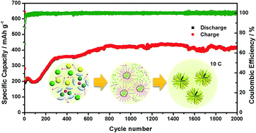
For diverse electrode materials, especially those reacting through a conversion mechanism, nanostructure engineering has proven to be one of the most effective strategies to improve their electrochemical performance. Typically, construction of three-dimensional (3D) nanostructures has offered a valid solution for the challenging issues involved in bulk transition metal based electrode materials for energy storage devices. Herein, we present a facile and effective solvothermal method to fabricate 3D hierarchical dendritic MnF2 nanostructures built from a bunch of radially oriented nanorods. Oleylamine (OAm) is adopted as the surfactant and structural-directing template, giving rise to continuous morphology evolution with different dosages. The electrochemical performance of the as-prepared MnF2 anode for rechargeable lithium batteries is investigated. Long term cycle performance at 10C for 2000 cycles was also obtained with an activated capacity as high as 420 mA h g−1. It is also noteworthy that the activation span was significantly shortened. Desirable MnF2 nanostructures are responsible for the excellent Li-storage capacity with satisfactory cycle performance and superior rate capability.


Introduction

Recently, continuous progress in electrode materials has been witnessed, bringing about improvements in performance and thus meeting the growing demands for better lithium ion batteries (LIBs).1–3 By moving beyond traditional intercalation reactions, transition metal oxides electrodes have drawn intensive interest into a novel reactivity concept referred to as “conversion reaction”.4,5 As one type of conversion electrodes, transition metal fluorides have also started to demonstrate promising potential as both alternative cathode and anode materials for LIBs.6–10 Generally, metal fluorides (MFx) can deliver high discharge capacities via multielectron transfer through the conversion process, where multiple Li+ could be accommodated by one transition metal with high valence.11,12

However, on account of the poor electronic conductivity, which is attributed to the high ionicity as well as large bandgap of the metal–fluoride bond, Li+ insertion/extraction kinetics of fluoride electrodes is always limited. Efforts in terms of introduction of conductive phase have been made to deal with the insulating electronic characters.13–16 More importantly, the development of nanotechnology over the past few decades has opened up possibilities for performance improvement of various electrode materials. Therefore, much progress has also been made for metal fluoride electrodes through particle-size reduction, recently.17–19 The general agreement on their enhanced electrochemical properties can be ascribed to the potential advantages of shortened Li+ solid diffusion pathways, along with increased reactivity.

Among those morphology tailoring strategies, constructing three-dimensional (3D) hierarchical architectures has received tremendous attention for LIBs,20–22 whose main merits for electrochemistry enhancement can be summed up as follows.23 Firstly, numerous nanosized primary particles shall provide shortened path length for electronic and Li+ transportation, beneficial to the kinetics of insulating electrodes; secondly, spontaneous but random agglomeration of nanoparticles due to their high surface energy can be restrained or avoided, giving rise to enlarged electrode/electrolyte active contact area; last but not least, with hierarchical nanostructures, accommodation for the strain of Li+ insertion/extraction during cycling are achieved, ensuring enhanced cycling stability. Recently, porous TiO2 hollow microspheres constructed by radially oriented nanorods chains was reported by Su's group, demonstrating excellent Li+ storage capacity with outstanding cycle performance at different rates over 700 cycles.24 Although more examples can be found for delicate hierarchical nanostructures,25–27 to develop any simple and reliable synthetic method, especially for MFx, still remains challenging. To be specific, issues concerning rational selection of environmentally friendly fluorine source, facile removal of hard template or surfactants, precise control of chemical components and morphology, etc., should be tackled comprehensively.

Emerging reports can be found for MnF2 with promising application for both LIB anode and supercapacitors.28–30 In particular, with the theoretical specific capacity of 577 mA h g−1, submicron MnF2 particles via solvothermal method delivered superior cycling life at 10C, which significantly enhanced Li-storage performance than MnF2 film by the pulsed laser deposition (PLD) process. However, microstructural design based on pure MnF2 has not been reported to our knowledge.

Herein, we demonstrate a facile and effective solvothermal method to synthesize 3D hierarchical dendrite MnF2 nanostructures built from a bunch of radially oriented nanorods. A green and safe ionic liquid (IL) was employed as the fluorine source, instead of the toxic and dangerous HF. Meanwhile, oleylamine (OAm) was adopted as the surfactant and structural-directing template. Despite the multiple roles of OAm as a surfactant, solvent, or reducing agent for the synthesis of various nanoparticle systems, its effect on the formation of hierarchical MnF2 nanostructures was revealed for the first time. As expected, the obtained nanostructured MnF2 anode delivered excellent Li-storage capacity with satisfactory cycle performance and superior rate capability. Long cycle performance at 10C for 2000 cycles was also obtained with an activated capacity as high as 420 mA h g−1. It is also noteworthy that the activation span was significantly shortened.

Results and discussion

3D hierarchical dendritic MnF2 nanostructures were prepared successfully via a facile solvothermal method for the first time. The one-pot fabrication route involves IL C10mimBF4 as the fluorine source, while Mn(NO3)2·4H2O was used as manganese source. The amount of OAm was carefully adjusted, which was adopted as the surfactant and structural-directing template. Experimental details can be found in the ESI and the amount of the nanostructured MnF2 produced was about 0.1 g. The phase purity and crystal structure of the as-prepared sample with the addition of 3 mL OAm (M-3.0) were examined by X-ray diffraction (XRD), as shown in Fig. 1a. All the diffraction peaks can be perfectly indexed to tetragonal MnF2 (JCPDS CARD no. 80-0926). The corresponding lattice planes have also been marked in the XRD pattern. As is displayed by the typical unit cell in Fig. 1b, MnF2 exhibits a tetragonal rutile structure (space group: P42/mnm), which is comprised of edge-sharing MnF6 octahedrons along the [001] direction.
image file: c6ra03351b-f1.tif
Fig. 1 (a) XRD pattern of the as-prepared manganese-based fluoride M-3.0 (b) schematic illustration of the MnF2 (rutile) structures. Typical (c) EDS elemental mapping and (d) TEM image of the sample M-3.0. (e and f) HRTEM images of the building units of the 3D dendritic MnF2 nanostructures in the form of nanorods, inset: corresponding SAED pattern.

The microscopic morphology of the obtained pure phase sample M-3.0 was further examined by scanning electron microscopy (SEM) and transmission electron microscopy (TEM). As can be seen from the SEM image, uniformly distributed 3D dendritic MnF2 nanostructures displayed an average diameter of ∼300 nm. EDS elemental mapping was also conducted for further understanding of the sample composition. A homogeneous distribution of elements Mn and F is demonstrated from Fig. 1c, while carbon is absent in the product. Hence, a pure MnF2 product is obtained without any carbon coating. In Fig. 1d, favorable 3D dendritic MnF2 nanostructures with nanorod building units can be observed from the TEM image.

The crystal structure and growth direction of the nanorods were then investigated by HRTEM analysis together with the corresponding selected area electron diffraction (SAED) pattern (Fig. 1e and f, and inset). For a typical nanorods unit, a diameter of about 20 nm and a length of 200 nm can be determined. The single-crystalline nature is also revealed by the SAED result and fast Fourier transform (FFT) pattern (Fig. S1). As evidenced by the HRTEM images, the lattice distance between the fringes perpendicular to the wall, i.e., d = 3.30 Å, corresponds to the interplane distance of (001) planes. Meanwhile, lattice fringes that run parallel to the nanorod wall is 3.45 Å, which can be assigned as the interplane distance of (110) planes. Therefore, the nanorod crystals of the 3D dendritic nanostructures grew along the [001] direction. Despite various literatures about TiO2, FeF3, and SnO2 electrodes achieved in the form of nanowire/-rod/-tube flower-like clusters via a wet-chemistry process,25,31–33 it is the first report for the 3D dendritic nanostructures of manganese-based fluoride, to the best of our knowledge.

A series of control experiments were conducted to reveal the possible role of OAm additive. To begin with, the amount of OAm used in the synthesis process was carefully adjusted. In order to distinguish between the obtained samples with different morphologies, the products are denoted as M–X, where M represents manganese-based fluoride and X represents the amount of OAm. As is indicated by XRD patterns in Fig. S2, tetragonal phase of the as-prepared manganese-based fluoride can be retained, which shall not be affected by the addition of OAm. While continuous evolution of morphology with the increase of OAm from 0 to 3 mL can be obtained by TEM images in Fig. S3. Furthermore, representative MnF2 morphologies with specific amount of OAm were characterized as shown in Fig. 2. When no OAm was added (Fig. 2a and b), inhomogeneous morphology can be observed. For one thing, due to its high surface energy, agglomeration of nanoparticles of 10–20 nm can be observed. At the same time, large particles of ∼100 nm existed, which can be ascribed to the local overgrowth of nuclei. When increasing amount of OAm surfactant was added, the proportion of single nanoparticles declined while more homogeneously large particles increased. With a proper amount of 1 mL for OAm, highly uniform polycrystalline particles of 200–300 nm can be obtained as Fig. 2c and d displayed. In particular, some are obviously submicron spheres assembled by nanoparticles, while some are irregular polyhedron with smooth facets exposed. Corresponding TEM and HRTEM results for M-1.0 can be found in Fig. S4. When the amount of OAm was further increased, initially aggregated MnF2 nanoclusters were observed to show sprouts in random directions (Fig. S3f and g). Obviously, these sprouts started to grow in preferential directions and transformed to nanorods. Finally, by adjusting the OAm to 3 mL (Fig. 2e and f), the MnF2 clusters grew significantly and the 3D dendritic nanostructures could be distinguished.


image file: c6ra03351b-f2.tif
Fig. 2 SEM images of the as-prepared manganese-based fluoride with controllable morphologies by adjusting the amount of OAm added: M-0 (a and b); M-1.0 (c and d); M-3.0 (e and f).

A summary for the synthetic route involving the possible role of OAm is illustrated in Scheme 1. The procedure starts by dissolving the manganese source and IL in ethanol to obtain a homogeneous solution. IL is principally responsible for the supply of BF4, which has been known to undergo hydrolysis with the aid of hydration water from Mn(NO3)2·4H2O and thus release F gently and slowly.34,35 Subsequently, MnF2 nuclei can be generated with the combination of Mn2+ and F. When suitable amount of OAm (1 mL) is used, more homogenous morphology with uniform size distribution can be obtained. This can be attributed to two major aspects as follows. On one hand, Ostwald ripening process or the coarsening process, involving the growth of larger particles at the expense of smaller ones until the equilibrium state, should be taken into account. On the other hand, nanoparticles strongly tend to form aggregated clusters to eliminate surface energy. Herein, OAm plays an important role as capping agent to constrain the nucleation and growth of the products, yielding well-dispersed MnF2 polyhedrons and initially aggregated MnF2 nanoclusters.36,37 By further increasing the amount of OAm (2 mL), another function of OAm as structural-directing template become distinct.38–40 Oriented growth of surface MnF2 nanoparticles can be distinguished, bringing about an early-stage dendritic morphology. It is speculated that OAm could be possibly adsorbed on the (110) plane of MnF2, guiding the 1D growth along the [001] direction into nanorods, which is common for plenty of rutile group crystals (SnO2, TiO2, β-MnO2) with 1D morphology.41–43 As OAm increased to 3 mL, the nanoparticles in the inner core further dissolve and recrystallize, leading to the formation of 3D dendritic nanostructures eventually. So far, the role of OAm as both the size confining agent and the structural-directing template has been confirmed. However, more detailed mechanism of the morphology evolution with the assistance of OAm needs further investigation.


image file: c6ra03351b-s1.tif
Scheme 1 Schematic illustration for the formation process of the as-prepared 3D dendritic MnF2 nanostructures.

In addition, the larger cation [C10mim]+ of IL, which contains not only an imidazole ring but also a long-chain alkyl group in the N1 position, also exhibits strong steric hindrance. Its negligible function for morphology control can also be confirmed by control experiments, where BmimBF4 with a smaller cation was used instead. As Fig. S5 indicated, less homogeneous morphology can be obtained, while similar evolution process with pure phase rutile MnF2 (Fig. S6) can be observed.

The electrochemical properties of the as-prepared manganese-based fluorides as anode for LIBs were investigated in detail. Fig. 3a shows galvanostatic discharge–charge curves of the M-3.0 sample in a voltage range of 0.01–3.0 V at 0.1C (1C = 577 mA g−1). A well-defined initial discharge voltage plateau around 0.7 V is mainly attributed to the lithiation reaction of MnF2 via 2 electron transfer, accompanied with the formation of metallic Mn within the LiF matrix. The discharge capacity of ca. 960 mA h g−1 was obtained for the first cycle, which is higher than the theoretical value of 577 mA h g−1 for the conversion reaction. The extra capacity can be ascribed to the formation of a solid–electrolyte interface (SEI) film on the electrode surface due to the decomposition of the electrolyte on one hand.44,45 Similar cases can also be found for transition metal oxides/fluorides electrodes.46,47 Also, extra lithium storage at low potentials was responsible since LiX (X = O or F) surface layers or the interfacial core could serve as hosts for extra Li.48,49 Typical electrochemical features of conversion reaction concerning the voltage evolutions in particular, could also be revealed by cyclic voltammograms in Fig. S7. Due to the inevitable capacity loss during the first cycle, a reversible capacity as high as 376 mA h g−1 from the 10th cycle was achieved.


image file: c6ra03351b-f3.tif
Fig. 3 Electrochemical characterization for the M-3.0 anode in LIBs. (a) Discharge–charge curves at 0.1C in the voltage range of 0.01–3.0 V at typical cycles. (b) Rate performance for different current rates from 0.1C to 5C. (c) Cycling stability with discharge capacity as a function of cycle number at various rates. (d) Discharge–charge curves of the 10th cycle at 1C for M-3.0, M-1.0 and M-0 anodes. (e) Long cycle performance at 10C for M-3.0, M-1.0 and M-0 anodes.

In order to evaluate the rate performance, the 3D dendritic MnF2 nanostructured anode were cycled for 10 times at different current rates each, as shown in Fig. 3b. Excellent rate capability can be obtained with reversible capacities of 340 mA h g−1, 315 mA h g−1, 306 mA h g−1, 290 mA h g−1, 275 mA h g−1 and 216 mA h g−1, corresponding to stepwise increases in rates of 0.1C, 0.2C, 0.5C, 1C, 2C and 5C, respectively. Overlapping of the discharge and charge capacities for each rate also demonstrated good reversibility of the MnF2 anode. Cycling stability of the as-prepared M-3.0 MnF2 anode was further examined at various rates. As can be seen from Fig. 3c, large retentive discharge capacities of 331 mA h g−1 at 2C was successfully achieved, higher than that of 294 mA h g−1 at the 10th cycle. Besides, more apparent capacity increase was discovered within 50 cycles, especially at relatively low current rates (0.1C and 1C). Possible explanations for similar capacity increase of metal oxides electrodes can be found, which have generally been attributed to organic the gel/polymer-like film generated during the conversion reaction.50,51

For comparison, electrochemical behaviors of M-0 and M-1.0 were also characterized, revealing the effect of morphology on Li-storage performance of MnF2 anodes. Fig. 3d displays representative discharge-charge curves of the 10th cycle at 1C for different samples. Larger discharge capacity of 356 mA h g−1 was achieved for M-3.0 (269 mA h g−1 for M-0 and 161 mA h g−1 for M-1.0) along with a better polarization behavior. Simultaneously, M-3.0 delivered superior cycling performance at 1C to M-0 and M-1.0, whose retentive capacities were 251 mA h g−1 and 162 mA h g−1 after 50 cycles as shown in Fig. S8. To be specific, the unsatisfactory electrochemical performance of M-0 was resulted from its inhomogeneous morphology, despite the existence of nanosized particles. As for M-1.0 with uniform submicron aggregates, undesirable larger particle size without interior nanostructures should be responsible for the poor performance.

In addition, long-term cycle performance of the MnF2 anodes was further investigated at 10C for 2000 cycles. Particularly, a distinct electrochemical activation process can be noticed, as illustrated by the abrupt capacity raise during the early 400 cycles. In this case, the activation process was greatly shortened, as compared to our previous reports on MnF2 anodes,30,52 wherein over 1000 cycles were usually necessary for the complete activation. Moreover, a stabilized capacity as high as ∼420 mA h g−1 was maintained afterwards for M-3.0, together with steady coulombic efficiency nearly 100% throughout the 2000 cycles. In contrast, the long cycling performance at such a high current rate for M-1.0 and M-0 was not as satisfactory. Both M-1.0 and M-0 anodes went through a rapid capacity fading process in the initial 200 cycles. Then, the retentive capacity of M-0 after 2000 cycles was merely 150 mA h g−1, while a capacity of only 47 mA h g−1 could be retained for M-1.0 at the 1000th cycle.

Since Li-storage properties are considered to be related to morphology and structural evolution of nanostructured electrodes, ex situ HRTEM was performed on cycled 3D dendritic MnF2 nanostructured anodes for further understanding of reaction mechanism. As shown by typical HRTEM results in Fig. 4a and b for one nanorod as representative building units, electrochemically generated metallic Mn embedded in LiF matrix are confirmed, which are also indexed to corresponding planes. At the same time, newly created MnF2 nanoparticles (domains of 2–3 nm) can be observed within the nanorods after the initial recharging process, as shown by HRTEM image in Fig. S9. In short, a typical conversion reaction took place during the initial discharge–charge process of the M-3.0 anode. The reaction scheme is shown as follows:

 
MnF2 + 2Li+ + 2e → Mn(nano)/LiF (discharge) (1)
 
Mn(nano) + LiF − 2Li+ → MnF2(nano) + 2e (charge) (2)


image file: c6ra03351b-f4.tif
Fig. 4 (a) and (b) Representative HRTEM images of the MnF2 anode at the first discharged state. (c) Representative TEM image and (d) HRTEM image of the MnF2 anode at the 400th discharged state.

The phase and microstructure of the M-3.0 anode were further characterized after ca. 400 cycles when the abrupt capacity improving process terminated. In Fig. 4d, at the discharged likewise, much smaller Mn nanodomains were detected, which tended to interconnect with each other. According to our previous work, formation of the continuous network of Mn within LiF on a microscopic scope was responsible for the long-term activation process of MnF2.30 Therefore, the accelerated activation process could be attributed to the completion of phase redistribution in advance, namely 400 cycles, along with the resultant self-improving electronic conductivity. Fig. 4c suggests that the 3D hierarchical dendritic architecture was generally reserved, further indicating the structural and electrochemical stabilities of the M-3.0 anode. Besides, the well preservation of the dendritic nanostructure after long cycle performance was further confirmed by ex situ FESEM images in Fig. S10.

Based on the above discussion, the reaction mechanism for the nanostructured MnF2 anode with 3D dendritic architecture was proposed and illustrated in Scheme 2. On one hand, the nanostructured MnF2 constructed by the loose packing of nanorods can significantly improve the interaction between the active material and electrolyte. On the other hand, volume change during the Li+ insertion/extraction process can be moderately buffered, ensuring the integrity of the nanostructured anode. Moreover, the 1D building blocks of nanorods grant good carrier access via effectively reduced radial pathways. Last but not least, from the intrinsic view for MnF2, the enhanced process of the formation of continuous Mn network should be noted, which leads to the long-term activation at high current rates. As a consequence of all the merits above, the novel 3D dendritic MnF2 nanostructured anode delivered enhanced electrochemical properties including excellent capacity retention and superior rate performance.


image file: c6ra03351b-s2.tif
Scheme 2 Schematic mechanism depicting the lithiation and delithiation.

Conclusions

In summary, a one-pot solvothermal method was developed in this work to fabricate 3D hierarchical dendritic MnF2 nanostructures, which were built from fine rutile nanorods. Based on a series of control experiments, a possible mechanism for the formation of 3D hierarchical architecture was proposed. It was believed that the amount of OAm played a key role for the morphology evolution. As expected, the obtained nanostructured MnF2 anode delivered excellent Li-storage capacity with satisfactory cycle performance and superior rate capability. Long cycle performance at 10C for 2000 cycles was also obtained with an activated capacity as high as 420 mA h g−1. It is also noteworthy that the activation span was significantly shortened. These desirable electrochemical properties can be attributed to the unique nanostructure of the 3D dendritic MnF2, in which the contact between the electrode material and the electrolyte was improved, while the volume change during the electrochemical cycles could be accommodated.

Acknowledgements

This work was supported by the National Natural Science Foundation of China (No. 51432010) and Key Fundamental Research Project from Science and Technology Commission of Shanghai Municipality (14JC1493000).

Notes and references

  1. J. M. Tarascon and M. Armand, Nature, 2001, 414, 359–367 CrossRef CAS PubMed.
  2. J. B. Goodenough and Y. Kim, Chem. Mater., 2010, 22, 587–603 CrossRef CAS.
  3. M. Armand and J. M. Tarascon, Nature, 2008, 451, 652–657 CrossRef CAS PubMed.
  4. J. Cabana, L. Monconduit, D. Larcher and M. R. Palacin, Adv. Mater., 2010, 22, E170–E192 CrossRef CAS PubMed.
  5. M. R. Palacin, Chem. Soc. Rev., 2009, 38, 2565–2575 RSC.
  6. Y. Li, B. Tan and Y. Wu, Nano Lett., 2008, 8, 265–270 CrossRef CAS PubMed.
  7. X. Xu, R. Cao, S. Jeong and J. Cho, Nano Lett., 2012, 12, 4988–4991 CrossRef CAS PubMed.
  8. F. Jiao and P. G. Bruce, Adv. Mater., 2007, 19, 657–660 CrossRef CAS.
  9. Y. Zhu, X. Fan, L. Suo, C. Luo, T. Gao and C. Wang, ACS nano, 2016, 10, 1529–1538 CrossRef CAS PubMed.
  10. S. Zhang, B. Chowdari, Z. Wen, J. Jin and J. Yang, ACS nano, 2015, 9, 12464–12472 CrossRef CAS PubMed.
  11. F. Badway, N. Pereira, F. Cosandey and G. Amatucci, 2002.
  12. H. Li, P. Balaya and J. Maier, J. Electrochem. Soc., 2004, 151, A1878–A1885 CrossRef CAS.
  13. M. A. Reddy, B. Breitung, V. S. K. Chakravadhanula, C. Wall, M. Engel, C. Kuebel, A. K. Powell, H. Hahn and M. Fichtner, Adv. Energy Mater., 2013, 3, 308–313 CrossRef CAS.
  14. F. Badway, F. Cosandey, N. Pereira and G. G. Amatucci, J. Electrochem. Soc., 2003, 150, A1318–A1327 CrossRef CAS.
  15. H. Song, G. Yang, H. Cui and C. Wang, J. Mater. Chem. A, 2015, 3, 19832–19841 CAS.
  16. X. Wang, W. Gu, J. T. Lee, N. Nitta, J. Benson, A. Magasinski, M. W. Schauer and G. Yushin, Small, 2015, 11, 5164–5173 CrossRef CAS PubMed.
  17. M. F. Oszajca, K. V. Kravchyk, M. Walter, F. Krieg, M. I. Bodnarchuk and M. V. Kovalenko, Nanoscale, 2015, 7, 16601–16605 RSC.
  18. S.-T. Myung, S. Sakurada, H. Yashiro and Y.-K. Sun, J. Power Sources, 2013, 223, 1–8 CrossRef CAS.
  19. F. Wang, S. W. Kim, D. H. Seo, K. Kang, L. Wang, D. Su, J. J. Vajo, J. Wang and J. Graetz, Nat Commun, 2015, 6, 6668 CrossRef CAS PubMed.
  20. S.-Z. Huang, Y. Cai, J. Jin, J. Liu, Y. Li, Y. Yu, H.-E. Wang, L.-H. Chen and B.-L. Su, Nano Energy, 2015, 12, 833–844 CrossRef CAS.
  21. S.-Z. Huang, J. Jin, Y. Cai, Y. Li, Z. Deng, J.-Y. Zeng, J. Liu, C. Wang, T. Hasan and B.-L. Su, Scientific Reports, 2015, 5, 14686 CrossRef CAS PubMed.
  22. Y. Li, S. Huang, Y. Cai, J. Jin, J. Liu, H.-E. Wang, L. Chen, T. Hasan and B.-L. Su, J. Mater. Chem. A, 2016, 4, 4264–4272 Search PubMed.
  23. Y. Guo, L. Yu, C.-Y. Wang, Z. Lin and X. W. D. Lou, Adv. Funct. Mater., 2015, 25, 5184–5189 CrossRef CAS.
  24. J. Jin, S.-Z. Huang, J. Shu, H.-E. Wang, Y. Li, Y. Yu, L.-H. Chen, B.-J. Wang and B.-L. Su, Nano Energy, 2015, 16, 339–349 CrossRef CAS.
  25. Y. Lu, Z. Wen, J. Jin, K. Rui and X. Wu, Phys. Chem. Chem. Phys., 2014, 16, 8556–8562 RSC.
  26. S. Ding and X. W. D. Lou, Nanoscale, 2011, 3, 3586–3588 RSC.
  27. B. Wang, J. S. Chen, H. B. Wu, Z. Wang and X. W. Lou, J. Am. Chem. Soc., 2011, 133, 17146–17148 CrossRef CAS PubMed.
  28. Y. Cui, M. Xue, K. Hu, D. Li and Z. Fu, J. Inorg. Mater., 2010, 25, 145–150 CrossRef CAS.
  29. R. Ma, Y. Zhou, L. Yao, G. Liu, Z. Zhou, J.-M. Lee, J. Wang and Q. Liu, J. Power Sources, 2016, 303, 49–56 CrossRef CAS.
  30. K. Rui, Z. Wen, Y. Lu, J. Jin and C. Shen, Adv. Energy Mater., 2014 DOI:10.1002/aenm.201401716.
  31. Z. Sun, J. H. Kim, Y. Zhao, F. Bijarbooneh, V. Malgras, Y. Lee, Y. M. Kang and S. X. Dou, J. Am. Chem. Soc., 2011, 133, 19314–19317 CrossRef CAS PubMed.
  32. J. Zhou, G. L. Zhao, B. Song and G. R. Han, Crystengcomm, 2011, 13, 2294–2302 RSC.
  33. L. Wang, J. Ren, X. Liu, G. Lu and Y. Wang, Mater. Chem. Phys., 2011, 127, 114–119 CrossRef CAS.
  34. R. J. Gillespie and J. S. Hartman, Can. J. Chem., 1967, 45, 2243–2246 CrossRef CAS.
  35. C. Zhang, J. Chen, Y. C. Zhou and D. Q. Li, J. Phys. Chem. C, 2008, 112, 10083–10088 CAS.
  36. Y. Cai, X. Li, L. Wang, H. Gao, Y. Zhao and J. Ma, J. Mater. Chem. A, 2015, 3, 1396–1399 CAS.
  37. S. Mourdikoudis and L. M. Liz-Marzán, Chem. Mater., 2013, 25, 1465–1476 CrossRef CAS.
  38. J. Xiao and L. Qi, Nanoscale, 2011, 3, 1383–1396 RSC.
  39. Z. Li, J. Tao, X. Lu, Y. Zhu and Y. Xia, Nano Lett., 2008, 8, 3052–3055 CrossRef CAS PubMed.
  40. N. Li, X. Zhang, S. Chen, X. Hou, Y. Liu and X. Zhai, Mater. Sci. Eng., B, 2011, 176, 688–691 CrossRef CAS.
  41. D. Su, H.-J. Ahn and G. Wang, NPG Asia Materials, 2013, 5, e70 CrossRef CAS.
  42. M. Q. Lv, D. J. Zheng, M. D. Ye, J. Xiao, W. X. Guo, Y. K. Lai, L. Sun, C. J. Lin and J. Zuo, Energy Environ. Sci., 2013, 6, 1615–1622 CAS.
  43. G. Xi and J. Ye, Inorg. Chem., 2010, 49, 2302–2309 CrossRef CAS PubMed.
  44. S. Grugeon, S. Laruelle, L. Dupont and J. M. Tarascon, Solid State Sci., 2003, 5, 895–904 CrossRef CAS.
  45. S. Laruelle, S. Grugeon, P. Poizot, M. Dolle, L. Dupont and J. Tarascon, J. Electrochem. Soc., 2002, 149, A627–A634 CrossRef CAS.
  46. Z. Bai, X. Zhang, Y. Zhang, C. Guo and B. Tang, J. Mater. Chem. A, 2014, 2, 16755–16760 CAS.
  47. G. Q. Jian, Y. H. Xu, L. C. Lai, C. S. Wang and M. R. Zachariah, J. Mater. Chem. A, 2014, 2, 4627–4632 CAS.
  48. P. Balaya, H. Li, L. Kienle and J. Maier, Adv. Funct. Mater., 2003, 13, 621–625 CrossRef CAS.
  49. Y. F. Zhukovskii, E. A. Kotomin, P. Balaya and J. Maier, Solid State Sci., 2008, 10, 491–495 CrossRef CAS.
  50. P. Poizot, S. Laruelle, S. Grugeon, L. Dupont and J. M. Tarascon, Nature, 2000, 407, 496–499 CrossRef CAS PubMed.
  51. L. Taberna, S. Mitra, P. Poizot, P. Simon and J. M. Tarascon, Nat. Mater., 2006, 5, 567–573 CrossRef PubMed.
  52. K. Rui, Z. Wen, X. Huang, Y. Lu, J. Jin and C. Shen, Phys. Chem. Chem. Phys., 2016, 18, 3780–3787 RSC.

Footnote

Electronic supplementary information (ESI) available. See DOI: 10.1039/c6ra03351b

This journal is © The Royal Society of Chemistry 2016
Click here to see how this site uses Cookies. View our privacy policy here.