The green reduction of graphene oxide

M. T. H. Aunkora, I. M. Mahbubul*b, R. Saidurb and H. S. C. Metselaar*a
aDepartment of Mechanical Engineering, Faculty of Engineering, University of Malaya, 50603 Kuala Lumpur, Malaysia. E-mail: h.metselaar@um.edu.my; Fax: +603 7967 5317; Tel: +603 7967 4451
bCenter of Research Excellence in Renewable Energy (CoRE-RE), Research Institute, King Fahd University of Petroleum & Minerals (KFUPM), Dhahran, 31261, Saudi Arabia. E-mail: mahbub_ipe@yahoo.com

Received 3rd February 2016 , Accepted 5th March 2016

First published on 8th March 2016


Abstract

Graphene is an ultra-thin material, which has received broad interest in many areas of science and technology because of its unique physical, chemical, mechanical and thermal properties. Synthesis of high quality graphene in an inexpensive and eco-friendly manner is a big challenge. Among various methods, chemical synthesis is considered the best because it is easy, scalable, facile, and inexpensive. Different kinds of chemical reducers have been used to produce graphene sheets. However, some chemicals are toxic, corrosive, and hazardous. For this reason, researchers have been using different environmentally friendly substances (termed green reducers) to produce functional graphene sheets. This paper presents an overview and discussion of the green reduction of graphene oxide (GO) to graphene. It also reviews the characterization of GO and its oxide reduction through the analysis of different spectroscopic and microscopic techniques such as Raman spectroscopy, Fourier transform infrared spectroscopy, X-ray photoelectron spectroscopy, X-ray diffraction, transmission electron microscopy, scanning electron microscopy, and atomic force microscopy.

1. Introduction

Graphene promises to be the most exciting material of the decade. It is a plane sheet of sp2 hybridized carbon atoms tightly confined into a honeycomb lattice. In 2004, the first discovery of graphene using a scotch tape peeling method brought a dramatic revolution, especially in the world of materials science.1 Recently, this single sheet of carbon has attracted huge interest among the scientific community owing to the two dimensional structure.2 It is proved that an ideal graphene sheet is highly ordered and shows several extraordinary behaviors including outstanding surface areas (2630 m2 g−1), high Young's modulus (1.0 TPa), high thermal conductivity (∼5000 W m−1 K−1) and strong chemical durability and high electron mobility (2.5 × 105 cm2 V−1 s−1).3 It also exhibits single-molecule gas detection sensitivity,4 quantum confinement in nano scale ribbon,5 long range ballistic transport at room temperature6 and high optical transmittance (∼97.7%).7,8 These unique characteristics of graphene opened a new way for its wide range of application at different branches of technology such as: lithium-ion batteries,9,10 catalyst engineering,11–15 chemically derived sensor,16–18 biosensor,19,20 anti-bacterial activities,21 flexible thin film transistor,22 delivery of drugs,23,24 solar cells,18,25,26 photovoltaic devices,27 imaging,28 p–n junction materials,29 super capacitor,30 touch panel,31–33 electromagnetic shielding application,34 water purification,35 absorption of different non aqueous liquid e.g., oils, alkenes, aromatic compounds, dyes, organic solvents, and ionic solutions.36–41

Large quantities of graphene materials are required to fulfil the huge demand of the aforementioned applications. Production of the best quality graphene in an inexpensive manner and on the desired scale is a big challenge of the time. There are a number of approaches have been designed to synthesis graphene from pristine graphite such as: epitaxial growth,42–44 vacuum thermal annealing,45,46 non-catalytic synthesis,13 scotch tape method,47 micromechanical cleavage,48 chemical vapour deposition (CVD),49,50 thermal exfoliation,51 carbon nanotubes cutting,52 liquid phase exfoliation,53,54 direct ultrasound sonication52 and reduction of graphene oxide.55 There are also some unconventional and hazardous methods of graphene production i.e. the chemical based detonation,56 arc discharging57 and carbon nano tube unzipping.58 Epitaxial growth is able to produce good quality multilayer graphene but it cannot isolate conductive single or bilayer that are suitable for device applications.48 A remarkable limitation of mechanical cleavage is repeated peeling during layer isolation and it is difficult to maintain the number of separated layers produced by this method.59 On the other hand, thermal exfoliation results in high production costs.48 In case of CVD, it is not easy to achieve layer separation without material damaging and successful exfoliation from the substrate has been a stumbling block. In contrast, graphene oxide (GO) reduction is considered a promising approach for the mass production of graphene. The product obtained by this method is commonly termed as reduced graphene oxide (RGO) or graphene nano sheet (GNS).

However, GO acts as a precursor to synthesis RGO or GNS. It is produced from graphite flakes by oxidative reaction, which heavily decorates the sheets with different oxygen moieties. The presence of abundant amounts of oxygen functional groups over the carbon basal plane not only makes GO electrically insulating but also provokes thermal instability.54,60 Therefore, reduction of oxygen molecules is the only key to resettle the π lattice. It helps to produce thermally stable graphene and regain electronic conductivity. Several reduction approaches of GO e.g., the electrochemical reduction,13,61–70 thermal reduction,71,72 photocatalytic reduction,73,74 and chemical reduction have been reported in recent time. Electrochemical reduction is unable to heal the defects and vacancies inherited from the precursor GO. Secondly, the oxygen content should be as low as possible in the GO precursor for exfoliation to single layer GO sheets. But the low oxygen content in GO may depress the interaction between oxygen functionalities. It could produce stable species like in-plane ether or out-of-plane carbonyl groups which are relatively difficult to remove by the electrochemical reduction method.75 Meanwhile, thermal deoxygenation is expensive. It is also a complicated phenomenon because of the thermal-energy induced multistep removal process of intercalated H2O and oxide groups of –OH (hydroxyl groups), –COOH (carboxyl groups) and [double bond splayed left]O (epoxy groups).76 Photocatalytic reduction requires active performance of light sensitive materials under continuous ultraviolet radiation.73,74 Hence, in comparison with the aforementioned reduction approaches, the chemical reduction is considered as the easiest way to produce ultrathin graphene sheet with large lateral dimension and area.77 In addition, this method is relatively fast, economic, facile, and suitable for subsequent processing and chemical modifications.

Scientists have applied different types of reducing agents to convert GO to graphene. Hydrazine, sodium borohydride, hydroquinone, etc. are toxic reducers. As alternatives, different kinds of organic acids, biochemical substances, amino acids, bacteria, fungus, plant extract, cellulosic compounds or metal powders have been applied to prepare graphene sheets from the GO. These reducing agents are termed “green reducers”. Since they are free from corrosion, carcinogenicity, and toxicity. There are few review papers available about the chemical or other reduction of GO.75,78 However, there is no compilation giving an overview of the green reduction of GO to graphene. Therefore, in this review, we summarize the reduction of GO using different environmentally friendly reducing agents in recent time. Herein, we also briefly discussed the selection of pristine graphite, synthesis of GO precursor and the exfoliation of GO. The results of different characterization techniques such as Raman spectroscopy, X-ray diffraction (XRD), X-ray photoelectron spectroscopy (XPS), Fourier transform infrared spectroscopy (FTIR), transmission electron microscopy (TEM), scanning electron microscopy (SEM), and atomic force microscopy (AFM) have also been summarized in this study. Obviously, this review will show a guideline to the researchers interested in the green synthesis of graphene.

2. Synthesis of graphene nano sheets

This is a two step procedure. The first step is the production of GO precursor from graphite and the following step is the reduction of oxygen from the GO sheets. In order to synthesise GO the selection of graphite is also important. Brief descriptions of the processes are presented in the following subsections.

2.1. Selection of parent graphite

Parent graphite is the source for GO production, which is exfoliated and later reduced to synthesise functional graphene sheets. It is experimentally investigated79 that the GO produced from natural graphite (NGrO) performs better than the synthetic graphite (SGrO) when exfoliating in aqueous solution. The probable reason is that NGrO possesses a larger crystalline structure. It also contains a large number of sp3 carbon to carbon bonds (NGrO ≈ 12.8% and SGrO ≈ 4.9%) and C–OH bonds (NGrO ≈ 36.2% and SGrO ≈ 1.8%). In contrast, GO produced from synthetic graphite (SGrO) possesses higher degree of carboxylic groups (NGrO ≈ 4.4% and SGrO ≈ 10.7%) and ring shaped epoxy groups (C–O–C) (NGrO ≈ 14.3% and SGrO ≈ 39.4%). This presence of large amounts of C–OH groups in the inward direction of the basal plane leads to easy exfoliation of NGrO. According to the same study the exfoliation yield of NGrO is nearly double that of SrGO.79 The size of graphite flake also plays important role in the oxidation of graphite. McAllister et al.80 observed that the small sized flakes (∼45 μm) are quickly oxidized and intercalated with a maximum reliability, while large sized flakes (∼400 μm) take more time to oxidize and it regularly fails to intercalate completely. It is noteworthy that the reduction of the size of starting flakes allows faster and more reliable oxidation without a significant difference in the final size of the GO sheets.80

Different types of graphite powder can be used as a starting material such as natural flaky graphite, artificial graphite, kish graphite and highly oriented pyrolytic graphite (HOPG) (please refer to Table 1 from the cited article).81 This selection of starting graphite is an important factor to pre-determine the number of layer in functional graphene produced upon the reduction of GO.82 Table 1 discusses the possible layer number of functional graphene obtained from different types of parent graphite. Significantly, graphite with low crystallinity and small lateral size is suitable to produce high quality single layer graphene with good electron conductivity ∼ 1 × 10³ S cm−1. On the other hand, the highly oriented pyrolytic graphite (HOPG) is appropriate to produce thick or multiple layer graphene (>10 sheets).

Table 1 The relationship between parent graphite and number of layers in graphene
Types of parent graphite Number of layers in graphene Possibility Reference
Artificial graphite Single layer ∼80% 82
Flake graphite Single and double layer
Kish graphite Double and triple layer
Natural flakes Few layers (4–10)
Highly-oriented pyrolytic graphite Thick graphene (>10)


2.2. Synthesis of graphene oxide from graphite flakes

The Hummers method (HM)83 and the modified Hummers method (MHM)84 are the commonly used method of graphene oxide synthesis. In Hummers method the oxidation of graphite is conducted with the assistance of sodium nitrate (NaNO3), potassium permanganate (KMnO4) and sulphuric acid (H2SO4). Hydrogen peroxide (H2O2) is added to terminate the reaction and remove residual permanganate and manganese dioxide producing a bright-yellow colour suspension. This colour ensures the successful conversion of GO from pristine graphite. Here, H2SO4 helps to create acidic reaction condition.85 And KMnO4 acts as a strong oxidizing agent specially in the acidic media.86 With the aid of this KMnO4, a complete intercalation of graphite with concentrated H2SO4 can be occurred. It develops graphite bisulfate in which each mono-layer of graphene is sandwiched by the layer of bisulphate ions.87,88 This successful intercalation confirms the efficacious penetration of KMnO4 into graphene layers for the oxidation of graphite.

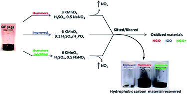
The modified Hummers method is either one step method or two step method. The one step modified Hummers method is developed by Chen et al.89 It is also called as “Improved Hummers” method where the graphite was oxidized without using NaNO3. In two step modified Hummers method the graphite powder is initially oxidized by H2SO4, K2S2O8 and P2O5. Then the pre-oxidized graphite was subjected to oxidation by Hummers method. Marcano and co-workers90 developed a new method which they called “Improved synthesis” of GO that can be seen in Fig. 1. They used KMnO4 in a 9[thin space (1/6-em)]:[thin space (1/6-em)]1 weight ratio with H2SO4 and H3PO4 mixture. In addition to, Xu et al.91 developed another method by reducing KMnO4/graphite ratio from 3[thin space (1/6-em)]:[thin space (1/6-em)]1 to 1[thin space (1/6-em)]:[thin space (1/6-em)]1. This approach is termed “mild oxidation”. A brief experimental procedure of all the aforementioned methods has been discussed below. In Table 2 a comparative study of all these methods has also been discussed.


image file: c6ra03189g-f1.tif
Fig. 1 Representation of GO synthesis by different methods with the production of under-oxidized hydrophobic carbon material.90
Table 2 A comparative study of GO synthesis
Method of oxidation Advantages Disadvantages
HM • One step method • Higher degrees of defect due to harsh oxidation
• High efficiency • Production of higher under-oxidized hydrophobic carbon materials
• High yielding • Generation of toxic gases (NO2, N2O4, ClO2)
• Free from the emission of acid fog • Production of residual Na+ and NO3 ions which are difficult to remove from the waste water
Two step modified HM • High yielding • Two step method
• Penetration of more oxygen functional groups throughout the sheets • Time consuming
• Formation of higher defects or disorders
One step modified HM/improved HM • Short oxidation period N/A
• Simple procedure
• High yielding
• Low cost
• Free from the production of toxic gases and residual Na+ and NO3 ions
• Production of very low under-oxidized hydrophobic carbon materials
Improved synthesis • One step method • Lengthy oxidation period
• High yield • Requires twice as much KMnO4 and 5.2 times as much H2SO4 as those applied by Hummers method
• Fewer defects • Introduction of H3PO4 as an additional oxidising agent in the reaction system
• Free from the production of toxic gases
• Production of lower under-oxidized hydrophobic carbon materials
Mild oxidation • One step method • Low yield
• Good water solubility • Production of higher unoxidized graphite in the form of aggregate
• Large π-conjugated structure
• Less lattice disorder
• Act as a low defect precursor to prepare highly conductive graphene upon chemical reduction


2.2.1. The original Hummers' method. The GO was synthesised by stirring 100 g of graphite flake and 50 g of NaNO3 into 2.3 liters of H2SO4. The ingredients were mixed in 15 liter battery jar and cooled to 0 °C in an ice bath. During the conduction of vigorous agitation 300 g of KMnO4 was added to the suspension. The rate of addition was managed carefully to keep the suspension temperature below 20 °C. After that the ice bath was removed and the solution was warmed at 35 ± 3 °C for 30 minutes. As the oxidation progressed, the mixture gradually thickened. At the end of 20 minutes the mixture became pasty with the release of small amounts of gas. The colour of the paste was brownish gray. At the end of 30 minutes, 4.6 liters of water was gradually added and stirred into the paste to dilute it. The dilute suspension was brown in color. During this addition of water a large exothermal heat was generated to 98 °C which is maintained for more 15 minutes through exothermal heating. The suspension was further diluted by pouring 14 liter of warm water and treated with 3% H2O2. After treating with peroxide, the suspension took brownish yellow color appearance. Later the suspension was filtered resulting a yellow-brown filter cake. The filter cake was washed 3 times and the obtaining residue was dispersed in 32 liter of water to yield 0.5% solids. The dry form of the product was obtained by centrifugation followed by vacuum dehydration at 40 °C.
2.2.2. The two step modified Hummers' method. The graphite flake (20 g) was added to a mixture of concentrated H2SO4 (30 mL), K2S2O8 (10 g) and P2O5 (10 g). After the admixture a dark blue color suspension was produced with large exothermal heat. The resultant suspension allowed to cool at ambient temperature over a period of 6 h. Distilled water was carefully added to the mixture to dilute it. Several filtration and washing was carried out until the pH of the rinse water became neutral. The product was dried in the room temperature for overnight. Thus the pre-oxidized graphite was prepared which is later subjected to oxidation by the Hummers' method. The pre-oxidized graphite powder (20 g) was put into cold concentrated H2SO4 (460 mL). KMnO4 (60 g) was slowly added while agitating and cooling the solution, during which the temperature of the solution was not allowed to exceed 20 °C. Then the mixture was stirred at 35 °C for 2 h with the addition distilled water (920 mL). Upon 15 min, the reaction was terminated by pouring a large amount of distilled water (2.8 L) and a 30% H2O2 solution (50 mL), after which the color of the mixture was turned into bright yellow. After that, the mixture was filtered and washed with a 1[thin space (1/6-em)]:[thin space (1/6-em)]10 HCl solution (5 L) to discard metal ions.
2.2.3. The improved Hummers' method/one step modified Hummers' method. The graphite flake (3 g) was added to a mixture of concentrated H2SO4 (70 mL) under the agitation in an ice bath. Under vigorous stirring, KMnO4 (9 g) was slowly added to keep the suspension temperature lower than 20 °C. Successively, the reaction mixture was shifted to a 40 °C oil bath and vigorously stirred for about 30 minutes. After that, 150 mL distilled water was poured and the solution was stirred for more 15 min at 95 °C. The reaction was terminated by pouring additional water (500 mL) and a 30% H2O2 solution (15 mL), after which the colour of the suspension was turned into yellow from brown. The mixture was then filtered and washed with a 1[thin space (1/6-em)]:[thin space (1/6-em)]10 HCl solution (200 mL) to discard metal ions.
2.2.4. The method of “improved synthesis” of GO. A 9[thin space (1/6-em)]:[thin space (1/6-em)]1 mixture of concentrated H2SO4/H3PO4 (360[thin space (1/6-em)]:[thin space (1/6-em)]40 mL) was poured into a mixture of graphite powder (3 g, 1 wt equiv.) and KMnO4 (18 g, 6 wt equiv.), producing a mild exotherm to 35–40 °C. Then the reaction mixture was warmed to 50 °C and stirred for 12 h. The mixture was cooled to room temperature and poured onto ice (∼400 mL) with 30% H2O2 (3 mL). For work-up, the mixture was sifted through a metal U.S. Standard testing sieve (W.S. Tyler, 300 μm) and then filtered through polyester fiber (Carpenter Co.) The filtrate was centrifuged at 4000 rpm for 4 h and the supernatant was removed. The remaining solid material was then washed in succession with 200 mL of water, 200 mL of 30% HCl and 200 mL of ethanol (2×). For each wash, the mixture was sifted through the U.S. standard testing sieve and then filtered through polyester fiber. Later the filtrate was centrifuged at 4000 rpm for 4 h and the supernatant was decanted away. The remaining material was removed by multiple-washing and the resulting suspension was filtered over a polytetrafluoroethylene membrane with a 0.45 μm pore size. The solid yield on the filter was vacuum-dried at room temperature for overnight to extract 5.8 g of GO.
2.2.5. The method of “mild oxidation”. Graphite (0.5 g, 1 wt equiv.) and KMnO4 (0.5 g, 1 wt equiv.) were successively mixed with concentrated sulphuric acid (15 mL) under stirring at room temperature. Then the reaction mixture was transferred to a 50 °C water bath for about 3 h. Then the mixture was poured into 150 mL water and the solution was agitated for another 15 min. The oxidation was terminated by the addition of 10 mL H2O2 (30%). The mixture was then filtered and washed with 1[thin space (1/6-em)]:[thin space (1/6-em)]10 HCl aqueous solution (50 mL) to discard metal ions followed by washing with water to remove the acid. The resulting solid was dispersed in 150 mL water by ultrasonication for 1 h to make an aqueous dispersion. The obtained dispersion was then centrifuged at 3000 rpm for 30 min to get a black-brown supernatant. The sediments at the bottom were again dispersed in 50 mL water by ultrasonication for 1 h and the dispersion was treated by centrifugation as above. The two parts of supernatants were mixed to get the aqueous dispersion of mildly oxidised GO. The yield of as-synthesised GO was calculated to be about 20% (the weight of mildly oxidized graphene divided by the weight of graphite powder).

The physical properties of GO including electron conductivity, surface charge, transparency, band gap energy etc. can be determined by the type and quantity of oxygen functionalities over the GO sheets. The lateral size of GO is affected by the degree of oxidation, oxygen moieties and defect sites. It can be increased by a high degree oxidation which can rupture the GO sheets during the exfoliation step.92 Lerf et al.93 proposed a new structural model of GO. They demonstrated that the hydroxyl and epoxy groups are the major or dominant oxygen functionalities of GO. These groups are located at the basal plane of the sheets. In contrast, the other oxygen moieties such as the carboxyl and carbonyl groups are typically found at the edge of the sheets.94 The quality of graphene oxide is evaluated by the carbon and oxygen (C/O) atomic ratio and the average size of the GO sheets. The increment of oxidation temperature decreases the C/O atomic ratio and the size of the sheets. It is attributed to the formation of more functional groups and defect sites due to the extensive oxidation.95 This relationship can be represented as follows in Fig. 2.


image file: c6ra03189g-f2.tif
Fig. 2 The relationship of oxidation temperature with C/O atomic ratio and average size of the sheets.

The thickness of GO is another important parameter to evaluate the successful production of GO. Typically it is increased after oxidation owing to the attachment of covalently bonded oxygen. It is briefly discussed in the AFM section of the study. A list of C/O atomic ratio and thickness of GO produced by different procedure has been tabulated in Table 3.

Table 3 Summary of C/O atomic ratio and thickness of GO samples
Method of GO synthesis Size of graphite flakes Time of oxidation Temperature of oxidation Ratio of C/O Thickness of GO (nm) Reference
a Modified Hummer method.
Hummer method <1 mm ∼2 hours 98 °C 2.30 ∼1.0–1.4 96
Hummer method 30 μm ∼4 hours 35 °C 2.33 ∼0.9–2.3 97
Hummer method 30 μm ∼2 hours 60 °C 2.28 ∼1.0 97
Hummer method 30 μm ∼2 hours 45 °C 2.25 ∼1.0 97
Two step MHMa 74 μm ∼8.5 hours Pre-oxi: 80 °C 1.18 ∼0.80 95
Oxi: 35 °C
Two step MHMa 74 μm ∼8.5 hours Pre-oxi: 80 °C 1.24 ∼0.89 95
Oxi: 27 °C
Two step MHMa 74 μm ∼8.5 hours Pre-oxi: 80 °C 1.26 ∼0.82 95
Oxi: 20 °C
One step MHM 325 mesh ∼1 hour 40 °C 2.36 ∼0.80 89
Improved synthesis 150 μm Over night 50 °C N/A ∼1.1 90
Mild oxidation 325 mesh 3 hours 50 °C 3.1 ∼0.90 91


2.3. Exfoliation of graphene oxide

The oxidation reaction helps to penetrate oxygen functionalities on both sides of the sheets. It widens the inter sheet distance and disbands the tightly packed sp2 hybridized atoms of carbon.98,99 Botas et al.100 pointed out a probable mechanism for the invasion of oxygen into the graphite flakes. They claimed that in the case of graphites with smaller crystal size (optical texture composed of mosaics) the invasion of oxygen occurs predominantly at the boundaries of the sheets, yielding small size GO sheets. In contrast, the more ordered graphites (optical texture composed of flow domains) are predominantly attacked in the basal plane where the epoxy groups are developed. It yields large size GO sheets. This study also revealed that GO derived from lower crystalline graphite contains a large number of carboxyl and hydroxyl groups and GO obtained from higher crystalline graphite contains abundant epoxy groups. As a result of the above mechanism, the material becomes hydrophilic facilitating the separation of individual GO sheet using sonication99 or magnetic stirring.95,101 This phenomenon is called exfoliation of GO. Water is the widely used solvent to disperse GO. Samulski et al.102 claimed, Water soluble graphene is similar to single graphene sheets peeled from pyrolytic graphite (0.9 nm thick). Besides water the dispersion of GO is also possible in several organic solvents such as: N-methyl pyrrolidine (NMP), ethylene glycol and N,N-dimethyl formamide (DMF).96 It has been observed that graphite could utilize the surface energy of NMP to exfoliate and produce single layer GO sheets.54,103 But solvents are expensive and their high boiling point limited their applications. Lotya et al.53 demonstrated that graphene exfoliation is possible in a surfactant–water solution (sodium dodecyl benzene sulfonate). The major benefit of surfactant is the prevention of sheet aggregation. In another study, Green and Hersam53 used sodium cholate to simultaneously extract single graphene sheets from the bulk graphite with the help of a density gradient centrifuge machine at different buoyant density.

Both ultrasonication102 and magnetic stirring101 can be used in graphene exfoliation. However, excessive sonication can reduce the sheet quality. Chhowalla et al.104 found that GO dispersed in DMF solution could laterally degrade after 10 hours of sonication. Over sonication may increase the number of sheet to sheet junctions which limits electron mobility.104 These types of sheets are not suitable for device applications. A higher amount of oxygen functionalities (epoxy and hydroxyl) can generate areas of weakness mainly consisting of cracks and fault lines. This defective zone leads to sheet breaking when ultrasonicated for a longer duration. It follows the mechanism of cooperative unzipping proposed by Li et al.105 In addition, erosion of ultrasound tip is an unavoidable outcome of prolonged ultrasonication.106 Furthermore, a recent investigation by Qi et al.107 showed that the first hour of sonication is the critical point. Because the exfoliation and sheet break up mainly occur in the very first hour, while later sonication facilitates sheet restacking. This suggests that when GO sheets are degraded in size, they would trend to intercalate or restack together. Therefore in order to address poor quality or hole defects of graphene higher sonication time should be avoided.108 It is better to continue until the whole solution becomes clear with no visible particulate matter and brilliant yellow in colour.99

2.4. The green reduction of graphene oxide

In a typical reduction, dispersed GO suspension and environmentally friendly reducing agents are mixed together to produce reaction solution first. Secondly, the reaction solution is kept under controlled temperature for different duration. In most of the previous studies, the reaction temperature and the concentration of GO have been observed 80–95 °C and 0.5–1 mg mL−1, respectively. In Table 4, the concentration of GO, temperature and time of GO reduction for different studies has been discussed. The adjustment of pH is an important parameter for the reaction solution. Because the reduction is possible in both acidic109 or alkaline environment.110,111 Notably, HCl,109,112–114 NH4OH115–118 and dilute NaOH52,119 are used to regulate the pH of the solution. According to Merino et al.120 alkaline condition is able to reduce the time of reduction. It can deoxygenate GO sheets without using any further reducer.110 It strongly stimulates the colloidal durability of GO with the help of electrostatic repulsion. Bosch et al.121 clearly illustrated the effects of different pH values on the GO sheets. According to their findings, acidic pH is responsible for a higher concentration of defects with sheet aggregation and size reduction. An acidic pH may turn the sheet like structure of graphene into other nano forms of graphite such as fullerene, nanoonion or multi-walled carbon nano tubes. This investigation suggests that the alkaline pH is more suitable to produce high quality or less defective graphene.122 After reduction the brownish yellow colour solution of GO turns into black. Graphene sheets are extracted from the black slurry by filtration113,119,123 or centrifugation.118 After centrifugation, the product should be washed and recentrifuged several times with deionised water, alcohol or 5% ammonia to remove all the unwanted materials (unreduced graphene, excess reducers, and reaction by-products). After completing the washing procedure, the pH of the product should be neutral (pH = 7).124,125
Table 4 A list of different green reducing agents used in graphene oxide reductiona
Reducing agents of GO Concentration of GO in aqueous solutions (mg mL−1) Temperature of reduction Time of reduction Ref.
a RT = room temperature.
Aluminum powder 1 RT 30 min 109
Aqueous phyto extracts 0.5 RT 5–8 h 130
Baking soda 0.1 90 °C 3 h 131
Benzylamine 0.5 90 °C 90 min 123
Benzyl alcohol 8 100 °C 5 days 132
Beta carotene 2 95 °C 24 h 133
Caffeic acid 0.1 95 °C   113
Carrot root 0.5 RT 48 h 134
Casein 1 90 °C 7 h 116
Cinnamon 1.6 N/A 45 min 135
Clove extract 1.6 100 °C 30 min 136
Coconut water 0.66 100 °C 12, 24, 36 h 137
Curcumin N/A 85 °C 2 h 138
E. coli 5 37 °C 48 h 139
Enhanced green fluorescent protein 1 90 °C 1 h 140
Formic acid 2.5 100 °C 18 h 141
G. biloba 0.5 30 °C 12 h and 24 h 142
Grape juice 0.6 95 °C 1 h, 3 h and 6 h 143
Gallic acid 4 RT and 95 °C 24 h and 7 h 115
Green tea polyphenol 1 80 °C 8 h 128
Green tea polyphenol + iron 0.1 40, 60, 80 °C 10 min 144
Glucosamine 0.496 90 °C 7 h 52
Glycine 0.25 95 °C 24 h 145
H. sabdariffa 0.4 N/A 1 h 146
Humanin 1 40 °C 1 h 147
Iron powder 0.5 RT 30 min 114
L-Ascorbic acid 0.1 23 °C N/A 148
L-Cysteine 0.5 26 ± 2 °C 12–72 h 124
L-Glutathione 0.1 50 °C 6 h 149
L-Lysine 1 90 °C 9 h 150
L-Valine 0.1 90 °C Several hours 118
Low temperature aluminum 4 100–200 °C 3 h 151
Melatonin 0.1 40, 60, 80 °C 3 h 152
Metallic zinc 0.5 RT 6 h 119
Metal salts 0.1 RT 1 h 153
Manganese powder 0.5 RT Till black sedimentation 154
Nascent hydrogen 1.0 RT 30 min, 20 min, 6 h 155
Natural cellulose + ionic liquid 0.8 80 °C 12 h 156
Oligothiophene 0.48 RT 6 h 126
Oxalic acid 2.5 75 °C 18 h 157
P. glutinosa 5 98 °C 24 h 158
PDDA 3 90 °C 5 h 159
Polyphenol 1 80 °C 8 h 128
Pomegranate juice 0.4 N/A 12 h, 18 h, 24 h 160
Potassium hydroxide 0.5–1 50–90 °C Few minutes 122
Pyrrole 0.5 95 °C 12 h 161
Rose water 7 95 °C 5 h 162
Reducing sugar 0.1 95 °C 1 h 117
Spinach 1.6 N/A 30 min 163
Shewanella sp. 300 RT 48 h 164
Sodium acetate trihydrate 1 95 °C 24 h 165
Sodium carbonate 2 80 °C 4 h 110
Sodium hydroxide 0.5–1 50–90 °C Few minutes 122
Supercritical alcohols 125 400 °C 2 h 127
T. chebula 1 90 °C 24 h 166
Tea solution 0.5 90 °C N/M 167
Thiourea dioxide (TUD) 1 90 °C Several hours 125
Tin powder 1 RT 0.5–3 h 112
Tannin N/A N/A Several hours 168
Vitamin C 0.1 95 °C 0.5–4 h 120
Vitamin C + amino acid 0.1 80 °C 24 h 129
Yeast 0.5 35–40 °C 72 h 169
Zn powder + ultrasonication N/A RT Till black sedimentation 170
Zn powder + acid 1 RT 30 min 171


But in case of filtration, after each filtering the filter cake should be dissolved in deionised water and resonicated for 10 minutes prior to next filtering.125 In different studies, 0.45 μm poly ether sulfonate paper,126 poly vinylidene fluoride filter,127 0.22 μm nylon membrane128,129 or 0.20 μm Whatman paper have been used to drain out the waste solution. Finally, the solid and black RGO sediment can be dried inside a vacuum oven at 50–100 °C.109,124 Xiao et al.115 also dried the graphene at room temperature but it took 2 days. Thus the functional graphene sheets are obtained as final product. A pictorial image of green reduction of GO using L-valine is shown in Fig. 3. Moreover, the quality of the functional graphene highly dependents on the following factors: (i) the properties of pristine graphite, (ii) method of oxidation, and (iii) final deoxygenation procedure of GO to graphene. A flow chart of green reduction of GO is shown in Fig. 4.


image file: c6ra03189g-f3.tif
Fig. 3 The green reduction of graphene oxide using L-valine.118

image file: c6ra03189g-f4.tif
Fig. 4 A schematic representation of green reduction of graphene oxide.

3. Characterization techniques to evaluate GO and RGO

GO and RGO expose unique set of spectroscopic and microscopic characteristics that differentiate them. The coming section will discuss the commonly used spectroscopic and microscopic techniques to evaluate GO and RGO.

3.1. Raman spectroscopy characterization

Raman spectroscopy is a most important tool to characterise graphene and other carbon related materials such as carbon nano tube,172 graphene oxide,64 and graphite.173 Its main purpose is to study the electronic and compositional characteristics of graphene. The spectrum of Raman displays two bands: the D band and the G band together with a weak 2D band.174 The D band is associated with the breathing mode of K point phonons of A1g symmetry. It mainly indicates the surface disorder and defects of graphene.175 And the G band is the result of first order scattering of E2g phonon from sp2 carbon. It expresses the graphitic composition of the materials.176 In pristine graphite, a sharp and intense G band observes at 1575 cm−1. It is related to the lattice structure of graphite. It also displays a weak D band at approximately 1355 cm−1 caused by the graphite edges.177 After the oxidation reaction the synthesized graphene oxide exhibits characteristic D band G band at around 1350 and 1580 cm−1, respectively.174 In GO the G band is broaden and blue shifted to higher wave number compared to the pristine graphite.177 On the other hand, the D band of the Raman spectrum of GO becomes prominent. It ensures the degradation of the in-plane sp2 domains in GO due to the extensive oxidation.99 While GO is being reduced, both of the D band and G band is blue shifted to lower wave number.178 It reveals the construction of new structure through reduction reaction. Structurally, the D band and G band of RGO are sharper and more intense in comparison with those of GO.177,179 Additionally, the weak 2D band of the Raman spectrum of RGO centres within the range of 2700 cm−1. It is ascribed to the second-order of the zone-boundary phonons generated by the double resonance Raman scattering with two-phonon emission. It can be used to see whether the RGO is single layer or multiple.174 Generally, the Lorentzian peak for the 2D band of single layer graphene is observed at 2679 cm−1. This peak is broadened and shifted to a higher wave number if multilayer graphene is formed.130 For example, Akhavan found this band at 2699 cm−1 for the bilayer graphene.180 In Fig. 5 the Raman spectroscopy of graphite, GO and RGO has been given.
image file: c6ra03189g-f5.tif
Fig. 5 Raman spectra of graphite, GO and RGO (here SRGO = baking soda reduced graphene oxide).131

The intensity ratio of band D and G ((ID/IG)) is used to investigate the degree of disorders in graphene. This ratio is unique to differentiate RGO from GO. Because it is either increase or decrease in RGO (Table 5). The value of both of the band is required to calculate this ratio through the Tuinstra–Koenig relation.173 After reduction two different types of regime can arise in RGO: the high defect density regime and the low defect density regime.59,181,182 In high defect density regime, the ratio starts to fall with the increment of defect density. This is ascribed to the larger amount of sp2 amorphous texture of carbon. In this case the ID/IG ratio is proportional to the square of the size of nanocrystalline graphitic domains.183

Table 5 ID/IG ratio of GO and RGO after different chemical reduction reactionsa
Reducing agents GO RGO Reference
a *the decrease of ID/IG ratio from GO to RGO.
Aluminium powder 0.96 1.81 109
Amino acid (L-cysteine) 0.94 1.17 124
Baking soda 1.47* 1.37* 131
Benzyl alcohol N/A 1.20 132
E. coli 1.37* 0.97* 139
Gallic acid 1.74 1.86 115
Carrot root 0.80 1.09 134
Glucose amine 1.43 1.50 52
Glycine 0.98 1.09 145
Humanin 1.4 2.3 147
Iron powder 0.96 1.02 114
Lithium aluminium hydride 0.96 1.30 191
Hydrazine hydrate 0.96 1.21 191
Metallic zinc 1.19 1.40 119
Manganese dioxide 1.44 1.68 194
Nascent hydrogen 1.01 1.36, 1.65, 1.88 155
Natural cellulose 1.32 1.53 156
Non aromatic amino acid 0.95 1.02, 1.05, 1.11 118
Oligothiophene* 1.19* 1.02* 126
Oxalic acid 1.2 1.3 157
Pyrrole* 1.3* 0.1* 161
Sodium acetate trihydrate 0.87 0.96 165
Sodium hydrosulphite 0.73 1 195
Sodium carbonate 0.81 1.04 110
Solvothermal 0.86 1 190
Sulfur coating compounds N/A 0.95–1.22 196
Supercritical alcohols 1.11 1.30 127
Tanin 0.97 1.18 168
Thiourea dioxide 0.83 >1 125
Tin powder 0.94 ≥1.30 112
Vitamin C + amino acid 1.56 1.75 129
Vitamin C/L-ascorbic acid 0.95 1.2 120
Yeast 0.80 1.44 169
Zinc powder + acid 0.89 1.61 171


On the other hand, in the low defect density regime the ID/IG ratio starts to increase. Here defect density is assorted with the generation of higher elastic scattering. This regime is regarded as nanocrystalline graphite.184–187 The reason of this increment is the degradation of overall size of sp2 domains.188,189 In this case the intensity ID/IG ratio is inversely proportional to the size of sp2 domains.190 Notably, in RGO the new graphitic domains are more numerous than those of GO.191 In order to explain this phenomenon Stankovich et al.99 pointed out that the reduction of GO increases the number of aromatic domains of smaller average size in graphene, which could lead to an increase of the ID/IG ratio. Additionally, the increase of ID/IG ratio is a proven sign of crystal defects, which are of two types. The first one is the edge defect, which does not cause structural rapture. It plays the main role to the increment of ID/IG ratio in RGO. And the second one is the basal defect responsible for structural disruption.145 Moreover, the ID/IG ratio of high temperature reduction is greater than that of room temperature, which indicates a lower degree of disorder at room temperature.119 But room temperature reduction is unable to increase the average size of the graphene domains.115

The Knights empirical equation is used to calculate the size of the sp2 carbon domains (designated as La) using the ID/IG ratio of the samples.192

 
La = 4.35(ID/IG)−1 (1)

In an investigation, Khai et al.193 calculated the size of sp2 domains to be approximately 5 and 4 nm for GO and RGO, respectively. This achievement suggests that the chemical reduction can cause nucleation of sp2 domains in the sp3 matrix. As a result, the density of small sized sp2 nuclei increases and the average size of sp2 domains decreases.

3.2. Fourier transforms infrared spectroscopy characterization

In graphene research, it is important to study what types of oxygen functional groups are attached to the basal plane. FTIR is the technique to identify the bonding configuration of various sorts of oxygen. It is also a complementary tool to Raman spectroscopy. The FTIR spectrum of pristine graphite exhibits no characteristic peak for the discernible functional groups.197 It only displays two peaks at approximately 1610 and 450 cm−1 attributed to the skeletal vibrations from graphite domains (the sp2 aromatic C[double bond, length as m-dash]C) and the vibration of adsorbed water molecules (the O–H stretching), respectively.52 After treating with oxidizing agents, the oxygenated graphene sheet could display a series of different absorption bands or characteristics peaks ranging from 900 to 3500 cm−1. These includes carboxyl peaks (1700–1750 cm−1) mainly present at the edges of the sheet, alkoxy stretching vibrations (1040–1170 cm−1), O–H stretching vibrations (3300–3500 cm−1), O–H deformation peaks (1300–1400 cm−1) and the stretching vibration of epoxy C–O groups (1000–1280 cm−1). Notably, the aromatic C[double bond, length as m-dash]C peak could be seen between the range of 1600–1650 cm−1. This peak is resulted from the sp2 domains of the unoxidized graphite125,165 and this vibration generated from the unoxidized region is termed skeletal vibration.102,126 However, in different GO samples, the above characteristic peaks are found in slightly different positions (Table 6). This may be the consequence of different reaction system and conditions.
Table 6 FTIR characterization of GO in different studiesa
Peak positions of oxygen functionalities for GO samples Agents of GO reduction Ref.
C[double bond, length as m-dash]O stretching Epoxy C–O Alkoxy C–O O–H stretching O–H deformation
a N/A = not available.
1746 1220 1053 3420 1395 Amino acid (L-cysteine) 124
1720 1204 1049 3400 N/M Aquatic phyto extract 130
1706 1219 1052 3395 1386 Baking soda 131
1728 1223 1051 N/M 1395 Carrot root 134
1720 1224 1048 3424 N/M Gallic acid 115
1727 1224 1065 3424 N/M Glycine 145
N/M 1250 1120 N/M N/M Glucose amine 52
1720 1220 1030 3400 1400 Humanin 147
1726 1226 1052 3395 1410 L-Ascorbic acid 148
1730 1064 1123 3430 1410 Metallic zinc 119
1730 1220 1050 N/A N/A Natural cellulose 156
1730 1224, 985 N/M N/A 1417 Oligothiophene 126
1740 N/M N/M N/A N/A Polyphenol 128
1739 N/M 1600 3403 1395 Reducing sugar 117
1730 1037 N/A 3420 1395 Sodium acetate trihydrate 165
1730 1222 1050 3387 N/A Sodium carbonate 110
1730 1220 1050 3380 N/A Super critical alcohol 127
1718 1050 N/A 3200 1380 Thiureadioxide 125
1725 1224 1054 3421 1398 Tin powder 198
1700 1600 N/A 3650 N/A Urea ammonium nitrate 199
1720 1220, 980 N/A 3000–3500 1300–1350 Vitamin C 120
1732 1231 1051 3408 1394 Yeast 169


While GO is being reduced by the chemical reducers, the abovementioned characteristic peaks are gradually weaker over the course of the reaction and some of them vanish completely. This indicates the successful removal of oxygen from the sheets. The spectra of RGO displays a sharp C[double bond, length as m-dash]C band within the range of 1600 cm−1.124,165 Additionally, It displays two new peaks in the region of 2922 and 2853 cm−1 attributed to the presence of –CH2 and –CH stretching vibrations. It is also explained as the adsorption of reducing agent by the sheet surface.126,134,169 These observations confirm that the atomic frame of sp2 carbon has formed in RGO. In a previous study, the alkoxy and epoxy vibration peaks remained undistorted in RGO although their position has been changed. This is ascribed to the partial reduction of GO.134 In another study, the absorption peaks of alkoxy and carboxyl groups are found to be weaker at higher temperature reduced graphene oxide (HT-RGO) than those of room temperature reduced graphene oxide (RT-RGO). This investigation suggests that higher degree of reduction is related to higher temperature (90–95 °C).115 Khai et al.193 also found in completely disappeared carboxyl acid and hydroxyl peaks after the solvothermal treatment of GO. This is attributed to the mild reduction procedure. Herein, Fig. 6 shows the FTIR spectra of GO and RGO and Table 6 shows the FTIR characterization of different GO samples.


image file: c6ra03189g-f6.tif
Fig. 6 FTIR spectra of GO and RGO (here, SRGO = baking soda reduced graphene oxide).131

3.3. X-ray photoelectron spectroscopy characterisation

It is very difficult to reduce hundred percent of the oxygen molecules from the sheets of GO.90,97,109,200 That is why elemental analysis is crucial to characterize the newly synthesised graphene. XPS is commonly used to determine the relative amounts of carbon, oxygen, and other functional groups present in graphene.201 The XPS spectrum of GO and RGO exhibit two major peaks (C1s and O1s) at the wide regions.109 The characteristic C1s peak is the result of sp2 carbon–carbon bonding and the O1s peak is associated with different sp3 carbon–oxygen bonding. The relative intensity of these two peaks varies with sample to sample, depending on the conditions of the reaction system.91 Both of the O1s and C1s spectra can be used to calculate the ratio of peak intensity between O1s and C1s i.e. the C/O atomic ratio. This ratio can be used to determine the oxygen moieties present in the carbon skeleton of graphene.202 Table 7 discusses the C/O atomic ratio of RGO produced using different reducing agents.
Table 7 C/O atomic ratio of RGO at different studies
Reducing agents Reaction time C/O atomic ratio References
Aluminium powder 30 min 18.6 109
Caffeic acid 24 h 7.15 113
Carrot root 72 h 11.9 134
Glycine 24 h 11.14 145
Gallic acid 6 h 5.82 115
Hibiscus sabdariffa 1 h 3.1 146
Hydroxyl amine 1 h 9.7 208
Iron powder 6 h 7.9 114
L-Ascorbic acid 48 h 5.7 148
Metallic zinc 7 h 3.76 119
Oligothiophene 6 h 6.12 126
Pyrrole 2 h 7.7 161
Sodium citrate 3 h 5.6 209
Sodium carbonate 4 h 8.15 110
Tea solution Several hours 3.1 167
Yeast 72 h 5.9 169
Zn powder + acid 30 min 8.2 171


Typically, in the wide region the C1s and O1s peak of GO and RGO can be observed at ∼530 eV and ∼284 eV (Fig. 7a). In order to conduct the quantitative analysis, both of the C1s and O1s spectra are needed to be separated into their individual components. This is performed by fitting the total spectrum into a linear superposition for all elements. Gaussian–Lorentzian peak shape method is used to fit the C1s and O1s curve after performing the Shirley background correction.112,202 This separation helps to determine the fractions of every single element present in the sheet. The software XPSPEAK41 is an useful tool for background subtraction, curve fitting, and peak separation.119,203


image file: c6ra03189g-f7.tif
Fig. 7 XPS pattern of graphene: (a) wide region (GO + RGO), (b) C1s region of GO, (c) C1s region of RGO109 and (d) O1s region of GO.202

The high resolution C1s signal of GO and RGO represents five different elemental peaks: C–C ≈ (BE = 284.6 eV), C–OH ≈ (BE = 285.6 eV), C–(epoxy group) ≈ (BE = 286.8 eV), C[double bond, length as m-dash]O ≈ (BE = 288.2 eV) and O–C[double bond, length as m-dash]O ≈ (BE = 289.4 eV), as seen in Fig. 7b and c; although the accurate position of these peaks as well as their exact binding energy (BE) is difficult to specify.202,204–206 On the other hand, the O1s spectrum of GO displays several individual component peaks which represent C[double bond, length as m-dash]O groups ≈ (BE = 531.20 eV), C–O groups ≈ (BE = 533 eV), O–C[double bond, length as m-dash]O groups ≈ (BE = 534.4–535.6 eV), respectively, as seen in Fig. 7d. All of this information allows to determine the fraction of epoxy, hydroxyl or carboxyl groups present in the basal plane.202,205 The information obtained by the analysis of O1s spectrum can complement the information obtained by the analysis of C1s spectrum. Because the photoelectron kinetic energies of O1s are lower than those of the C1s, the O1s sampling depth is smaller, and therefore the O1s spectra are slightly more surface specific.202

After the reduction of GO the intensity of the peak related to the C[double bond, length as m-dash]O, C–O and O–C[double bond, length as m-dash]O groups decreases to a much lower value (Fig. 7c and d). This phenomenon suggests that most of the oxygen functional groups have been disappeared after the reduction of GO.66 Secondly, the pick intensity of the sp2 carbon to carbon bonding considerably increases in the C1s spectrum of RGO. This result suggests the restoration of new sp2 network of graphene.66,207 Besides these two peaks some additional peaks might be observed in the spectra of RGO such as sodium peak,124 nitrogen peak,123 iron peak,114 sulphur peak,126 tin peak,112 phosphate peak.169 These are probably the residual components of the reaction mixture caused by other side reactions. These components clearly show the involvement of the reducing agent in the reaction.145

The elemental analysis on the basis of C1s spectrum indicated that GO contained approximately 69.63 at% carbon and 30.37 at% oxygen in the C–C/C[double bond, length as m-dash]C and C–O bonds.146 Upon reduction the atomic percentage of carbon is increased in RGO. According to several studies the better range of carbon percentage is found to be 90–95%.109,134,198 Meanwhile, the C/O atomic ratio of GO is around 2.3, as mentioned in Table 3. After the reduction of GO the C/O atomic ratio is also increased in RGO, as discussed in Table 7. Temperature is an important factor to control the graphene C/O atomic ratio. Yang et al.119 and Zhang et al.122 demonstrated that the graphene produced by the room temperature reduction contains large amount of residual oxygen than that of higher temperature. Reduction time is another important factor. It is established that the increment of reduction time is proportional to the removal of oxygen groups from the plane of graphene.113,123,203

3.4. X-ray diffraction characterization

The inter sheet gap is the most significant parameter to access the structural information of graphene. XRD is the technique which deals with the layer to layer space and crystalline structure of graphene. In this study, we highlighted the interlayer distance of GO before and after reduction. The XRD spectra of primitive graphite revels more intense and sharp peak in comparison to GO and RGO.124 Typically, this graphitic peak is found at 2θ ≈ 26° corresponding to a d-spacing of 0.34 nm.90 After oxidation, this sharp and strong graphitic peak becomes invisible and a new peak observes in the range of 2θ ≈ 9–11°.210,211 It has been reported that the value of d-spacing is found within the range of 8–9 Å upon different method of oxidation such as: Hummer method (8 Å), modified Hummer method (9.5 Å), and improved synthesis (9 Å).90 This result strongly indicates that the d-spacing of GO is directly increased by oxidation due to the penetration of epoxy, hydroxyl, and carboxyl groups on both side of the sheets.212,213 From this result, it can be said that the inter layer spacing of graphene material is proportional to the degree of oxidation. After treating with the green reducing agents the XRD peak of GO vanishes and a broad, prominent and new pick can be seen near 2θ ≈ 26°. It is related to the opening of epoxy rings and removal of other oxygen moieties. This result confirms the successful restoration of new graphitic network in RGO. Table 8 shows the interlayer distance of GO and RGO after different reduction reactions (Fig. 8).
Table 8 Interlayer distance of different GO and RGO samplesa
Reducing agents GO (2θ) d-Spacing of GO (nm) RGO (2θ) d-Spacing of RGO (nm) Ref.
a N/A – not available.
Aluminium powder 10.3° 0.80 nm 23.4° 0.375 nm 109
L-Cysteine 11.3° 0.78 nm 26.2° 0.370 nm 124
Phytoextract 9.75° 0.906 nm 25° 0.360 nm 130
Calcium carbonate N/A N/A 26.3° N/A 214
Caffeic acid 10.02° 0.880 nm 24.79° 0.359 nm 113
Carrot root 11.2° 0.79 nm 23.96 N/A 134
Gallic acid 9.8° 0.90 nm 26° N/A 115
Glucose amine 11.2° 0.79 nm N/M N/A 52
Glycine 11.05° 0.7997 nm 23.9° 0.3718 nm 145
Iron powder 10.3° N/A 24.3° N/A 114
Metallic zinc 11.6° 0.779 nm 24° 0.368 nm 119
Cellulose 11.9° 0.74 nm 22.5° N/A 156
Oxalic acid 11.9° 0.743 nm 21.9° 0.405 nm 157
PDDA 11.4° 0.78 nm 20° N/A 159
Pyrrole 10.1° 0.87 nm 20.1° N/A 161
Sodium acetate 11.2° 0.790 nm 25.4° 0.350 nm 165
Supercritical alcohol 11.76° 0.752 nm 23–25° 0.358–0.381 nm 127
Tannin 9.7° 0.90 nm 26.3° 0.340 nm 168
Tin powder 11° N/A 26.6° N/A 112
Tea solution 9.6° 0.92 nm 24.6° 0.340 nm 167
Yeast 11.32° 0.7816 nm 23.5° N/A 169


image file: c6ra03189g-f8.tif
Fig. 8 XRD spectra of (a) GO, (b–d) aquatic phytoextract reduced graphene oxides and (e) graphite.130

3.5. Microscopy characterization

Microscopy is essential to study morphology of graphene. The TEM, SEM and AFM are commonly used in this purpose. A brief of the above techniques are discussed below:
3.5.1. Transmission electron microscopy. TEM acts on the basis of diffraction patterns emitted by the samples.215,216 To generate this diffraction pattern the highly energetic electrons beams are transmitted through the sheet. The scattered electrons are captured and processed by an electromagnetic lens to display an image. The low magnification TEM image is quite useful in sheet visualization, but they are unable to provide complete information about the internal atomic structure of the sheets. For the structural characterization, the selected area electron diffraction (SAED) patterns are often obtained from the individual sheets.

The two dimensional (2D) GO and RGO nano sheets show transparent, corrugated or wrinkled structure under the TEM.148,217 It is also explained as ultrathin silk veil morphology with fold and scroll on its edges.218 It is attributed to the intrinsic nature of graphene.169,219 Yu et al.219 claimed that the corrugation can be present not only at the edge but also in the middle of the nanosheets. It is responsible for the sheets to look folded and coiled.119,124,134,219 Notably, high resolution transmission electron microscopy (HR-TEM) is able to show the number of layers at different locations of the sheets.134,145 With this technique, the measured lattice spacing of single layer graphene is 0.345 nm.214 Some TEM and HR-TEM images of RGO are portrayed in Fig. 9.


image file: c6ra03189g-f9.tif
Fig. 9 TEM image of GO and RGO at lower magnification (a and b) over a TEM grid with lacey carbon support.131 HR-TEM image (c) of RGO is showing the formation of single layer graphene.214

The structure of GO is different from RGO particularly in the SAED analysis. This illustrates the crystalline structure or hexagonal symmetry of the graphene layer. After chemical reduction, strong and well-defined diffraction spots are observed in the polycrystalline ring of SAED (Fig. 10b). This result clearly confirms the destruction of inter layer coherence and random orientation of oxygen free sheets.127,145,148,151,159,169,220 The SAED patterns of GO and RGO are depicted in Fig. 10.


image file: c6ra03189g-f10.tif
Fig. 10 SAED pattern of (a) GO and (b) RGO displaying six fold symmetry.118
3.5.2. Scanning electron microscopy. The SEM is commonly used to study the surface morphology of graphene, GO and graphite. Generally, graphite sheets appears to be heaped up in thick cakes.221 It also exhibits flaky appearance for the strong sp2 carbon to carbon bonding in the plane.131 In contrast, the GO nanosheets show wavy wrinkled appearance under the SEM where the surface are hairy and coarse, and the edges of the sheets are blurry.221 At higher concentration the surface of the GO displays soft carpet like morphology which may be the attachment of residual water molecules, carboxyl and hydroxyl groups with the sheets.147 In RGO the nanosheets display thin and wrinkled texture. It is caused by the stacking of individual sheets by various self-assembly techniques. This is attributed to the intrinsic properties of graphene.75 However, the average lateral size of giant GO sheets have been reported to be 18.5 μm with a relatively wide size distribution ranging from one to several micrometres.222 Gurunathan et al.147 also used the SEM image to count the average lateral size of GO and RGO. They found the average lateral size of GO and RGO sheets are 8 and 10 μm, respectively, although a wide range of sizes (between 2 and 20 μm) were analysed in the study. The SEM images of graphite, GO and RGO are portrayed in Fig. 11.
image file: c6ra03189g-f11.tif
Fig. 11 SEM image of (a) graphite, (b) GO131 and (c) RGO.214

3.6. Atomic force microscopy

AFM is a promising technique to measure the atomic steps present on a surface. It is used to determine the morphology and texture of the graphene materials. It also helps to determine the exact Z-height and number of layers in the specimen. The well-known van der Waals thickness of a pristine graphene sheet is ∼0.34 nm and it is atomically flat. After oxidation the graphene oxide sheets are expected to be thicker due to the presence of covalently bound oxygen on either side of the sheets.99 The typical thickness of single layer GO sheets is ∼0.8 nm which is ∼0.44 nm thicker than that of pristine graphene.180 In the case of bi-layer graphene oxide, the thickness was found to be 1.7 nm.134

Upon the reduction of GO the smaller thickness of RGO sheets is a clear proof of oxygen elimination from the graphene oxide.122,145,148,165,194 A AFM image of GO and RGO is depicted in Fig. 12. But in several studies the thickness of RGO was found to be higher than that of GO. It is attributed to either the binding of capping agents with the sheets or restacking of sheets in the absence of stabilizer molecules.134,168 For example, Akhavan et al.144 observed an increased thickness of graphene caused by the attachment of oxidized green tea polyphenols (GTPs) on both sides of the RGO. Some of the similar types of results have been reported in Table 9. Therefore, it is reasonable to conclude that the stabilizer or capping reagent play an important role to increase the thickness of graphene sheet, although most of the oxygen-containing functional groups are vanished after the reduction.117 Notably, the stabilization mechanisms between the reducer and the graphene layer are proposed to be π–π interactions or hydrogen bonding.128,138 Here, the capped molecules supply steric hindrance to stabilize the graphene sheets.128,168 Beside thickness analysis AFM has also been applied to analyse the lateral dimension of GO and RGO. In one of our previous work Mehrali and co-workers measured the average lateral dimension of GO and RGO around 3.88 ± 0.99 μm and 2.37 ± 0.65 μm.178 This finding also pointed out that the RGO sheets are laterally smaller than the GO sheet.


image file: c6ra03189g-f12.tif
Fig. 12 AFM image of (a) GO and (b) RGO.134
Table 9 A comparative study of the thickness of GO and RGO
Reducing agents Thickness of GO (nm) Number of layers Thickness of RGO (nm) Number of layers Attachment of molecules on both side of the RGO sheets References
Alkaline conditions 1.2 Single 0.881 Single N/A 122
Caffeic acid 1.116 Single 0.846 Single N/A 113
Calcium carbonate + magnesium N/A N/A 2.5 4–10 N/A 214
Carrot root 1.7 Bilayer 2.5 Restacked N/A 134
Curcumin 0.8 Single 1.5 Single Curcumin molecules 138
E. coli 1 Single N/A N/A N/A 223
Gallic acid 1.0 Single 1.0 and 1.4 Single N/A 115
Green tea polyphenol 1.19 Single 1.39 N/A Oxidized tea polyphenol (TP) 128
Green tea polyphenol + iron 1.2 Single Average 1.8, considered one layer as 0.80 nm Single Oxidized greentea polyphenol (GTP) 144
Humanin N/A N/A Between 1 and 3 N/A N/A 147
Iron powder 1 Single 1–5 Multiple N/A 114
L-Ascorbic acid 1.2 Single N/A N/A N/A 148
L-Cysteine 1 Single 0.8 Single   124
L-Glutathione N/A N/A 8 N/A Capping agent 149
L-Valine 1 Single 1 Single N/A 118
Melatonin 0.9 Single 1.8 (considered one layer as 0.80 nm) Single N/A 152
Metallic zinc 1.22 N/A 0.93 Single N/A 119
Metal salts 1.50 N/A 0.86 Single N/A 153
Natural cellulose + ionic liquids 1 Single 1.9 N/A Cellulose chains 156
Pomegranate juice N/A N/A 0.67 Single N/A 160
Pyrrole 1.3 Single 2.7 N/A Stabilizer molecules 161
Reducing sugar 0.97 Single 1.1 Single Capping agent 117
Sodium acetate trihydrate 0.97 Single 0.79 Single N/A 165
Tannin 1.1 Single 1.5 Single Tannic acid (TA) 168
Tea solution 1 Single Average 2 N/A Tea polyphenol (TP) 167
Vitamin C 1 Single 1 Single N/A 120
Vitamin C + amino acid N/A N/A Average 2 N/A L-Tryptophan 129
Yeast 0.8 Single 1.2 Bilayer NADPH 169
Zn powder 1 Single 0.55 Single N/A 170
Zn powder + acid 1.2 Single N/A N/A N/A 171


4. Conclusion and suggestions

The chemical reduction of GO is found to be very promising technique to produce large scale functional graphene. The use of eco-friendly reducing agents to synthesis graphene achieves a great interest of the scientific community, evidenced by the continuous increment of research publications on the topic. In this review, the authors described some of the recent achievements in the green reduction of GO. It is considered as one of the most versatile method. These types of reagents are seemed to be an alternate of hydrazine or other poisonous reducing agents. They are also safe to handle and the reaction coproduces are biocompatible. But green reducing agents are not out of limitations. Sometimes one agent cannot fully deoxygenate the GO alone, but it needs the aid of a supporting reducing agent or surface modifier. There is also a big possibility for the attachment of addition functional groups or capping agents with the sheets supplied by the reducers. This phenomenon can hamper the electron conductivity of the sheets. Notably, The irreversible aggregation, lower electron conductivity, increased thickness, substrate specificity, chance of product contamination e.g. mixing of metallic impurities when reduced with metal powders, limited solubility, several downstream processing e.g. repeated centrifugation or filtration to remove all the reaction by-products and maintenance of complex or light sensitive reaction system e.g. bacterial reduction of GO can be considered as the limitations of the approaches. These drawbacks limited their application to the laboratory and they are not yet encouraged in the industrial batch production of graphene. To overcome all the drawbacks is a challenging task for the researchers in the field.

The replacement of the toxic and harsh chemical agents with mild reagents will be a big achievement of this study. More studies can help to understand the exact reduction mechanism of GO using most of the reported green reducing agents. Theoretical calculations, modeling the interaction between GO and reducing molecules could offer some solutions.

Acknowledgements

“The authors are thankful to University of Malaya for financial support under the High Impact Research MoE Grant: UM.C/625/1/HIR/MoE/ENG/40 (D000040-16001) from the Ministry of Education Malaysia.”

References

  1. K. S. Novoselov, A. K. Geim, S. Morozov, D. Jiang, Y. Zhang and S. A. Dubonos, et al., Electric field effect in atomically thin carbon films, Science, 2004, 306(5696), 666–669 CrossRef CAS PubMed .
  2. A. K. Geim and K. S. Novoselov, The rise of graphene, Nat. Mater., 2007, 6(3), 183–191 CrossRef CAS PubMed .
  3. V. Chabot, D. Higgins, A. Yu, X. Xiao, Z. Chen and J. Zhang, A review of graphene and graphene oxide sponge: material synthesis and applications to energy and the environment, Energy Environ. Sci., 2014, 7(5), 1564–1596 CAS .
  4. M. Katsnelson, F. Guinea and A. Geim, Scattering of electrons in graphene by clusters of impurities, Phys. Rev. B: Condens. Matter Mater. Phys., 2009, 79(19), 195426 CrossRef .
  5. Z. Chen, Y.-M. Lin, M. J. Rooks and P. Avouris, Graphene nano-ribbon electronics, Phys. E, 2007, 40(2), 228–232 CrossRef CAS .
  6. D. A. Areshkin, D. Gunlycke and C. T. White, Ballistic transport in graphene nanostrips in the presence of disorder: importance of edge effects, Nano Lett., 2007, 7(1), 204–210 CrossRef CAS PubMed .
  7. W. Cai, Y. Zhu, X. Li, R. D. Piner and R. S. Ruoff, Large area few-layer graphene/graphite films as transparent thin conducting electrodes, Appl. Phys. Lett., 2009, 95(12), 123115 CrossRef .
  8. X. Li, Y. Zhu, W. Cai, M. Borysiak, B. Han and D. Chen, et al., Transfer of large-area graphene films for high-performance transparent conductive electrodes, Nano Lett., 2009, 9(12), 4359–4363 CrossRef CAS PubMed .
  9. Z.-S. Wu, W. Ren, L. Wen, L. Gao, J. Zhao and Z. Chen, et al., Graphene anchored with Co3O4 nanoparticles as anode of lithium ion batteries with enhanced reversible capacity and cyclic performance, ACS Nano, 2010, 4(6), 3187–3194 CrossRef CAS PubMed .
  10. G. Wang, X. Shen, J. Yao and J. Park, Graphene nanosheets for enhanced lithium storage in lithium ion batteries, Carbon, 2009, 47(8), 2049–2053 CrossRef CAS .
  11. Ö. Metin, S. F. Ho, C. Alp, H. Can, M. N. Mankin and M. S. Gültekin, et al., Ni/Pd core/shell nanoparticles supported on graphene as a highly active and reusable catalyst for Suzuki–Miyaura cross-coupling reaction, Nano Res., 2013, 6(1), 10–18 CrossRef .
  12. R. Wang, Z. Wu, C. Chen, Z. Qin, H. Zhu and G. Wang, et al., Graphene-supported Au–Pd bimetallic nanoparticles with excellent catalytic performance in selective oxidation of methanol to methyl formate, Chem. Commun., 2013, 49(74), 8250–8252 RSC .
  13. D. Wang, W. Yan, S. H. Vijapur and G. G. Botte, Electrochemically reduced graphene oxide–nickel nanocomposites for urea electrolysis, Electrochim. Acta, 2013, 89, 732–736 CrossRef CAS .
  14. Y.-H. Lu, M. Zhou, C. Zhang and Y.-P. Feng, Metal-embedded graphene: A possible catalyst with high activity, J. Phys. Chem. C, 2009, 113(47), 20156–20160 CAS .
  15. R. Kou, Y. Shao, D. Wang, M. H. Engelhard, J. H. Kwak and J. Wang, et al., Enhanced activity and stability of Pt catalysts on functionalized graphene sheets for electrocatalytic oxygen reduction, Electrochem. Commun., 2009, 11(5), 954–957 CrossRef CAS .
  16. Y. Lu, B. R. Goldsmith, N. J. Kybert and A. C. Johnson, DNA-decorated graphene chemical sensors, Appl. Phys. Lett., 2010, 97(8), 083107 CrossRef .
  17. J. D. Fowler, M. J. Allen, V. C. Tung, Y. Yang, R. B. Kaner and B. H. Weiller, Practical chemical sensors from chemically derived graphene, ACS Nano, 2009, 3(2), 301–306 CrossRef CAS PubMed .
  18. Y. Dan, Y. Lu, N. J. Kybert, Z. Luo and A. C. Johnson, Intrinsic response of graphene vapor sensors, Nano Lett., 2009, 9(4), 1472–1475 CrossRef CAS PubMed .
  19. Y. Shao, J. Wang, H. Wu, J. Liu, I. A. Aksay and Y. Lin, Graphene based electrochemical sensors and biosensors: a review, Electroanalysis, 2010, 22(10), 1027–1036 CrossRef CAS .
  20. Y. Liu, D. Yu, C. Zeng, Z. Miao and L. Dai, Biocompatible graphene oxide-based glucose biosensors, Langmuir, 2010, 26(9), 6158–6160 CrossRef CAS PubMed .
  21. W. Hu, C. Peng, W. Luo, M. Lv, X. Li and D. Li, et al., Graphene-based antibacterial paper, ACS Nano, 2010, 4(7), 4317–4323 CrossRef CAS PubMed .
  22. Q. He, S. Wu, S. Gao, X. Cao, Z. Yin and H. Li, et al., Transparent, flexible, all-reduced graphene oxide thin film transistors, ACS Nano, 2011, 5(6), 5038–5044 CrossRef CAS PubMed .
  23. X. Sun, Z. Liu, K. Welsher, J. T. Robinson, A. Goodwin and S. Zaric, et al., Nano-graphene oxide for cellular imaging and drug delivery, Nano Res., 2008, 1(3), 203–212 CrossRef CAS PubMed .
  24. Z. Liu, J. T. Robinson, X. Sun and H. Dai, PEGylated nanographene oxide for delivery of water-insoluble cancer drugs, J. Am. Chem. Soc., 2008, 130(33), 10876–10877 CrossRef CAS PubMed .
  25. C. X. Guo, M. Wang, T. Chen, X. W. Lou and C. M. Li, A Hierarchically Nanostructured Composite of MnO2/Conjugated Polymer/Graphene for High-Performance Lithium Ion Batteries, Adv. Energy Mater., 2011, 1(5), 736–741 CrossRef CAS .
  26. G. Zhou, D.-W. Wang, F. Li, L. Zhang, N. Li and Z.-S. Wu, et al., Graphene-wrapped Fe3O4 anode material with improved reversible capacity and cyclic stability for lithium ion batteries, Chem. Mater., 2010, 22(18), 5306–5313 CrossRef CAS .
  27. C. X. Guo, H. B. Yang, Z. M. Sheng, Z. S. Lu, Q. L. Song and C. M. Li, Layered graphene/quantum dots for photovoltaic devices, Angew. Chem., Int. Ed., 2010, 49(17), 3014–3017 CrossRef CAS PubMed .
  28. C. Peng, W. Hu, Y. Zhou, C. Fan and Q. Huang, Intracellular Imaging with a Graphene-Based Fluorescent Probe, Small, 2010, 6(15), 1686–1692 CrossRef CAS PubMed .
  29. J. Williams, L. DiCarlo and C. Marcus, Quantum Hall effect in a gate-controlled pn junction of graphene, Science, 2007, 317(5838), 638–641 CrossRef CAS PubMed .
  30. C. XianáGuo and C. MingáLi, A self-assembled hierarchical nanostructure comprising carbon spheres and graphene nanosheets for enhanced supercapacitor performance, Energy Environ. Sci., 2011, 4(11), 4504–4507 Search PubMed .
  31. S. Bae, H. Kim, Y. Lee, X. Xu, J.-S. Park and Y. Zheng, et al., Roll-to-roll production of 30-inch graphene films for transparent electrodes, Nat. Nanotechnol., 2010, 5(8), 574–578 CrossRef CAS PubMed .
  32. F. Bonaccorso, Z. Sun, T. Hasan and A. Ferrari, Graphene photonics and optoelectronics, Nat. Photonics, 2010, 4(9), 611–622 CrossRef CAS .
  33. G. Jo, M. Choe, S. Lee, W. Park, Y. H. Kahng and T. Lee, The application of graphene as electrodes in electrical and optical devices, Nanotechnology, 2012, 23(11), 112001 CrossRef PubMed .
  34. V. Eswaraiah, V. Sankaranarayanan and S. Ramaprabhu, Graphene-based engine oil nanofluids for tribological applications, ACS Appl. Mater. Interfaces, 2011, 3(11), 4221–4227 CAS .
  35. Z. Sui, Q. Meng, X. Zhang, R. Ma and B. Cao, Green synthesis of carbon nanotube–graphene hybrid aerogels and their use as versatile agents for water purification, J. Mater. Chem., 2012, 22(18), 8767–8771 RSC .
  36. Y. Zhao, C. Jiang, C. Hu, Z. Dong, J. Xue and Y. Meng, et al., Large-scale spinning assembly of neat, morphology-defined, graphene-based hollow fibers, ACS Nano, 2013, 7(3), 2406–2412 CrossRef CAS PubMed .
  37. H. Sun, Z. Xu and C. Gao, Multifunctional, ultra-flyweight, synergistically assembled carbon aerogels, Adv. Mater., 2013, 25(18), 2554–2560 CrossRef CAS PubMed .
  38. H. Bi, X. Xie, K. Yin, Y. Zhou, S. Wan and L. He, et al., Spongy graphene as a highly efficient and recyclable sorbent for oils and organic solvents, Adv. Funct. Mater., 2012, 22(21), 4421–4425 CrossRef CAS .
  39. X. Dong, J. Chen, Y. Ma, J. Wang, M. B. Chan-Park and X. Liu, et al., Superhydrophobic and superoleophilic hybrid foam of graphene and carbon nanotube for selective removal of oils or organic solvents from the surface of water, Chem. Commun., 2012, 48(86), 10660–10662 RSC .
  40. J. Zhao, W. Ren and H.-M. Cheng, Graphene sponge for efficient and repeatable adsorption and desorption of water contaminations, J. Mater. Chem., 2012, 22(38), 20197–20202 RSC .
  41. J. Shen, B. Yan, T. Li, Y. Long, N. Li and M. Ye, Study on graphene-oxide-based polyacrylamide composite hydrogels, Composites, Part A, 2012, 43(9), 1476–1481 CrossRef CAS .
  42. T. Ohta, A. Bostwick, T. Seyller, K. Horn and E. Rotenberg, Controlling the electronic structure of bilayer graphene, Science, 2006, 313(5789), 951–954 CrossRef CAS PubMed .
  43. C. Berger, Z. Song, X. Li, X. Wu, N. Brown and C. Naud, et al., Electronic confinement and coherence in patterned epitaxial graphene, Science, 2006, 312(5777), 1191–1196 CrossRef CAS PubMed .
  44. P. W. Sutter, J.-I. Flege and E. A. Sutter, Epitaxial graphene on ruthenium, Nat. Mater., 2008, 7(5), 406–411 CrossRef CAS PubMed .
  45. V. C. Tung, M. J. Allen, Y. Yang and R. B. Kaner, High-throughput solution processing of large-scale graphene, Nat. Nanotechnol., 2008, 4(1), 25–29 CrossRef PubMed .
  46. H. Chen, M. B. Müller, K. J. Gilmore, G. G. Wallace and D. Li, Mechanically strong, electrically conductive, and biocompatible graphene paper, Adv. Mater., 2008, 20(18), 3557–3561 CrossRef CAS .
  47. K. S. Novoselov, A. K. Geim, S. Morozov, D. Jiang, Y. Zhang and S. Dubonos, et al., Electric field effect in atomically thin carbon films, Science, 2004, 306(5696), 666–669 CrossRef CAS PubMed .
  48. B. Jayasena and S. Subbiah, A novel mechanical cleavage method for synthesizing few-layer graphenes, Nanoscale Res. Lett., 2011, 6(1), 95–101 CrossRef PubMed .
  49. Z. G. Cambaz, G. Yushin, S. Osswald, V. Mochalin and Y. Gogotsi, Noncatalytic synthesis of carbon nanotubes, graphene and graphite on SiC, Carbon, 2008, 46(6), 841–849 CrossRef CAS .
  50. X. Li, W. Cai, J. An, S. Kim, J. Nah and D. Yang, et al., Large-area synthesis of high-quality and uniform graphene films on copper foils, Science, 2009, 324(5932), 1312–1314 CrossRef CAS PubMed .
  51. M. Jin, H.-K. Jeong, T.-H. Kim, K. P. So, Y. Cui and W. J. Yu, et al., Synthesis and systematic characterization of functionalized graphene sheets generated by thermal exfoliation at low temperature, J. Phys. D: Appl. Phys., 2010, 43(27), 275402 CrossRef .
  52. C. Li, X. Wang, Y. Liu, W. Wang, J. Wynn and J. Gao, Using glucosamine as a reductant to prepare reduced graphene oxide and its nanocomposites with metal nanoparticles, J. Nanopart. Res., 2012, 14(6), 1–11 Search PubMed .
  53. A. A. Green and M. C. Hersam, Solution Phase Production of Graphene with Controlled Thickness via Density Differentiation, Nano Lett., 2009, 9(12), 4031–4036 CrossRef CAS PubMed .
  54. Y. Hernandez, V. Nicolosi, M. Lotya, F. M. Blighe, Z. Sun and S. De, et al., High-yield production of graphene by liquid-phase exfoliation of graphite, Nat. Nanotechnol., 2008, 3(9), 563–568 CrossRef CAS PubMed .
  55. S. Pei, J. Zhao, J. Du, W. Ren and H.-M. Cheng, Direct reduction of graphene oxide films into highly conductive and flexible graphene films by hydrohalic acids, Carbon, 2010, 48(15), 4466–4474 CrossRef CAS .
  56. G. Sun, X. Li, Y. Qu, X. Wang, H. Yan and Y. Zhang, Preparation and characterization of graphite nanosheets from detonation technique, Mater. Lett., 2008, 62(4), 703–706 CrossRef CAS .
  57. K. Subrahmanyam, L. Panchakarla, A. Govindaraj and C. Rao, Simple method of preparing graphene flakes by an arc-discharge method, J. Phys. Chem. C, 2009, 113(11), 4257–4259 CAS .
  58. I. Janowska, O. Ersen, T. Jacob, P. Vennégues, D. Bégin and M.-J. Ledoux, et al., Catalytic unzipping of carbon nanotubes to few-layer graphene sheets under microwaves irradiation, Appl. Catal., A, 2009, 371(1), 22–30 CrossRef CAS .
  59. M. L. Acik, G. Mattevi, C. Pirkle, A. Wallace, R. M. Chhowalla, M. Cho and K. C. Yves, The role of oxygen during thermal reduction of graphene oxide studied by infrared absorption spectroscopy, J. Phys. Chem. C, 2011, 115(40), 19761–19781 CAS .
  60. W. Lv, D.-M. Tang, Y.-B. He, C.-H. You, Z.-Q. Shi and X.-C. Chen, et al., Low-temperature exfoliated graphenes: vacuum-promoted exfoliation and electrochemical energy storage, ACS Nano, 2009, 3(11), 3730–3736 CrossRef CAS PubMed .
  61. K.-Q. Deng, J.-H. Zhou and X.-F. Li, Direct electrochemical reduction of graphene oxide and its application to determination of L-tryptophan and L-tyrosine, Colloids Surf., B, 2013, 101, 183–188 CrossRef CAS PubMed .
  62. J. Yang, S. Deng, J. Lei, H. Ju and S. Gunasekaran, Electrochemical synthesis of reduced graphene sheet–AuPd alloy nanoparticle composites for enzymatic biosensing, Biosens. Bioelectron., 2011, 29(1), 159–166 CrossRef CAS PubMed .
  63. X.-Y. Peng, X.-X. Liu, D. Diamond and K. T. Lau, Synthesis of electrochemically-reduced graphene oxide film with controllable size and thickness and its use in supercapacitor, Carbon, 2011, 49(11), 3488–3496 CrossRef CAS .
  64. G. K. Ramesha and S. Sampath, Electrochemical reduction of oriented graphene oxide films: an in situ Raman spectroelectrochemical study, J. Phys. Chem. C, 2009, 113(19), 7985–7989 CAS .
  65. W. Li, J. Liu and C. Yan, Reduced graphene oxide with tunable C/O ratio and its activity towards vanadium redox pairs for an all vanadium redox flow battery, Carbon, 2013, 55, 313–320 CrossRef CAS .
  66. Y. Shao, J. Wang, M. Engelhard, C. Wang and Y. Lin, Facile and controllable electrochemical reduction of graphene oxide and its applications, J. Mater. Chem., 2010, 20(4), 743–748 RSC .
  67. M. Zhou, Y. Wang, Y. Zhai, J. Zhai, W. Ren and F. Wang, et al., Controlled Synthesis of Large-Area and Patterned Electrochemically Reduced Graphene Oxide Films, Chem.–Eur. J., 2009, 15(25), 6116–6120 CrossRef CAS PubMed .
  68. Z. Wang, X. Zhou, J. Zhang, F. Boey and H. Zhang, Direct electrochemical reduction of single-layer graphene oxide and subsequent functionalization with glucose oxidase, J. Phys. Chem. C, 2009, 113(32), 14071–14075 CAS .
  69. H.-L. Guo, X.-F. Wang, Q.-Y. Qian, F.-B. Wang and X.-H. Xia, A green approach to the synthesis of graphene nanosheets, ACS Nano, 2009, 3(9), 2653–2659 CrossRef CAS PubMed .
  70. M. A. Raj and S. A. John, Simultaneous determination of uric acid, xanthine, hypoxanthine and caffeine in human blood serum and urine samples using electrochemically reduced graphene oxide modified electrode, Anal. Chim. Acta, 2013, 771, 14–20 CrossRef CAS PubMed .
  71. X. Liu, H. Kim and L. J. Guo, Optimization of thermally reduced graphene oxide for an efficient hole transport layer in polymer solar cells, Org. Electron., 2013, 14(2), 591–598 CrossRef CAS .
  72. X. Gao, J. Jang and S. Nagase, Hydrazine and thermal reduction of graphene oxide: reaction mechanisms, product structures, and reaction design, J. Phys. Chem. C, 2009, 114(2), 832–842 Search PubMed .
  73. G. Williams, B. Seger and P. V. Kamat, TiO2-graphene nanocomposites. UV-assisted photocatalytic reduction of graphene oxide, ACS Nano, 2008, 2(7), 1487–1491 CrossRef CAS PubMed .
  74. O. Akhavan, Photocatalytic reduction of graphene oxides hybridized by ZnO nanoparticles in ethanol, Carbon, 2011, 49(1), 11–18 CrossRef CAS .
  75. S. Y. Toh, K. S. Loh, S. K. Kamarudin and W. R. W. Daud, Graphene production via electrochemical reduction of graphene oxide: Synthesis and characterisation, Chem. Eng. J., 2014, 251, 422–434 CrossRef CAS .
  76. S. H. Huh, Thermal reduction of graphene oxide, INTECH Open Access Publisher, 2011 Search PubMed .
  77. F. W. Low, C. W. Lai and S. B. A. Hamid, Easy preparation of ultrathin reduced graphene oxide sheets at a high stirring speed, Ceram. Int., 2015, 41(4), 5798–5806 CrossRef CAS .
  78. S. Pei and H.-M. Cheng, The reduction of graphene oxide, Carbon, 2012, 50(9), 3210–3228 CrossRef CAS .
  79. C. Botas, A. M. Pérez-Mas, P. Álvarez, R. Santamaría, M. Granda and C. Blanco, et al., Optimization of the size and yield of graphene oxide sheets in the exfoliation step, Carbon, 2013, 63, 576–578 CrossRef CAS .
  80. M. J. McAllister, J.-L. Li, D. H. Adamson, H. C. Schniepp, A. A. Abdala and J. Liu, et al., Single sheet functionalized graphene by oxidation and thermal expansion of graphite, Chem. Mater., 2007, 19(18), 4396–4404 CrossRef CAS .
  81. M. Inagaki, Y. Kim and M. Endo, Graphene: preparation and structural perfection, J. Mater. Chem., 2011, 21(10), 3280–3294 RSC .
  82. Z.-S. Wu, W. Ren, L. Gao, B. Liu, C. Jiang and H.-M. Cheng, Synthesis of high-quality graphene with a pre-determined number of layers, Carbon, 2009, 47(2), 493–499 CrossRef CAS .
  83. Y. Wang, Y. Li, L. Tang, J. Lu and J. Li, Application of graphene-modified electrode for selective detection of dopamine, Electrochem. Commun., 2009, 11(4), 889–892 CrossRef CAS .
  84. N. I. Kovtyukhova, P. J. Ollivier, B. R. Martin, T. E. Mallouk, S. A. Chizhik and E. V. Buzaneva, et al., Layer-by-layer assembly of ultrathin composite films from micron-sized graphite oxide sheets and polycations, Chem. Mater., 1999, 11(3), 771–778 CrossRef CAS .
  85. L. Sun and B. Fugetsu, Mass production of graphene oxide from expanded graphite, Mater. Lett., 2013, 109, 207–210 CrossRef CAS .
  86. D. R. Dreyer, S. Park, C. W. Bielawski and R. S. Ruoff, The chemistry of graphene oxide, Chem. Soc. Rev., 2010, 39(1), 228–240 RSC .
  87. V. Avdeev, L. Monyakina, I. Nikol'Skaya, N. Sorokina and K. Semenenko, The choice of oxidizers for graphite hydrogenosulfate chemical synthesis, Carbon, 1992, 30(6), 819–823 CrossRef CAS .
  88. N. Sorokina, M. Khaskov, V. Avdeev and I. Nikol'Skaya, Reaction of graphite with sulfuric acid in the presence of KMnO4, Russ. J. Gen. Chem., 2005, 75(2), 162–168 CrossRef CAS .
  89. J. Chen, B. Yao, C. Li and G. Shi, An improved Hummers method for eco-friendly synthesis of graphene oxide, Carbon, 2013, 64, 225–229 CrossRef CAS .
  90. D. C. Marcano, D. V. Kosynkin, J. M. Berlin, A. Sinitskii, Z. Sun and A. Slesarev, et al., Improved synthesis of graphene oxide, ACS Nano, 2010, 4(8), 4806–4814 CrossRef CAS PubMed .
  91. Y. Xu, K. Sheng, C. Li and G. Shi, Highly conductive chemically converted graphene prepared from mildly oxidized graphene oxide, J. Mater. Chem., 2011, 21(20), 7376–7380 RSC .
  92. D. Kang and H. S. Shin, Control of size and physical properties of graphene oxide by changing the oxidation temperature, Carbon Lett., 2012, 13(1), 39–43 CrossRef .
  93. A. Lerf, H. He, M. Forster and J. Klinowski, Structure of graphite oxide revisited, J. Phys. Chem. B, 1998, 102(23), 4477–4482 CrossRef CAS .
  94. H. He, J. Klinowski, M. Forster and A. Lerf, A new structural model for graphite oxide, Chem. Phys. Lett., 1998, 287(1), 53–56 CrossRef CAS .
  95. D.-W. Kang and H.-S. Shin, Control of size and physical properties of graphene oxide by changing the oxidation temperature, Carbon Lett., 2012, 13, 39–43 CrossRef .
  96. J. Paredes, S. Villar-Rodil, A. Martinez-Alonso and J. Tascon, Graphene oxide dispersions in organic solvents, Langmuir, 2008, 24(19), 10560–10564 CrossRef CAS PubMed .
  97. G. Shao, Y. Lu, F. Wu, C. Yang, F. Zeng and Q. Wu, Graphene oxide: the mechanisms of oxidation and exfoliation, J. Mater. Sci., 2012, 47(10), 4400–4409 CrossRef CAS .
  98. A. Buchsteiner, A. Lerf and J. Pieper, Water dynamics in graphite oxide investigated with neutron scattering, J. Phys. Chem. B, 2006, 110(45), 22328–22338 CrossRef CAS PubMed .
  99. S. Stankovich, D. A. Dikin, R. D. Piner, K. A. Kohlhaas, A. Kleinhammes and Y. Jia, et al., Synthesis of graphene-based nanosheets via chemical reduction of exfoliated graphite oxide, Carbon, 2007, 45(7), 1558–1565 CrossRef CAS .
  100. C. Botas, P. Álvarez, C. Blanco, R. Santamaría, M. Granda and P. Ares, et al., The effect of the parent graphite on the structure of graphene oxide, Carbon, 2012, 50(1), 275–282 CrossRef CAS .
  101. T.-Y. Zhang and D. Zhang, Aqueous colloids of graphene oxide nanosheets by exfoliation of graphite oxide without ultrasonication, Bull. Mater. Sci., 2011, 34(1), 25–28 CrossRef CAS .
  102. Y. Si and E. T. Samulski, Synthesis of water soluble graphene, Nano Lett., 2008, 8(6), 1679–1682 CrossRef CAS PubMed .
  103. P. Blake, P. D. Brimicombe, R. R. Nair, T. J. Booth, D. Jiang and F. Schedin, et al., Graphene-based liquid crystal device, Nano Lett., 2008, 8(6), 1704–1708 CrossRef PubMed .
  104. G. Eda and M. Chhowalla, Graphene-based composite thin films for electronics, Nano Lett., 2009, 9(2), 814–818 CrossRef CAS PubMed .
  105. J.-L. Li, K. N. Kudin, M. J. McAllister, R. K. Prud'homme, I. A. Aksay and R. Car, Oxygen-driven unzipping of graphitic materials, Phys. Rev. Lett., 2006, 96(17), 176101 CrossRef PubMed .
  106. I. M. Mahbubul, R. Saidur, M. A. Amalina, E. B. Elcioglu and T. Okutucu-Ozyurt, Effective ultrasonication process for better colloidal dispersion of nanofluid, Ultrason. Sonochem., 2015, 26, 361–369 CrossRef CAS PubMed .
  107. G.-Q. Qi, J. Cao, R.-Y. Bao, Z.-Y. Liu, W. Yang and B.-H. Xie, et al., Tuning the structure of graphene oxide and the properties of poly(vinyl alcohol)/graphene oxide nanocomposites by ultrasonication, J. Mater. Chem. A, 2013, 1(9), 3163–3170 CAS .
  108. S. Wang, M. Tambraparni, J. Qiu, J. Tipton and D. Dean, Thermal expansion of graphene composites, Macromolecules, 2009, 42(14), 5251–5255 CrossRef CAS .
  109. Z. Fan, K. Wang, T. Wei, J. Yan, L. Song and B. Shao, An environmentally friendly and efficient route for the reduction of graphene oxide by aluminum powder, Carbon, 2010, 48(5), 1686–1689 CrossRef CAS .
  110. Y. Jin, S. Huang, M. Zhang, M. Jia and D. Hu, A green and efficient method to produce graphene for electrochemical capacitors from graphene oxide using sodium carbonate as a reducing agent, Appl. Surf. Sci., 2013, 268, 541–546 CrossRef CAS .
  111. J. Zhang, J. Jiang and X. Zhao, Synthesis and capacitive properties of manganese oxide nanosheets dispersed on functionalized graphene sheets, J. Phys. Chem. C, 2011, 115(14), 6448–6454 CAS .
  112. N. H. Kim, P. Khanra, T. Kuila, D. Jung and J. H. Lee, Efficient reduction of graphene oxide using Tin-powder and its electrochemical performances for use as an energy storage electrode material, J. Mater. Chem. A, 2013, 1(37), 11320–11328 CAS .
  113. Z. Bo, X. Shuai, S. Mao, H. Yang, J. Qian and J. Chen, et al., Green preparation of reduced graphene oxide for sensing and energy storage applications, Sci. Rep., 2014, 4, 4684 Search PubMed .
  114. Z.-J. Fan, W. Kai, J. Yan, T. Wei, L.-J. Zhi and J. Feng, et al., Facile synthesis of graphene nanosheets via Fe reduction of exfoliated graphite oxide, ACS Nano, 2010, 5(1), 191–198 CrossRef PubMed .
  115. J. Li, G. Xiao, C. Chen, R. Li and D. Yan, Superior dispersions of reduced graphene oxide synthesized by using gallic acid as a reductant and stabilizer, J. Mater. Chem. A, 2013, 1(4), 1481–1487 CAS .
  116. S. B. Maddinedi, B. K. Mandal, R. Vankayala, P. Kalluru, S. K. Tammina and H. Kiran Kumar, Casein mediated green synthesis and decoration of reduced graphene oxide, Spectrochim. Acta, Part A, 2014, 126, 227–231 CrossRef CAS PubMed .
  117. C. Zhu, S. Guo, Y. Fang and S. Dong, Reducing sugar: new functional molecules for the green synthesis of graphene nanosheets, ACS Nano, 2010, 4(4), 2429–2437 CrossRef CAS PubMed .
  118. D. N. Tran, S. Kabiri and D. Losic, A green approach for the reduction of graphene oxide nanosheets using non-aromatic amino acids, Carbon, 2014, 76, 193–202 CrossRef CAS .
  119. S. Yang, W. Yue, D. Huang, C. Chen, H. Lin and X. Yang, A facile green strategy for rapid reduction of graphene oxide by metallic zinc, RSC Adv., 2012, 2(23), 8827–8832 RSC .
  120. M. Fernandez-Merino, L. Guardia, J. Paredes, S. Villar-Rodil, P. Solis-Fernandez and A. Martinez-Alonso, et al., Vitamin C is an ideal substitute for hydrazine in the reduction of graphene oxide suspensions, J. Phys. Chem. C, 2010, 114(14), 6426–6432 CAS .
  121. C. Bosch-Navarro, E. Coronado, C. Martí-Gastaldo, J. Sánchez-Royo and M. G. Gómez, Influence of the pH on the synthesis of reduced graphene oxide under hydrothermal conditions, Nanoscale, 2012, 4(13), 3977–3982 RSC .
  122. X. Fan, W. Peng, Y. Li, X. Li, S. Wang and G. Zhang, et al., Deoxygenation of exfoliated graphite oxide under alkaline conditions: a green route to graphene preparation, Adv. Mater., 2008, 20(23), 4490–4493 CrossRef CAS .
  123. S. Liu, J. Tian, L. Wang and X. Sun, A method for the production of reduced graphene oxide using benzylamine as a reducing and stabilizing agent and its subsequent decoration with Ag nanoparticles for enzymeless hydrogen peroxide detection, Carbon, 2011, 49(10), 3158–3164 CrossRef CAS .
  124. D. Chen, L. Li and L. Guo, An environment-friendly preparation of reduced graphene oxide nanosheets via amino acid, Nanotechnology, 2011, 22(32), 325601 CrossRef PubMed .
  125. C. K. Chua, A. Ambrosi and M. Pumera, Graphene oxide reduction by standard industrial reducing agent: thiourea dioxide, J. Mater. Chem., 2012, 22(22), 11054–11061 RSC .
  126. M. S. H. Ahmed and H. S. J. Seungwon, One-step chemical reduction of graphene oxide with oligothiophene for improved electrocatalytic oxygen reduction reactions, Carbon, 2013, 61, 164–172 CrossRef CAS .
  127. M. Seo, D. Yoon, K. S. Hwang, J. W. Kang and J. Kim, Supercritical alcohols as solvents and reducing agents for the synthesis of reduced graphene oxide, Carbon, 2013, 64, 207–218 CrossRef CAS .
  128. R. Liao, Z. Tang, Y. Lei and B. Guo, Polyphenol-reduced graphene oxide: mechanism and derivatization, J. Phys. Chem. C, 2011, 115(42), 20740–20746 CAS .
  129. J. Gao, F. Liu, Y. Liu, N. Ma, Z. Wang and X. Zhang, Environment-friendly method to produce graphene that employs vitamin C and amino acid, Chem. Mater., 2010, 22(7), 2213–2218 CrossRef CAS .
  130. S. Thakur and N. Karak, Green reduction of graphene oxide by aqueous phytoextracts, Carbon, 2012, 50(14), 5331–5339 CrossRef CAS .
  131. M. Aunkor, I. Mahbubul, R. Saidur and H. S. C. Metselaar, Deoxygenation of graphene oxide using household baking soda as a reducing agent: a green approach, RSC Adv., 2015, 5(86), 70461–70472 RSC .
  132. D. R. Dreyer, S. Murali, Y. Zhu, R. S. Ruoff and C. W. Bielawski, Reduction of graphite oxide using alcohols, J. Mater. Chem., 2011, 21(10), 3443–3447 RSC .
  133. R. M. Zaid, F. C. Chong, E. Y. L. Teo, E.-P. Ng and K. F. Chong, Reduction of graphene oxide nanosheets by natural beta carotene and its potential use as supercapacitor electrode, Arabian J. Chem., 2014, 8(4), 560–569 CrossRef .
  134. T. Kuila, S. Bose, P. Khanra, A. K. Mishra, N. H. Kim and J. H. Lee, A green approach for the reduction of graphene oxide by wild carrot root, Carbon, 2012, 50(3), 914–921 CrossRef CAS .
  135. D. Suresh, M. P. Kumar, H. Nagabhushana and S. Sharma, Cinnamon supported facile green reduction of graphene oxide, its dye elimination and antioxidant activities, Mater. Lett., 2015, 151, 93–95 CrossRef CAS .
  136. D. Suresh, H. Nagabhushana and S. Sharma, Clove extract mediated facile green reduction of graphene oxide, its dye elimination and antioxidant properties, Mater. Lett., 2015, 142, 4–6 CrossRef CAS .
  137. B. Kartick and S. Srivastava, Green synthesis of graphene, J. Nanosci. Nanotechnol., 2013, 13(6), 4320–4324 CrossRef CAS PubMed .
  138. S. Hatamie, O. Akhavan, S. K. Sadrnezhaad, M. M. Ahadian, M. M. Shirolkar and H. Q. Wang, Curcumin-reduced graphene oxide sheets and their effects on human breast cancer cells, Mater. Sci. Eng., C, 2015, 55, 482–489 CrossRef CAS PubMed .
  139. O. G. Akhavan and E. Ghaderi, Escherichia coli bacteria reduce graphene oxide to bactericidal graphene in a self-limiting manner, Carbon, 2012, 50(5), 1853–1860 CrossRef CAS .
  140. S. Gurunathan, J. W. Han, E. Kim, D.-N. Kwon, J.-K. Park and J.-H. Kim, Enhanced green fluorescent protein-mediated synthesis of biocompatible graphene, J. Nanobiotechnol., 2014, 12(1), 41 CrossRef PubMed .
  141. M. Mitra, K. Chatterjee, K. Kargupta, S. Ganguly and D. Banerjee, Reduction of graphene oxide through a green and metal-free approach using formic acid, Diamond Relat. Mater., 2013, 37, 74–79 CrossRef CAS .
  142. S. Gurunathan, J. W. Han, J. H. Park, V. Eppakayala and J.-H. Kim, Ginkgo biloba: a natural reducing agent for the synthesis of cytocompatible graphene, Int. J. Nanomed., 2014, 9, 363 CrossRef PubMed .
  143. R. K. Upadhyay, N. Soin, G. Bhattacharya, S. Saha, A. Barman and S. S. Roy, Grape extract assisted green synthesis of reduced graphene oxide for water treatment application, Mater. Lett., 2015, 160, 355–358 CrossRef CAS .
  144. O. Akhavan, M. Kalaee, Z. Alavi, S. Ghiasi and A. Esfandiar, Increasing the antioxidant activity of green tea polyphenols in the presence of iron for the reduction of graphene oxide, Carbon, 2012, 50(8), 3015–3025 CrossRef CAS .
  145. S. Bose, T. Kuila, A. K. Mishra, N. H. Kim and J. H. Lee, Dual role of glycine as a chemical functionalizer and a reducing agent in the preparation of graphene: an environmentally friendly method, J. Mater. Chem., 2012, 22(19), 9696–9703 RSC .
  146. H.-J. Chu, C.-Y. Lee and N.-H. Tai, Green reduction of graphene oxide by Hibiscus sabdariffa L. to fabricate flexible graphene electrode, Carbon, 2014, 80, 725–733 CrossRef CAS .
  147. S. Gurunathan, J. Han and J. H. Kim, Humanin: a novel functional molecule for the green synthesis of graphene, Colloids Surf., B, 2013, 111, 376–383 CrossRef CAS PubMed .
  148. J. Zhang, H. Yang, G. Shen, P. Cheng, J. Zhang and S. Guo, Reduction of graphene oxide via L-ascorbic acid, Chem. Commun., 2010, 46(7), 1112–1114 RSC .
  149. T. A. Pham, J. S. Kim, J. S. Kim and Y. T. Jeong, One-step reduction of graphene oxide with L-glutathione, Colloids Surf., B, 2011, 384(1), 543–548 CrossRef CAS .
  150. J. Ma, X. Wang, Y. Liu, T. Wu, Y. Liu and Y. Guo, et al., Reduction of graphene oxide with L-lysine to prepare reduced graphene oxide stabilized with polysaccharide polyelectrolyte, J. Mater. Chem. A, 2013, 1(6), 2192–2201 CAS .
  151. D. Wan, C. Yang, T. Lin, Y. Tang, M. Zhou and Y. Zhong, et al., Low-temperature aluminum reduction of graphene oxide, electrical properties, surface wettability, and energy storage applications, ACS Nano, 2012, 6(10), 9068–9078 CrossRef CAS PubMed .
  152. A. Esfandiar, O. Akhavan and A. Irajizad, Melatonin as a powerful bio-antioxidant for reduction of graphene oxide, J. Mater. Chem., 2011, 21(29), 10907–10914 RSC .
  153. Q. Zhuo, J. Gao, M. Peng, L. Bai, J. Deng and Y. Xia, et al., Large-scale synthesis of graphene by the reduction of graphene oxide at room temperature using metal nanoparticles as catalyst, Carbon, 2013, 52, 559–564 CrossRef CAS .
  154. X. Li, X. Xu, F. Xia, L. Bu, H. Qiu and M. Chen, et al., Electrochemically active MnO2/RGO nanocomposites using Mn powder as the reducing agent of GO and the MnO2 precursor, Electrochim. Acta, 2014, 130, 305–313 CrossRef CAS .
  155. V. H. Pham, H. D. Pham, T. T. Dang, S. H. Hur, E. J. Kim and B. S. Kong, et al., Chemical reduction of an aqueous suspension of graphene oxide by nascent hydrogen, J. Mater. Chem., 2012, 22(21), 10530–10536 RSC .
  156. H. Peng, L. Meng, L. Niu and Q. Lu, Simultaneous reduction and surface functionalization of graphene oxide by natural cellulose with the assistance of the ionic liquid, J. Phys. Chem. C, 2012, 116(30), 16294–16299 CAS .
  157. P. Song, X. Zhang, M. Sun, X. Cui and Y. Lin, Synthesis of graphene nanosheets via oxalic acid-induced chemical reduction of exfoliated graphite oxide, RSC Adv., 2012, 2(3), 1168–1173 RSC .
  158. M. Khan, A. H. Al-Marri, M. Khan, N. Mohri, S. F. Adil and A. Al-Warthan, et al., Pulicaria glutinosa plant extract: a green and eco-friendly reducing agent for the preparation of highly reduced graphene oxide, RSC Adv., 2014, 4(46), 24119–24125 RSC .
  159. S. Zhang, Y. Shao, H. Liao, M. H. Engelhard, G. Yin and Y. Lin, Polyelectrolyte-induced reduction of exfoliated graphite oxide: a facile route to synthesis of soluble graphene nanosheets, ACS Nano, 2011, 5(3), 1785–1791 CrossRef CAS PubMed .
  160. F. Tavakoli, M. Salavati-Niasari and F. Mohandes, Green synthesis and characterization of graphene nanosheets, Mater. Res. Bull., 2015, 63, 51–57 CrossRef CAS .
  161. C. A. H. Amarnath, C. E. Kim, N. H. Ku, B.-C. Kuila, T. Lee and J. Hee, Efficient synthesis of graphene sheets using pyrrole as a reducing agent, Carbon, 2011, 49(11), 3497–3502 CrossRef CAS .
  162. B. Haghighi and M. A. Tabrizi, Green-synthesis of reduced graphene oxide nanosheets using rose water and a survey on their characteristics and applications, RSC Adv., 2013, 3(32), 13365–13371 RSC .
  163. D. Suresh, P. Nethravathi, H. Nagabhushana and S. Sharma, Spinach assisted green reduction of graphene oxide and its antioxidant and dye absorption properties, Ceram. Int., 2015, 41(3), 4810–4813 CrossRef CAS .
  164. G. Wang, F. Qian, C. W. Saltikov, Y. Jiao and Y. Li, Microbial reduction of graphene oxide by Shewanella, Nano Res., 2011, 4(6), 563–570 CrossRef CAS .
  165. X. Zhang, K. Li, H. Li, J. Lu, Q. Fu and Y. Chu, Graphene nanosheets synthesis via chemical reduction of graphene oxide using sodium acetate trihydrate solution, Synth. Met., 2014, 193, 132–138 CrossRef CAS .
  166. S. B. Maddinedi, B. K. Mandal, R. Vankayala, P. Kalluru and S. R. Pamanji, Bioinspired reduced graphene oxide nanosheets using Terminalia chebula seeds extract, Spectrochim. Acta, Part A, 2015, 145, 117–124 CrossRef CAS PubMed .
  167. Y. Wang, Z. Shi and J. Yin, Facile synthesis of soluble graphene via a green reduction of graphene oxide in tea solution and its biocomposites, ACS Appl. Mater. Interfaces, 2011, 3(4), 1127–1133 CAS .
  168. Y. Lei, Z. Tang, R. Liao and B. Guo, Hydrolysable tannin as environmentally friendly reducer and stabilizer for graphene oxide, Green Chem., 2011, 13(7), 1655–1658 RSC .
  169. P. Khanra, T. Kuila, N. H. Kim, S. H. Bae, D.-S. Yu and J. H. Lee, Simultaneous bio-functionalization and reduction of graphene oxide by baker's yeast, Chem. Eng. J., 2012, 183, 526–533 CrossRef CAS .
  170. X. Mei and J. Ouyang, Ultrasonication-assisted ultrafast reduction of graphene oxide by zinc powder at room temperature, Carbon, 2011, 49(15), 5389–5397 CrossRef CAS .
  171. P. Liu, Y. Huang and L. Wang, A facile synthesis of reduced graphene oxide with Zn powder under acidic condition, Mater. Lett., 2013, 91, 125–128 CrossRef CAS .
  172. M. S. Dresselhaus, G. Dresselhaus, R. Saito and A. Jorio, Raman spectroscopy of carbon nanotubes, Phys. Rep., 2005, 409(2), 47–99 CrossRef .
  173. F. Tuinstra and J. L. Koenig, Raman spectrum of graphite, J. Chem. Phys., 1970, 53(3), 1126–1130 CrossRef CAS .
  174. A. Ferrari, J. Meyer, V. Scardaci, C. Casiraghi, M. Lazzeri and F. Mauri, et al., Raman spectrum of graphene and graphene layers, Phys. Rev. Lett., 2006, 97(18), 187401 CrossRef CAS PubMed .
  175. H. Wang, T. Maiyalagan and X. Wang, Review on recent progress in nitrogen-doped graphene: synthesis, characterization, and its potential applications, ACS Catal., 2012, 2(5), 781–794 CrossRef CAS .
  176. G. Eda and M. Chhowalla, Chemically Derived Graphene Oxide: Towards Large-Area Thin-Film Electronics and Optoelectronics, Adv. Mater., 2010, 22(22), 2392–2415 CrossRef CAS PubMed .
  177. K. N. Kudin, B. Ozbas, H. C. Schniepp, R. K. Prud'Homme, I. A. Aksay and R. Car, Raman spectra of graphite oxide and functionalized graphene sheets, Nano Lett., 2008, 8(1), 36–41 CrossRef CAS PubMed .
  178. M. Mehrali, E. Moghaddam, S. F. S. Shirazi, S. Baradaran, M. Mehrali and S. T. Latibari, et al., Synthesis, Mechanical Properties, and in vitro Biocompatibility with Osteoblasts of Calcium Silicate–Reduced Graphene Oxide Composites, ACS Appl. Mater. Interfaces, 2014, 6(6), 3947–3962 CAS .
  179. D. Graf, F. Molitor, K. Ensslin, C. Stampfer, A. Jungen and C. Hierold, et al., Spatially resolved Raman spectroscopy of single- and few-layer graphene, Nano Lett., 2007, 7(2), 238–242 CrossRef CAS PubMed .
  180. O. Akhavan, Bacteriorhodopsin as a superior substitute for hydrazine in chemical reduction of single-layer graphene oxide sheets, Carbon, 2015, 81, 158–166 CrossRef CAS .
  181. D. Mattia, M. Rossi, B. Kim, G. Korneva, H. Bau and Y. Gogotsi, Effect of graphitization on the wettability and electrical conductivity of CVD-carbon nanotubes and films, J. Phys. Chem. B, 2006, 110(20), 9850–9855 CrossRef CAS PubMed .
  182. A. C. Ferrari, Raman spectroscopy of graphene and graphite: disorder, electron–phonon coupling, doping and nonadiabatic effects, Solid State Commun., 2007, 143(1), 47–57 CrossRef CAS .
  183. A. C. Ferrari and J. Robertson, Raman spectroscopy of amorphous, nanostructured, diamond–like carbon, and nanodiamond, Philos. Trans. R. Soc., A, 2004, 1824(362), 2477–2512 CrossRef PubMed .
  184. W.-W. Liu, S.-P. Chai, A. R. Mohamed and U. Hashim, Synthesis and characterization of graphene and carbon nanotubes: A review on the past and recent developments, J. Ind. Eng. Chem., 2014, 20(4), 1171–1185 CrossRef CAS .
  185. A. Obraztsov, E. Obraztsova, A. Tyurnina and A. Zolotukhin, Chemical vapor deposition of thin graphite films of nanometer thickness, Carbon, 2007, 45(10), 2017–2021 CrossRef CAS .
  186. M. M. Lucchese, F. Stavale, E. Ferreira, C. Vilani, M. Moutinho and R. B. Capaz, et al., Quantifying ion-induced defects and Raman relaxation length in graphene, Carbon, 2010, 48(5), 1592–1597 CrossRef CAS .
  187. T. Gokus, R. Nair, A. Bonetti, M. Bohmler, A. Lombardo and K. Novoselov, et al., Making graphene luminescent by oxygen plasma treatment, ACS Nano, 2009, 3(12), 3963–3968 CrossRef CAS PubMed .
  188. S. Guo, D. Wen, Y. Zhai, S. Dong and E. Wang, Platinum nanoparticle ensemble-on-graphene hybrid nanosheet: one-pot, rapid synthesis, and used as new electrode material for electrochemical sensing, ACS Nano, 2010, 4(7), 3959–3968 CrossRef CAS PubMed .
  189. R. Saito, M. Hofmann, G. Dresselhaus, A. Jorio and M. Dresselhaus, Raman spectroscopy of graphene and carbon nanotubes, Adv. Phys., 2011, 60(3), 413–550 CrossRef CAS .
  190. Z. Lin, Y. Yao, Z. Li, Y. Liu, Z. Li and C.-P. Wong, Solvent-assisted thermal reduction of graphite oxide, J. Phys. Chem. C, 2010, 114(35), 14819–14825 CAS .
  191. C. H. A. Wong and M. Pumera, Highly conductive graphene nanoribbons from the reduction of graphene oxide nanoribbons with lithium aluminium hydride, J. Mater. Chem. C, 2014, 2(5), 856–863 RSC .
  192. F.-C. Tai, C. Wei, S. Chang and W. Chen, Raman and X-ray diffraction analysis on unburned carbon powder refined from fly ash, J. Raman Spectrosc., 2010, 41(9), 933–937 CrossRef CAS .
  193. T. Van Khai, D. S. Kwak, Y. J. Kwon, H. Y. Cho, T. N. Huan and H. Chung, et al., Direct production of highly conductive graphene with a low oxygen content by a microwave-assisted solvothermal method, Chem. Eng. J., 2013, 232, 346–355 CrossRef .
  194. Y. Qian, S. Lu and F. Gao, Synthesis of manganese dioxide/reduced graphene oxide composites with excellent electrocatalytic activity toward reduction of oxygen, Mater. Lett., 2011, 65(1), 56–58 CrossRef CAS .
  195. T. Zhou, F. Chen, C. Tang, H. Bai, Q. Zhang and H. Deng, et al., The preparation of high performance and conductive poly(vinyl alcohol)/graphene nanocomposite via reducing graphite oxide with sodium hydrosulfite, Compos. Sci. Technol., 2011, 71(9), 1266–1270 CrossRef CAS .
  196. W. Chen, L. Yan and P. Bangal, Chemical reduction of graphene oxide to graphene by sulfur-containing compounds, J. Phys. Chem. C, 2010, 114(47), 19885–19890 CAS .
  197. X. Geng, Y. Guo, D. Li, W. Li, C. Zhu and X. Wei, et al., Interlayer catalytic exfoliation realizing scalable production of large-size pristine few-layer graphene, Sci. Rep., 2013, 3, 1134 Search PubMed .
  198. E. C. Salas, Z. Sun, A. Luttge and J. M. Tour, Reduction of graphene oxide via bacterial respiration, ACS Nano, 2010, 4(8), 4852–4856 CrossRef CAS PubMed .
  199. V. Dilimon and S. Sampath, Electrochemical preparation of few layer-graphene nanosheets via reduction of oriented exfoliated graphene oxide thin films in acetamide–urea–ammonium nitrate melt under ambient conditions, Thin Solid Films, 2011, 519(7), 2323–2327 CrossRef CAS .
  200. S. Stankovich, R. D. Piner, S. T. Nguyen and R. S. Ruoff, Synthesis and exfoliation of isocyanate-treated graphene oxide nanoplatelets, Carbon, 2006, 44(15), 3342–3347 CrossRef CAS .
  201. O. C. Compton, B. Jain, D. A. Dikin, A. Abouimrane, K. Amine and S. T. Nguyen, Chemically active reduced graphene oxide with tunable C/O ratios, ACS Nano, 2011, 5(6), 4380–4391 CrossRef CAS PubMed .
  202. D. Yang, A. Velamakanni, G. Bozoklu, S. Park, M. Stoller and R. D. Piner, et al., Chemical analysis of graphene oxide films after heat and chemical treatments by X-ray photoelectron and Micro-Raman spectroscopy, Carbon, 2009, 47(1), 145–152 CrossRef CAS .
  203. Z. Xu, M. Yue, L. Chen, B. Zhou, M. Shan and J. Niu, et al., A facile preparation of edge etching, porous and highly reactive graphene nanosheets via ozone treatment at a moderate temperature, Chem. Eng. J., 2014, 240, 187–194 CrossRef CAS .
  204. F. Zeng, Z. Sun, X. Sang, D. Diamond, K. T. Lau and X. Liu, et al., In Situ One-Step Electrochemical Preparation of Graphene Oxide Nanosheet-Modified Electrodes for Biosensors, ChemSusChem, 2011, 4(11), 1587–1591 CrossRef CAS PubMed .
  205. C. Mattevi, G. Eda, S. Agnoli, S. Miller, K. A. Mkhoyan and O. Celik, et al., Evolution of electrical, chemical, and structural properties of transparent and conducting chemically derived graphene thin films, Adv. Funct. Mater., 2009, 19(16), 2577–2583 CrossRef CAS .
  206. D.-Q. Yang and E. Sacher, Carbon 1s X-ray photoemission line shape analysis of highly oriented pyrolytic graphite: the influence of structural damage on peak asymmetry, Langmuir, 2006, 22(3), 860–862 CrossRef CAS PubMed .
  207. M. Fang, Z. Chen, S. Wang and H. Lu, The deposition of iron and silver nanoparticles in graphene–polyelectrolyte brushes, Nanotechnology, 2012, 23(8), 085704 CrossRef PubMed .
  208. X. Zhou, J. Zhang, H. Wu, H. Yang, J. Zhang and S. Guo, Reducing graphene oxide via hydroxylamine: a simple and efficient route to graphene, J. Phys. Chem. C, 2011, 115(24), 11957–11961 CAS .
  209. X. Huang, Z. Zeng, Z. Fan, J. Liu and H. Zhang, Graphene-Based Electrodes, Adv. Mater., 2012, 24(45), 5979–6004 CrossRef CAS PubMed .
  210. B. Seger and P. V. Kamat, Electrocatalytically active graphene-platinum nanocomposites. Role of 2-D carbon support in PEM fuel cells, J. Phys. Chem. C, 2009, 113(19), 7990–7995 CAS .
  211. N. M. Huang, H. Lim, C. Chia, M. Yarmo and M. Muhamad, Simple room-temperature preparation of high-yield large-area graphene oxide, Int. J. Nanomed., 2011, 6, 3443 CrossRef CAS PubMed .
  212. C. Chen, Q. H. Yang, Y. Yang, W. Lv, Y. Wen and P. X. Hou, et al., Self-Assembled Free-Standing Graphite Oxide Membrane, Adv. Mater., 2009, 21(29), 3007–3011 CrossRef CAS .
  213. Z. Gao, W. Yang, J. Wang, H. Yan, Y. Yao and J. Ma, et al., Electrochemical synthesis of layer-by-layer reduced graphene oxide sheets/polyaniline nanofibers composite and its electrochemical performance, Electrochim. Acta, 2013, 91, 185–194 CrossRef CAS .
  214. J. Zhao, Y. Guo, Z. Li, Q. Guo, J. Shi and L. Wang, et al., An approach for synthesizing graphene with calcium carbonate and magnesium, Carbon, 2012, 50(13), 4939–4944 CrossRef CAS .
  215. S. Stankovich, D. A. Dikin, G. H. Dommett, K. M. Kohlhaas, E. J. Zimney and E. A. Stach, et al., Graphene-based composite materials, Nature, 2006, 442(7100), 282–286 CrossRef CAS PubMed .
  216. N. R. Wilson, P. A. Pandey, R. Beanland, R. J. Young, I. A. Kinloch and L. Gong, et al., Graphene oxide: structural analysis and application as a highly transparent support for electron microscopy, ACS Nano, 2009, 3(9), 2547–2556 CrossRef CAS PubMed .
  217. V. Štengl, J. Henych, J. Bludská, P. Ecorchard and M. Kormunda, A green method of graphene preparation in an alkaline environment, Ultrason. Sonochem., 2015, 24, 65–71 CrossRef PubMed .
  218. L. Tang, Y. Wang, Y. Li, H. Feng, J. Lu and J. Li, Preparation, structure, and electrochemical properties of reduced graphene sheet films, Adv. Funct. Mater., 2009, 19(17), 2782 CrossRef CAS .
  219. W. Yu, H. Xie, X. Wang and X. Wang, Significant thermal conductivity enhancement for nanofluids containing graphene nanosheets, Phys. Lett. A, 2011, 375(10), 1323–1328 CrossRef CAS .
  220. G. Eda, G. Fanchini and M. Chhowalla, Large-area ultrathin films of reduced graphene oxide as a transparent and flexible electronic material, Nat. Nanotechnol., 2008, 3(5), 270–274 CrossRef CAS PubMed .
  221. H. He and C. Gao, General approach to individually dispersed, highly soluble, and conductive graphene nanosheets functionalized by nitrene chemistry, Chem. Mater., 2010, 22(17), 5054–5064 CrossRef CAS .
  222. Z. Xu, H. Sun, X. Zhao and C. Gao, Ultrastrong fibers assembled from giant graphene oxide sheets, Adv. Mater., 2013, 25(2), 188–193 CrossRef CAS PubMed .
  223. O. Akhavan and E. Ghaderi, Escherichia coli bacteria reduce graphene oxide to bactericidal graphene in a self-limiting manner, Carbon, 2012, 50(5), 1853–1860 CrossRef CAS .

This journal is © The Royal Society of Chemistry 2016