Solvent-controlled formation of a reduced graphite oxide gel via hydrogen bonding†
Yang Liuab,
Cheng-Lu Lianga,
Jing-jie Wub,
Rui-Ying Baoa,
Guo-Qiang Qia,
Yu Wangc,
Wei Yang*a,
Bang-Hu Xiea and
Ming-Bo Yanga
aCollege of Polymer Science and Engineering, Sichuan University, State Key Laboratory of Polymer Materials Engineering, Chengdu, 610065, Sichuan, China. E-mail: weiyang@scu.edu.cn
bDepartment of Materials Science and NanoEngineering, Rice University, Houston, Texas 77005, USA
cSchool of Mechanical and Materials Engineering, Washington State University, Pullman, 99164, USA
First published on 11th March 2016
Abstract
As a promising material with broad applications, reduced graphite oxide (rGO) hydrogels have attracted more and more great attention recently. However, most reports on rGO hydrogels focused on their applications, while the formation mechanism has not been paid enough attention. For the first time, we demonstrated the higher the ability of the solvents to form hydrogen bonds with the rGO sheets, the better the structural stability and properties of gel are. This study indicates that hydrogen bonding between solvent molecules and the oxygen-containing functional groups on rGO sheets is vital to achieve high-performance gels.
Introduction
Graphene, a two-dimensional (2D) single layer of carbon atoms patterned in a honeycomb lattice form, has attracted great attention for its unique electronic, thermal and mechanical properties with intensive promising applications in nanoelectronic devices, sensors and nanocomposites.1–3 Among the materials constructed from 2D monolayer graphene, graphite-based gels, consisting of a three-dimensional (3D) cross-linked network, exhibit lots of advantages such as high porosity,4 excellent electronic5 and mechanical properties,6 good thermal stability7 and good biocompatibility.8,9 As a result, the graphite-based gels have been of great interest in various research communities,10,11 such as biomolecules and tissue engineering,12 supercapacitors,13 drug delivery.14 Therefore, understanding the formation mechanism of rGO gel provides the guideline to rationally design rGO gel-based materials which could show higher performance than before.In spite of the increasingly great potential applications of reduced graphite oxide (rGO) gels,15–19 little attention has been paid to the formation mechanism. Xu and coworkers obtained 3D self-assembled graphene hydrogel by hydrothermal process and conjectured that the combination of hydrophobic and π–π interactions between rGO sheets caused the 3D random stacking of flexible graphene sheets.20 Although this viewpoint has been widely accepted,21–25 there is no strong evidence clearly showing these interactions so far. However, to the best of our knowledge, hydrogen bonding is a crucial interaction during the formation of GO composites26 and rGO gel27,28 and revealing the formation mechanism of rGO gel is vital for preparing higher quality rGO gels with controllable microstructures and functions.
In this study, the role of the ability of solvent to form hydrogen bonds with the oxygen-containing functional groups on rGO sheets in controlling the structures and properties of gel has been investigated. The microstructure and properties of as-prepared gels were characterized in detail. It was found that the rGO gels which were formed by stronger hydrogen bonding perform much more perfect gel network, better structural stability and higher electrical conductivity. These findings experimentally confirm that the hydrogen bond between the solvents and residual oxygen-containing groups of rGO plays an important role in the self-assembly of rGO gel for the first time.
Experimental
1. Materials
Natural graphite flakes (NG) with an average particle size of 200 meshes and a purity of over 99.9% were purchased from Shenghua Research Institute (Changsha, China). Distilled water (H2O), N,N-dimethylformamide (DMF), N-methyl pyrrolidone (NMP), ethylene glycol (EG), dimethylsulfoxide (DMSO), ethanol, xylene, acetone, trichloromethane (THMS), concentrate sulfuric acid (H2SO4), potassium permanganate (KMnO4), hydrogen peroxide (H2O2), vitamin C (VC), potassium persulfate (K2S2O8) and phosphorus pentoxide (P2O5) supplied by Haihong Chemical Reagents Company (Chengdu, China) were used as received. All the reagents are of analytical grade. Graphite was dried at 60 °C in a vacuum oven for 24 h before use.2. Synthesis of rGO gels
GO was prepared form NG powder using a two-step oxidation method which was a facile way to obtain an appreciated nano structure of GO for the formation of rGO hydrogel as confirmed in our previous study.29 Then 500 mg freeze-dried GO was added to a 100 ml beaker with 50 ml distilled water and sonicated for 30 min at 25 °C under continuous stirring. After that, 2.5 g VC powder was added into the beaker slowly and the mixture was kept sonication for another 5 min. The solution was kept at 40 °C for 24 h to complete the reduction of GO and further self-assembly of rGO sheets into rGO hydrogel. According to related reports,30–32 the reaction time and temperature are more than enough for the formation of rGO hydrogel. GO suspension in three other solvents with different degree of ability to form hydrogen bonds, NMP, DMF and EG, were reduced by the same method as GO in water. The rGO gels prepared in different solvents were coded as rGO–NMP, rGO–DMF, rGO–EG and rGO–H2O. After the formation of gels network, the residual VC in various gels was washed by corresponding solvents. Then the samples were ready to be characterized.3. Characterizations
Dynamic rheological tests were carried out at 25 °C using a rotational rheometer (AR2000EX, TA instruments, USA) with parallel plates (25 mm diameter), and a resistance measurement device, Keithley 6517B, was attached to the rotational rheometer. The resistance (R) was measured accompanying rheology test at a dc voltage of 0.1 V.Freeze-dried GO and rGO aerogels which kept their primary structures in the gels were characterized by the following methods. The morphologies of the samples were characterized using an INSPECT F SEM and TEM (Tecnai G2F20 S-TWIN, FEI, USA). XPS measurements were performed on an XSAM 800 instrument (Kratos Company, UK) with AlKα radiation (hν = 1486.6 eV) and XPSpeak 41 software (Chemistry, CUHK) was used to calculate the atomic concentrations. FTIR was performed over the wave number range of 4000–400 cm−1 using a Nicolet 6700 FTIR spectrometer (Nicolet Instrument Company, USA).
Results and discussion
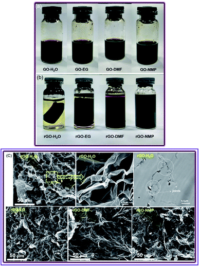
Four solvents, NMP, DMF, EG and water, were chosen to prepare rGO gels and discuss the interaction in the gels. Firstly, these four solvents could achieve long-term stability and similar initial suspension of GO as shown in Fig. 1(a) while other solvents could not as shown in Fig. S1.† Furthermore, the ability of the four solvents to form hydrogen bond is different according to “Hansen solubility parameters” (Table 1).33| Solvent | Water | EG | DMF | NMP |
| δH | 42.3 | 26.0 | 11.3 | 7.2 |
According to the value of hydrogen bonding cohesion parameter (δH), water shows the highest hydrogen bonding ability with the residual oxygen-containing groups on rGO sheets after the reduction of GO by VC, while NMP shows the lowest among these four solvents. The ability to form hydrogen bonds with the oxygen-containing functional groups on rGO sheets for EG and DMF is between that of water and NMP. As a consequence, the hydrogen bonding effect in water–rGO system shows the highest strength while the hydrogen bonding effect in NMP–rGO shows the lowest strength. Different degree of structural stability and mechanical strength in the as formed rGO gels were obtained in these four solvents due to the different degree of hydrogen bonding effect in Fig. 1(b). The hydrogel network of rGO–H2O hydrogel is strong enough to form a solid-like cylinder suspended in solution. Although a hydrogen bond network also formed in rGO–EG, the strength of the gel is not very high and the cylinder can be easily broken. In contrast, the rGO sheets in rGO–DMF and rGO–NMP didn't form any stable gel network. The photos of samples indicate the formation of some network related to hydrogen bonding.
Similarly, the cross-linked network of rGO–H2O hydrogel can be directly seen in microstructure of gels by scanning electron microscope (SEM) and transmission electron microscopy (TEM) images (Fig. 1(c)) and more SEM images of four gels are shown in ESI (Fig. S2†). As for rGO–H2O, a porous network of rGO–H2O hydrogel could be seen clearly. The porous network was formed from the overlapping of graphene sheets which was shown in the amplification of image of rGO–H2O. Then rGO–H2O sample was ultramicrotomed perpendicular to the graphite sheets, so we can see the joints between layers in TEM images as well. Compared with rGO–H2O, the porous structure of rGO–EG was not formed completely and the gel network was not tight as that of rGO–H2O. However, there is no obvious porous structure in rGO–DMF and rGO–NMP gel. The SEM images of rGO–DMF and rGO–NMP show a smooth surface instead of a porous network structure indicating that the rGO gel network was not formed in DMF and NMP. The results of SEM and TEM images for these different gels are consistent with the photos of four as-prepared gels. These results indicate that rGO sheets can form a stronger 3D gel network in solvents which show higher hydrogen bonding ability.
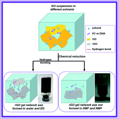
The construction scheme of rGO gel can be understood as shown in Scheme 1. The self-assembly process is driven by the hydrogen bond between solvents and the residual oxygen-containing groups of rGO which can be confirmed by XPS measurements (Table 2) and dehydroascorbic acid (DHA), the oxidation product of VC.34 At the beginning of the self-assembly of rGO gels, GO sheets were suspended uniformly in different solvents by hydrophilic interaction.35 After the addition of VC to GO suspension, the reduction of GO occurred. The sp3 hybridization C–C bond was reduced to sp2 hybridization C![[double bond, length as m-dash]](https://www.rsc.org/images/entities/char_e001.gif) C bond. The spatial structures of C
C bond. The spatial structures of C![[double bond, length as m-dash]](https://www.rsc.org/images/entities/char_e001.gif) C sp2 hybridization structure which were π–π conjugated are flat. The large-area flat structure of rGO sheets were crimped and overlapped by hydrogen bonds between residual oxygen-containing groups of rGO with water and DHA. So an integral self-assembly network of rGO–H2O hydrogel was formed successfully. As for rGO–EG, the gel network of that was constructed by hydrogen bond between hydroxyl of EG with residual oxygen-containing groups of rGO, but the strength of rGO–EG gel network is lower than that of rGO–H2O because the δH value of EG is lower than H2O. In contrast, the rGO gel networks in DMF and NMP hardly formed because the ability of DMF and NMP to form hydrogen bond with the residual oxygen-containing groups of rGO is too weak to obtain gel networks. The flexible rGO sheets stacked spontaneously and formed wrinkled rGO aggregates in DMF and NMP as shown in Fig. 1(c). Therefore, the properties and microstructures of these gels further confirm that the hydrogen bonding is an important factor during the formation of rGO gels.
C sp2 hybridization structure which were π–π conjugated are flat. The large-area flat structure of rGO sheets were crimped and overlapped by hydrogen bonds between residual oxygen-containing groups of rGO with water and DHA. So an integral self-assembly network of rGO–H2O hydrogel was formed successfully. As for rGO–EG, the gel network of that was constructed by hydrogen bond between hydroxyl of EG with residual oxygen-containing groups of rGO, but the strength of rGO–EG gel network is lower than that of rGO–H2O because the δH value of EG is lower than H2O. In contrast, the rGO gel networks in DMF and NMP hardly formed because the ability of DMF and NMP to form hydrogen bond with the residual oxygen-containing groups of rGO is too weak to obtain gel networks. The flexible rGO sheets stacked spontaneously and formed wrinkled rGO aggregates in DMF and NMP as shown in Fig. 1(c). Therefore, the properties and microstructures of these gels further confirm that the hydrogen bonding is an important factor during the formation of rGO gels.
 | ||
| Scheme 1 The formation of self-assembled 3D network for rGO gels during the reduction of GO by VC in various solvents. | ||
| Sample | Pristine G | GO | rGO–H2O | rGO–EG | rGO–DMF | rGO–NMP |
| C/O | 119 | 2.4 | 4.0 | 4.0 | 3.5 | 4.3 |
In order to confirm the existence of residual oxygen-containing groups and eliminate the effect of reduction degree of GO on the gel stability, the reduction degree of GO for the four gels was characterized by X-ray photoelectron spectroscopy (XPS, Table 2 and Fig. S3†) and Fourier-transform infrared spectroscopy (FTIR, Fig. S4 in ESI†), Raman (Fig. S5 and Table S1 in ESI†). The samples were freeze-dried before characterizations. After freeze-drying, the rGO–H2O and rGO–EG aerogel become brittle because of the removal of solvents wrapped in rGO sheets, which destroyed the integrity of the hydrogen bond network. The phenomenon also indicates that hydrogen bonding is the crucial interaction during the self-assembly of rGO gels.
Table 2 shows the C/O ratio value of pristine G, GO and the as-prepared rGO gels reduced in different solvents. The C/O ratio values of all the four rGO gels are similar and increase markedly after the reduction of GO. It indicates an efficient deoxidization of GO and the reduction degree of rGO gels in different solvents are nearly the same. So the formation of a strong gel network in water and EG is not determined by a higher reduction degree of GO but rather the ability to form hydrogen bond between solvents and the oxygen-containing functional groups on rGO sheets. Otherwise, the C/O ratio values of rGO aerogels are far lower than that of pristine G. It demonstrates that there were a number of residual oxygen-containing groups on rGO sheets which is confirmed by C1s XPS spectra (Fig. S4†) as well. So hydrogen bonds between the residual oxygen-containing groups of rGO with solvents and DHA are not only existent but also the key to achieve a more integrate gel network and higher-performance gels.
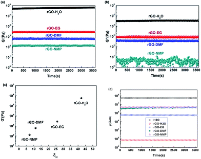
The mechanical strength and structural stability of the gels could further confirm the different degree of gel network of as-prepared gels in different solvent. In Fig. 2(a) and (b), the mechanical strength of the four rGO gels with time was tested by dynamic time sweep. The value of G′ (storage modulus) for rGO–H2O hydrogel is almost three orders of magnitude higher than the second high G′ value of rGO–EG gel, indicating that the rGO–H2O hydrogel network is the strongest among the four gels. And the weaker network of rGO–DMF and rGO–NMP gels than rGO–H2O and rGO–EG resulted in lower G′ value of that as expected. The values of G′ for the four gels were higher than that of the G′′ (loss modulus), also indicating the formation of gel network.36,37 The correlation between the hydrogen bonding cohesion parameters and the structural stability and mechanical strength of different gels (in Fig. 2(c)) revealed that hydrogen bonding actuate the self-assembly of rGO gels.
A stable self-assembled network of rGO gels will lead to a stable electric conductive pathway,38 so the electrical resistances of the gels were also measured during time sweep as shown in Fig. 2(d). The value of electrical resistance of rGO–H2O is 105 times lower than that of water, indicating that the hydrogel network of rGO–H2O provided a conductive pathway for electron even the pure water will impede the transportation of electron. In contrast, the electrical resistance of rGO–EG, rGO–DMF and rGO–NMP gels are much higher than that of rGO–H2O hydrogel. The electrical conductivity of rGO–DMF and rGO–NMP are similar and lower than that of rGO–EG. It is revealed that the gel network of rGO–DMF and rGO–NMP is less integral than that of rGO–EG gel and rGO–H2O hydrogel. The results of electrical resistance of the gels also indicate that the higher ability to form hydrogen bond of solvents, such as EG and water, the more integral network of rGO gels for electron conduction.
Conclusions
In summary, for the first time, we demonstrated that hydrogen bonding plays an important role in generating high-performance rGO gels in detail. rGO gels were prepared in different solvents (water, EG, NMP and DMF) with different ability to form hydrogen bond. According to the hydrogen bonding cohesion parameters of these four solvents, rGO sheets in water and EG can construct a much more integral gel network than that in DMF and NMP. The microstructure of rGO aerogel and the macroscopic properties of the four rGO gels are consistent with the hydrogen bonding cohesion parameters of that as expected. In particular, rGO–H2O hydrogel shows the highest mechanical strength and lowest electrical resistance while rGO–NMP gel performs the lowest mechanical strength and highest electrical resistance. The results of our study demonstrate hydrogen bonding is the key factor controlling the formation of rGO gel, which will be significant for the fabrication of high-performance rGO gels.Acknowledgements
This work was supported by the National Natural Science Foundation of China (NNSFC Grants 51422305 and 51421061), Sichuan Provincial Science Fund for Distinguished Young Scholars (2015JQO003), the Innovation Team Program of Science & Technology Department of Sichuan Province (Grant 2014TD0002) and State Key Laboratory of Polymer Materials Engineering (Grant No. sklpme 2014-2-02). We also thank the support of China Scholarship Council.Notes and references
- K. S. Novoselov, A. K. Geim, S. V. Morozov, D. Jiang, Y. Zhang, S. V. Dubonos, I. V. Grigorieva and A. A. Firsov, Science, 2004, 306, 666–669 CrossRef CAS PubMed.
- K. S. Novoselov, A. K. Geim, S. V. Morozov, D. Jiang, M. I. Katsnelson, I. V. Grigorieva, S. V. Dubonos and A. A. Firsov, Nature, 2005, 438, 197–200 CrossRef CAS PubMed.
- P. Kundu, E. A. Anumol and N. Ravishankar, Nanoscale, 2013, 5, 5215–5224 RSC.
- S. Chen, J. Duan, Y. Tang and S. Z. Qiao, Chem.–Eur. J., 2013, 19, 7118–7124 CrossRef CAS PubMed.
- J. Liu, W. Lv, W. Wei, C. Zhang, Z. Li, B. Li, F. Kang and Q.-H. Yang, J. Mater. Chem. A, 2014, 2, 3031 CAS.
- X. Zhang, Z. Sui, B. Xu, S. Yue, Y. Luo, W. Zhan and B. Liu, J. Mater. Chem., 2011, 21, 6494–6497 RSC.
- H.-P. Cong, X.-C. Ren, P. Wang and S.-H. Yu, ACS Nano, 2012, 6, 2693–2703 CrossRef CAS PubMed.
- F. Wang, B. Liu, A. C. F. Ip and J. Liu, Adv. Mater., 2013, 25, 4087–4092 CrossRef CAS PubMed.
- E. A. Appel, J. del Barrio, X. J. Loh and O. A. Scherman, Chem. Soc. Rev., 2012, 41, 6195–6214 RSC.
- P. M. Sudeep, T. N. Narayanan, A. Ganesan, M. M. Shaijumon, H. Yang, S. Ozden, P. K. Patra, M. Pasquali, R. Vajtai, S. Ganguli, A. K. Roy, M. R. Anantharaman and P. M. Ajayan, ACS Nano, 2013, 7, 7034–7040 CrossRef CAS PubMed.
- X. Y. Xie, K. W. Hu, D. D. Fang, L. H. Shang, S. D. Tran and M. Cerruti, Nanoscale, 2015, 7, 7992–8002 RSC.
- H. N. Lim, N. M. Huang, S. S. Lim, I. Harrison and C. H. Chia, Int. J. Nanomed., 2011, 6, 1817–1823 CrossRef CAS PubMed.
- H. Sun, Z. Xu and C. Gao, Adv. Mater., 2013, 25, 2554–2560 CrossRef CAS PubMed.
- Y. Xu, Q. Wu, Y. Sun, H. Bai and G. Shi, ACS Nano, 2010, 4, 7358–7362 CrossRef CAS PubMed.
- Y. Wu, N. Yi, L. Huang, T. Zhang, S. Fang, H. Chang, N. Li, J. Oh, J. A. Lee, M. Kozlov, A. C. Chipara, H. Terrones, P. Xiao, G. Long, Y. Huang, F. Zhang, L. Zhang, X. Lepró, C. Haines, M. D. Lima, N. P. Lopez, L. P. Rajukumar, A. L. Elias, S. Feng, S. J. Kim, N. T. Narayanan, P. M. Ajayan, M. Terrones, A. Aliev, P. Chu, Z. Zhang, R. H. Baughman and Y. Chen, Nat. Commun., 2015, 6, 6041 CrossRef PubMed.
- X. Yang, L. Zhang, F. Zhang, T. Zhang, Y. Huang and Y. Chen, Carbon, 2014, 72, 381–386 CrossRef CAS.
- H. Gao, Y. Sun, J. Zhou, R. Xu and H. Duan, ACS Appl. Mater. Interfaces, 2012, 5, 425–432 Search PubMed.
- C. Li and G. Shi, Adv. Mater., 2014, 26, 3992–4012 CrossRef CAS PubMed.
- Z. Zhao, X. Wang, J. Qiu, J. Lin, D. Xu, C. a. Zhang, M. Lv and X. Yang, Rev. Adv. Mater. Sci., 2014, 36, 137–151 Search PubMed.
- Y. Xu, K. Sheng, C. Li and G. Shi, ACS Nano, 2010, 4, 4324–4330 CrossRef CAS PubMed.
- C. Hou, Q. Zhang, Y. Li and H. Wang, J. Hazard. Mater., 2012, 205, 229–235 CrossRef PubMed.
- J. N. Tiwari, K. Mahesh, N. H. Le, K. C. Kemp, R. Timilsina, R. N. Tiwari and K. S. Kim, Carbon, 2013, 56, 173–182 CrossRef CAS.
- L. Zhang and G. Shi, J. Phys. Chem. C, 2011, 115, 17206–17212 CAS.
- L. Li, Z. Hu, Y. Yang, P. Liang, A. Lu, H. Xu, Y. Hu and H. Wu, Chin. J. Chem., 2013, 31, 1290–1298 CrossRef CAS.
- Z. Sui, X. Zhang, Y. Lei and Y. Luo, Carbon, 2011, 49, 4314–4321 CrossRef CAS.
- N. V. Medhekar, A. Ramasubramaniam, R. S. Ruoff and V. B. Shenoy, ACS Nano, 2010, 4, 2300–2306 CrossRef CAS PubMed.
- X. Jiang, Y. Ma, J. Li, Q. Fan and W. Huang, J. Phys. Chem. C, 2010, 114, 22462–22465 CAS.
- R. Du, J. Wu, L. Chen, H. Huang, X. Zhang and J. Zhang, Small, 2014, 10, 1387–1393 CrossRef CAS PubMed.
- Y. Liu, G.-Q. Qi, C.-L. Liang, R.-Y. Bao, W. Yang, B.-H. Xie and M.-B. Yang, J. Mater. Chem. C, 2014, 2, 3846–3854 RSC.
- Y. Liu, C.-L. Liang, R.-Y. Bao, G.-Q. Qi, W. Yang, B.-H. Xie and M.-B. Yang, RSC Adv., 2014, 5, 10–15 RSC.
- K.-x. Sheng, Y.-x. Xu, C. Li and G.-q. Shi, New Res. Carbon Mater., 2011, 26, 9–15 CrossRef CAS.
- Z. Li, J. Shen, H. Ma, X. Lu, M. Shi, N. Li and M. Ye, Soft Matter, 2012, 8, 3139–3145 RSC.
- C. M. Hansen, Hansen Solubility Parameters: A Users Handbook, CRC Press, 2007 Search PubMed.
- I. Saguy, I. J. Kopelman and S. Mizrahl, J. Food Process Eng., 1978, 2, 213–225 CrossRef CAS.
- J. I. Paredes, S. Villar-Rodil, A. Martinez-Alonso and J. M. D. Tascon, Langmuir, 2008, 24, 10560–10564 CrossRef CAS PubMed.
- F. Delbecq, H. Endo, F. Kono, A. Kikuchi and T. Kawai, Polymer, 2013, 54, 1064–1071 CrossRef CAS.
- S. K. Samanta, A. Pal, S. Bhattacharya and C. N. R. Rao, J. Mater. Chem., 2010, 20, 6881–6890 RSC.
- X. Yang, F. Zhang, L. Zhang, T. Zhang, Y. Huang and Y. Chen, Adv. Funct. Mater., 2013, 23, 3353–3360 CrossRef CAS.
Footnote |
| † Electronic supplementary information (ESI) available: More SEM images of gels prepared in different solvent, XPS, FT-IR and Raman analysis of the reduction degree of GO. See DOI: 10.1039/c6ra02942f |
| This journal is © The Royal Society of Chemistry 2016 |


