DOI:
10.1039/C6RA02932A
(Communication)
RSC Adv., 2016,
6, 54087-54091
Dopamine carbon nanodots as effective photothermal agents for cancer therapy
Received
1st February 2016
, Accepted 23rd May 2016
First published on 24th May 2016
Abstract
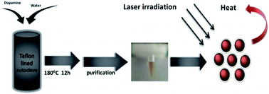
Dopamine carbon nanodots (DA CNDs) with an average diameter of approximately 23 nm were prepared through a facile hydrothermal method without adding any passivating agents. The as-prepared DA CNDs have high photothermal conversion efficiency (35%), excellent photostability and thermal stability. More importantly, DA CNDs exhibit significant photothermal therapeutic effects toward human cervical cancer (HeLa) cells under laser irradiation, while no appreciable dark cytotoxicity was observed even in high concentrations of DA CNDs aqueous solution. These results indicate that DA CNDs possess great potential to be effective photothermal agents for cancer therapy.
Introduction
Photothermal therapy (PTT) has attracted great attention for its minimal invasiveness, low toxicity to normal tissues and high specificity to tumor tissue in cancer therapy,1–7 when compared with other cancer therapy approaches like surgery, radiotherapy, and chemotherapy.8–14 An outstanding photothermal agent should not only have the characteristics of high extinction coefficient, photothermal conversion efficiency, good biocompatibility and clinical safety, but also can be applied through a minimally invasive way to target cancerous cells, without damaging surrounding healthy tissue.15–19
Nowadays, photothermal agents have been explored from diverse nanomaterials like graphene photothermal materials,6,20–26 black phosphorus,1 copper sulfide nanoparticles,27–29 gold nanoparticle30,31 and polymer PTT materials such as polypyrrole (PPy) nanomaterials,32–35 polyaniline.36,37 Although these agents hold the character of high photothermal conversion efficiency, but the drawbacks of poor photostability and complicated synthesis hinder their further applications.38
Carbon nanodots (CNDs) are emerging carbonaceous materials that have attracted enormous attention due to their prominent optical, chemical, electronic properties, and good biocompatibility, which have wide applications in catalysts,39,40 bioimaging,41 ion detection,42 and antitumor therapy.10 Until now, various CNDs with different photoluminescent properties and sizes were synthesised through top-down or bottom-up approaches. The top-down methods refer to break down bulk carbon materials into nanoparticles by laser ablation, acid oxidation, electrochemical oxidation and hydrothermal process,43–46 while bottom-up methods that fabricate unique structure precursor from small molecules to build up CNDs by thermal pyrolysis, microwave irradiation, and so on.47,48 Recently, Ge et al. used polythiophene derivatives as organic precursors for preparing CNDs with the capacity of PTT.49–51 However, in order to synthesize these special precursors a complicated and time-consuming reaction is indispensable. Thus, it is urgent to develop a simple method to CNDs-based PTT agent.
In this work, dopamine hydrochloride, a naturally occurring catecholamine was used as carbon source to synthesize dopamine carbon nanodots (DA CNDs) via a convenient and facile hydrothermal process. DA CNDs are monodispersed spherical nanoparticles with the average diameter of 23 nm. A series of experiments were conducted to evaluate photothermal capability of DA CNDs, the results demonstrate that DA CNDs possess excellent photostability, thermal stability and high photothermal conversion efficiency, which are promising PTT agents for cancer therapy.
Results and discussion
Synthesis and characterization of dopamine carbon nanodots
We synthesized DA CNDs through a facile hydrothermal process, as illustrated in Scheme 1. Dopamine hydrochloride was dissolved in distilled water, stirred for 5 minutes, then transferred into Teflon-lined autoclave and the solution was heated to 180 °C and kept for 12 h. After naturally cooled to room temperature, the crude product was centrifuged at 5000 rpm for 10 min to remove the precipitated particles, then the supernatant was subjected to dialysis (cutoff Mn: 3.0 kDa) for 2 d, the final product was freeze-dried to get brown powder. Fig. 1 shows the transmission electron microscopy (TEM) image of DA CNDs, the result indicate that the DA CNDs are well monodispersed in aqueous solution with an average diameter of approximately 23 nm (based on statistical analysis of the height of about one hundred nanoparticles).
 |
| | Scheme 1 Schematic diagram of the synthesis and photothermal conversion of DA CNDs. | |
 |
| | Fig. 1 TEM image of DA CNDs. Inset: size distribution of DA CNDs. | |
The UV-vis-NIR absorption and photoluminescence (PL) spectra of DA CNDs are shown in Fig. 2. The UV-vis-NIR absorption spectrum (Fig. 2a) illustrates that DA CNDs have a broad range of optical absorption (from ultraviolet to near infrared) without characteristic peaks, which endow them with the ability of converting NIR irradiation to heat. Weak emission is observed in the PL spectra of DA CNDs (Fig. 2b), the emission band is centred at 450 nm.
 |
| | Fig. 2 (a) UV-vis-NIR absorption spectrum of DA CNDs (b) PL spectrum of DA CNDs (λex = 320 nm). Inserts are photos of DA CNDs solution taken under natural light and UV irradiation. | |
X-ray photoelectron spectra (XPS) were used to analyse the element components of DA CNDs. The full scan XPS spectrum of DA CNDs (Fig. 3a) shows two primary peaks at 284.67 eV and 532 eV, which correspond to C1s and O1s, respectively. It can be presumed that high degree carbonization of DA CNDs contributes to the breakage of dopamine aliphatic chain, which terminated with amino-group, leaving the surface of the carbon cores at high temperatures. So we deduce that the high degree carbonization of dopamine could be the main reason that make DA CNDs convert photo energy to heat, that is to say the carbonization state of precursor largely influences the applicability of the formed materials, without any doping or passive agents.52,53
 |
| | Fig. 3 (a) The full scan XPS spectrum of DA CNDs, (b) high-resolution C1s XPS spectrum, (c) high-resolution O1s XPS spectrum and (d) FTIR spectrum of DA CNDs in the solid state. | |
The high resolution XPS spectrum of C1s (Fig. 3b) show three peaks at 284.64 eV, 286.12 eV, 288.65 eV, which are attributed to C
C, CO and C–OH, respectively. The O1s spectrum (Fig. 3c) indicate two peaks at 532.00 eV and 533.65 eV, corresponding to C
O and C–OH. Fourier transform infrared (FTIR) spectrum was applied to analyze the functional groups on the surface of DA CNDs. As illustrated in Fig. 3d, the stretching vibration of 3207.60 cm−1 belongs to –OH, while the C–OH and C
O bending vibrations locate at 1605.26 cm−1, 1289.47 cm−1. The surface components of the DA CNDs determined by the FT-IR results are in consistent with XPS. Meanwhile, the zeta-potential of DA CNDs is determined to be −20.4 mV, further confirms the existence of negatively charged groups on the surface of DA CNDs.
Photothermal evaluation
To further study the photothermal capability of DA CNDs, series of experiments were conducted. The temperature change of 0.3 mL DA CNDs solutions with different concentrations was monitored as a function of time and NIR laser irradiation power. As we can see in Fig. 4a, an obvious concentration-dependent increase of temperature was observed, at the same time, solution temperature increased significantly when increase the output power gradually at the concentration of 50 μg mL−1 (Fig. 4b). While deionized water as a control show little increase of temperature under the same irradiation condition.
 |
| | Fig. 4 (a) DA CNDs photothermal heating curves under 808 nm laser irradiation at various concentrations (1.5 W cm−2). (b) At the concentration of 50 μg mL−1 aqueous solution irradiated at different laser power. (c) DA CNDs under irradiation for 5 min then naturally cooling to room temperature after laser off at different laser power. (d) Photothermal cycle curve of DA CNDs under laser irradiation for 5 min (808 nm, 1.5 W cm−2). | |
A quantitative investigation of the photothermal performance and reusability of DA CNDs was carried out. Temperature vs. time curves were recorded for DA CNDs upon irradiation for 5 min following by natural cooling to room temperature as shown in Fig. 4c. The photothermal conversion efficiency (η value) was 35%, which was higher than that of Au nanorods (22%) and previously reported thiadiazole PTT agents (32%).54 The heating and cooling cycles were repeated more than 20 times, and the temperature elevation of each cycle was more than 30 °C, no obvious deterioration was observed (Fig. 4d). These results indicate the DA CNDs possess good reproducibility and high photothermal conversion efficiency.
Cytotoxicities
Photothermal capability is an essential standard for PTT agent to be applied in biomedicine. Therefore, PTT cytotoxicity of DA CNDs are investigated in detail. The standard methyl thiazolyl tetrazolium (MTT) assay toward HeLa cells was conducted.
After incubating with different concentrations (10–50 μg mL−1) of DA CNDs solution for 5 h, the HeLa cells were exposed to an NIR laser (808 nm, 1.5 W cm−2) for 5 min, after illumination, the cells were incubated for another 24 h. The MTT assays shown that a majority of the cells were ablated (Fig. 5), only 13% of cells remain alive at a concentration of 50 μg mL−1. In contrast, cells in the absence of laser irradiation exhibit high viability. These results demonstrate DA CNDs hold great promise as a PTT agent for cancer therapy.
 |
| | Fig. 5 Relative cell viabilities of HeLa cells incubated with different concentrations of DA CNDs in the absence or presence of irradiation. All the results were repeated three times and presented as mean ± SD (808 nm, 1.5 W cm−2, 5 min). | |
Experimental
Chemicals
Dopamine hydrochloride (99% purity) was purchased from Acros Organic. Solvents for chemical synthesis were purified by distillation. Ultrapure water was prepared from a Milli-Q system (Millipore, USA).
Spectroscopic and microscopic measurements
Ultraviolet-visible-NIR (UV-vis-NIR) absorption spectra were conducted on Shimadzu UV-2450 PC UV-vis spectrophotometer. Fluorescence emission spectra were recorded on a LS-55 fluorophotometer. TEM images were recorded with a FEI-TECNAI G2 transmission electron microscope operating at 200 kV. In the preparation of the specimen for TEM, a drop of DA CNDs solution was deposited onto a copper grid with a carbon coating. The specimen was air-dried and measured at room temperature. Fourier transform infrared (FTIR) spectra were recorded on a Bruker Vertex 70 spectrometer. X-ray photoelectron spectra (XPS) were obtained on a Thermo Scientific ESCALAB 250 Multitechnique Surface Analysis. Zeta potential and size distribution are obtained by a Malvern Zeta-sizer nano dynamic light scattering (DLS).
The synthesis and purification of dopamine carbon nanodots (DA CNDs)
DA CNDs were prepared through a facile one-pot hydrothermal process of dopamine aqueous solution as illustrated in Scheme 1, dopamine hydrochloride (100 mg) was added into 50 mL Teflon lined autoclave containing 30 mL distilled water, and the solution was sonicated for 5 min at room temperature then heated at 180 °C for 12 h. The reaction solution was centrifugated at 5000 rpm for 10 min in order to remove large particles, then the supernate was transferred into a dialysis bag (cutoff Mn: 3.0 kDa) and dialyzed against water for 2 d to remove small molecules. The water was replaced every 6 h, at last freeze-drying the DA CNDs solution for further use. The yield was calculated to be ca. 2.8%.
Cell culture, cell ablation study
Hela cells harvested in a logarithmic growth phase were seeded in 96-well plates at a density of 105 cells per well and incubated in DMEM (Dulbecco's modified Eagle's medium) for 24 h at 37 °C in a humidified 5% CO2 atmosphere, the culture medium was changed once every day. In the cell ablation study, the seeded cells were incubated with various concentration of DA CNDs (0, 10, 15, 20, 30, 40 and 50 μg mL−1) for 4 h and followed by an NIR laser irradiation (808 nm, 1.5 W cm−2) for 5 min, after illumination, the cells were incubated for another 24 h. The dark control group was performed under the same experimental procedures except for laser irradiation. Then 20 μL of MTT solution in PBS with the concentration of 5 mg mL−1 was added and the plates were incubated for another 4 h at 37 °C, followed by removal of the culture medium containing MTT and addition of 150 μL of DMSO to each well to dissolve the formed formazan crystals. Finally, the plates were shaken for 10 min, and the absorbance of formazan product was measured at 490 nm by a microplate reader.
Conclusions
In summary, an effective PTT agent (DA CNDs) were prepared from dopamine through a facile one-pot way. The as-synthesized DA CNDs are well dispersed in aqueous solution, which exhibit high photothermal conversion efficiency and can efficiently kill tumour cells under the laser irradiation at 808 nm. This work demonstrates that DA CNDs have considerable potential to be an effective photothermal agent for cancer treatment.
Acknowledgements
Financial support was provided by the Jilin Province Science and Technology Research Project (No. 20160101292JC), the Research Project of Science and Technology of the Education Department of Jilin Province During the 13th Five-year Plan Period (No. 2016322).
Notes and references
- Z. Sun, H. Xie, S. Tang, X.-F. Yu, Z. Guo, J. Shao, H. Zhang, H. Huang, H. Wang and P. K. Chu, Angew. Chem., 2015, 127, 11688–11692 CrossRef.
- J. Geng, C. Sun, J. Liu, L. D. Liao, Y. Yuan, N. Thakor, J. Wang and B. Liu, Small, 2015, 11, 1603–1610 CrossRef CAS PubMed.
- P. C. Ray, S. A. Khan, A. K. Singh, D. Senapati and Z. Fan, Chem. Soc. Rev., 2012, 41, 3193–3209 RSC.
- L. Dykman and N. Khlebtsov, Chem. Soc. Rev., 2012, 41, 2256–2282 RSC.
- K. Mebrouk, F. Chotard, C. Le Goff-Gaillard, Y. Arlot-Bonnemains, M. Fourmigué and F. Camerel, Chem. Commun., 2015, 51, 5268–5270 RSC.
- J. T. Robinson, S. M. Tabakman, Y. Liang, H. Wang, H. Sanchez Casalongue, D. Vinh and H. Dai, J. Am. Chem. Soc., 2011, 133, 6825–6831 CrossRef CAS PubMed.
- H. Liu, D. Chen, L. Li, T. Liu, L. Tan, X. Wu and F. Tang, Angew. Chem., 2011, 123, 921–925 CrossRef.
- M. Zheng, S. Liu, X. Guan and Z. Xie, ACS Appl. Mater. Interfaces, 2015, 7, 22181–22187 CAS.
- W. Wang, S. Liu, Y. Huang, X. Jing and Z. Xie, J. Mater. Chem. B, 2015, 3, 5753–5759 RSC.
- M. Zheng, S. Liu, J. Li, D. Qu, H. Zhao, X. Guan, X. Hu, Z. Xie, X. Jing and Z. Sun, Adv. Mater., 2014, 26, 3554–3560 CrossRef CAS PubMed.
- X. Hu, J. Li, W. Lin, Y. Huang, X. Jing and Z. Xie, RSC Adv., 2014, 4, 38405 RSC.
- W. Lin, T. Sun, M. Zheng, Z. Xie, Y. Huang and X. Jing, RSC Adv., 2014, 4, 25114 RSC.
- Q. Pei, X. Hu, Z. Li, Z. Xie and X. Jing, RSC Adv., 2015, 5, 81499–81501 RSC.
- T. Sun, X. Guan, M. Zheng, X. Jing and Z. Xie, ACS Med. Chem. Lett., 2015, 6, 430–433 CrossRef CAS PubMed.
- C. Nolsøe, S. Torp-Pedersen, F. Burcharth, T. Horn, S. Pedersen, N. Christensen, E. Olldag, P. H. Andersen, S. Karstrup and T. Lorentzen, Radiology, 1993, 187, 333–337 CrossRef PubMed.
- A. Vogel and V. Venugopalan, Chem. Rev., 2003, 103, 577–644 CrossRef CAS PubMed.
- A. C. Balazs, T. Emrick and T. P. Russell, Science, 2006, 314, 1107–1110 CrossRef CAS PubMed.
- A. M. Gobin, M. H. Lee, N. J. Halas, W. D. James, R. A. Drezek and J. L. West, Nano Lett., 2007, 7, 1929–1934 CrossRef CAS PubMed.
- C. M. Hessel, V. P. Pattani, M. Rasch, M. G. Panthani, B. Koo, J. W. Tunnell and B. A. Korgel, Nano Lett., 2011, 11, 2560–2566 CrossRef CAS PubMed.
- H. Hong, K. Yang, Y. Zhang, J. W. Engle, L. Feng, Y. Yang, T. R. Nayak, S. Goel, J. Bean and C. P. Theuer, ACS Nano, 2012, 6, 2361–2370 CrossRef CAS PubMed.
- H. Moon, D. Kumar, H. Kim, C. Sim, J.-H. Chang, J.-M. Kim, H. Kim and D.-K. Lim, ACS Nano, 2015, 9, 2711–2719 CrossRef CAS PubMed.
- C. Li, S. Bolisetty, K. Chaitanya, J. Adamcik and R. Mezzenga, Adv. Mater., 2013, 25, 1010–1015 CrossRef CAS PubMed.
- K. Yang, L. Feng, X. Shi and Z. Liu, Chem. Soc. Rev., 2013, 42, 530–547 RSC.
- K. Yang, S. Zhang, G. Zhang, X. Sun, S. T. Lee and Z. Liu, Nano Lett., 2010, 10, 3318–3323 CrossRef CAS PubMed.
- S. Shi, K. Yang, H. Hong, F. Chen, H. F. Valdovinos, S. Goel, T. E. Barnhart, Z. Liu and W. Cai, Biomaterials, 2015, 39, 39–46 CrossRef CAS PubMed.
- X. Tu, L. Wang, Y. Cao, Y. Ma, H. Shen, M. Zhang and Z. Zhang, Carbon, 2016, 97, 35–44 CrossRef CAS.
- Q. Tian, F. Jiang, R. Zou, Q. Liu, Z. Chen, M. Zhu, S. Yang, J. Wang, J. Wang and J. Hu, ACS Nano, 2011, 5, 9761–9771 CrossRef CAS PubMed.
- Q. Tian, M. Tang, Y. Sun, R. Zou, Z. Chen, M. Zhu, S. Yang, J. Wang, J. Wang and J. Hu, Adv. Mater., 2011, 23, 3542–3547 CrossRef CAS PubMed.
- K. Dong, Z. Liu, Z. Li, J. Ren and X. Qu, Adv. Mater., 2013, 25, 4452–4458 CrossRef CAS PubMed.
- Z. Wang, Z. Chen, Z. Liu, P. Shi, K. Dong, E. Ju, J. Ren and X. Qu, Biomaterials, 2014, 35, 9678–9688 CrossRef CAS PubMed.
- E. Ju, K. Dong, Z. Liu, F. Pu, J. Ren and X. Qu, Adv. Funct. Mater., 2015, 25, 1574–1580 CrossRef CAS.
- J. Y. Hong, H. Yoon and J. Jang, Small, 2010, 6, 679–686 CrossRef CAS PubMed.
- W.-K. Oh, H. Yoon and J. Jang, Biomaterials, 2010, 31, 1342–1348 CrossRef CAS PubMed.
- P. M. George, A. W. Lyckman, D. A. LaVan, A. Hegde, Y. Leung, R. Avasare, C. Testa, P. M. Alexander, R. Langer and M. Sur, Biomaterials, 2005, 26, 3511–3519 CrossRef CAS PubMed.
- J. M. Fonner, L. Forciniti, H. Nguyen, J. D. Byrne, Y.-F. Kou, J. Syeda-Nawaz and C. E. Schmidt, Biomed. Mater., 2008, 3, 034124 CrossRef PubMed.
- J. Yang, J. Choi, D. Bang, E. Kim, E. K. Lim, H. Park, J. S. Suh, K. Lee, K. H. Yoo and E. K. Kim, Angew. Chem., 2011, 123, 461–464 CrossRef.
- J. Zhou, Z. Lu, X. Zhu, X. Wang, Y. Liao, Z. Ma and F. Li, Biomaterials, 2013, 34, 9584–9592 CrossRef CAS PubMed.
- Z. Sheng, D. Hu, M. Xue, M. He, P. Gong and L. Cai, Nano-Micro Lett., 2013, 5, 145–150 CrossRef.
- D. Qu, M. Zheng, P. Du, Y. Zhou, L. Zhang, D. Li, H. Tan, Z. Zhao, Z. Xie and Z. Sun, Nanoscale, 2013, 5, 12272–12277 RSC.
- J. Liu, S. Z. Qiao, H. Liu, J. Chen, A. Orpe, D. Zhao and G. Q. Lu, Angew. Chem., Int. Ed., 2011, 50, 5947–5951 CrossRef CAS PubMed.
- M. Zheng, S. Ruan, S. Liu, T. Sun, D. Qu, H. Zhao, Z. Xie, H. Gao, X. Jing and Z. Sun, ACS Nano, 2015, 9, 11455–11461 CrossRef CAS PubMed.
- M. Zheng, Z. Xie, D. Qu, D. Li, P. Du, X. Jing and Z. Sun, ACS Appl. Mater. Interfaces, 2013, 5, 13242–13247 CAS.
- D. Pan, J. Zhang, Z. Li and M. Wu, Adv. Mater., 2010, 22, 734–738 CrossRef CAS PubMed.
- J. Peng, W. Gao, B. K. Gupta, Z. Liu, R. Romero-Aburto, L. Ge, L. Song, L. B. Alemany, X. Zhan and G. Gao, Nano Lett., 2012, 12, 844–849 CrossRef CAS PubMed.
- H. Tian, M. Saunders, A. Dodd, K. O'Donnell, M. Jaroniec, S. Liu and J. Liu, J. Mater. Chem. A, 2016, 4, 3721–3727 CAS.
- J. Liu, N. P. Wickramaratne, S. Z. Qiao and M. Jaroniec, Nat. Mater., 2015, 14, 763–774 CrossRef CAS PubMed.
- S. Zhu, Q. Meng, L. Wang, J. Zhang, Y. Song, H. Jin, K. Zhang, H. Sun, H. Wang and B. Yang, Angew. Chem., 2013, 125, 4045–4049 CrossRef.
- L. Tang, R. Ji, X. Cao, J. Lin, H. Jiang, X. Li, K. S. Teng, C. M. Luk, S. Zeng and J. Hao, ACS Nano, 2012, 6, 5102–5110 CrossRef CAS PubMed.
- L. Guo, J. Ge, W. Liu, G. Niu, Q. Jia, H. Wang and P. Wang, Nanoscale, 2015, 8, 729–734 RSC.
- J. Ge, M. Lan, B. Zhou, W. Liu, L. Guo, H. Wang, Q. Jia, G. Niu, X. Huang, H. Zhou, X. Meng, P. Wang, C. S. Lee, W. Zhang and X. Han, Nat. Commun., 2014, 5, 4596 CAS.
- J. Ge, Q. Jia, W. Liu, L. Guo, Q. Liu, M. Lan, H. Zhang, X. Meng and P. Wang, Adv. Mater., 2015, 27, 4169–4177 CrossRef CAS PubMed.
- Y. Song, S. Zhu, S. Zhang, Y. Fu, L. Wang, X. Zhao and B. Yang, J. Mater. Chem. C, 2015, 3, 5976–5984 RSC.
- S. N. Baker and G. A. Baker, Angew. Chem., 2010, 49, 6726–6744 CrossRef CAS PubMed.
- T. Sun, J. Qi, M. Zheng, Z. Xie, Z. Wang and X. Jing, Colloids Surf., B, 2015, 136, 201–206 CrossRef CAS PubMed.
|
| This journal is © The Royal Society of Chemistry 2016 |
Click here to see how this site uses Cookies. View our privacy policy here.