Towards the understanding of non-thermal air plasma action: effects on bacteria and fibroblasts

Oleg Lunov*a, Vitalii Zablotskiia, Olexander Churpitaa, Ales Jägera, Leoš Polívkaa, Eva Sykováb, Natalia Terebovac, Andrei Kulikovc, Šárka Kubinováab and Alexandr Dejnekaa
aInstitute of Physics of the Czech Academy of Sciences, Prague, Czech Republic. E-mail: lunov@fzu.cz
bInstitute of Experimental Medicine AS CR, Prague, Czech Republic
cSt Petersburg State University of Information Technologies, Mechanics and Optics, St Petersburg, Russia

Received 26th January 2016 , Accepted 27th February 2016

First published on 2nd March 2016


Abstract

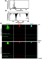
Non-thermal plasma research has put a growing focus on the bacteria inactivation problem. In this article we show how low temperature atmospheric plasma destroys Gram-positive and Gram-negative bacteria and discuss the mechanisms of plasma bactericidal effects and a discrepancy in the plasma-triggered effects and ozone (which is a component of air plasma gases). The proven safety of air plasma for fibroblasts is a key factor for the medical applications of plasma.


The last decade was marked by a large number of studies on the various biomedical applications of non-thermal (cold) atmospheric pressure plasmas.1 This burst of research activity on non-thermal plasma demonstrated the rapid evolution and great potential of this new interdisciplinary field.1–3 Primarily, the deactivation of microorganisms by non-thermal plasma attracted the most attention in the scientific community.4 Indeed, microorganism resistance represents, nowadays, an unprecedentedly rising problem.5,6 As eradication of multiresistant so-called ‘superbugs’ has become a clinical challenge,7 non-thermal plasmas provide an alternative approach for effectively killing multidrug resistant (MDR) strains.3,4,8 For biomedical applications non-thermal plasma (with a gas temperature less than 40 °C in the plasma jet) is generated under atmospheric pressure using different working gases, which are accompanied by reactive molecules, ions and radicals. Thus, plasma composition is very complex and represents a mixture of charged particles, electronically excited atoms and molecules (including ozone – O3), radicals, and UV irradiation due to ion recombination.9,10 In fact, it is possible to relatively easily control and vary plasma density and composition, producing distinct active chemical species within the plasma itself.11 Non-thermal plasmas can be applied on the skin without any harmful effects.12,13 Moreover, non-thermal plasmas have shown great potential and hold out great promise for eliminating bacterial colonization and facilitating chronic wound healing in several clinical trials.14–16

Taken together, the recent studies show that the bactericidal effects of non-thermal plasmas are undisputed.3 However, there is still a lack of knowledge on cellular targets as well as the molecular mechanisms of plasma action. Generally, the accumulation of reactive oxygen (ROS) and nitrogen (RNS) species has been implicated to explain the underlying biological effects of non-thermal plasma.17–19 Indeed, non-thermal plasma has been shown to generate many kinds of ROS and RNS.20,21 In our previous works we showed that depending on the exposure time, helium plasma induces either direct physical destruction of bacteria or triggers programmed cell death via accumulation of intra-bacterial ROS.11,22 In this work we focused our research on the bactericidal effects of air plasma. Interestingly, non-thermal air plasma has been shown to produce significant amounts of ozone,9,10 which is known to have very aggressive effects on cells.23,24

Therefore, the aim of our study was to investigate and compare the effects of chemically different non-thermal plasmas and ozone on the survival and functionality of different bacterial strains.

We used a designed discharge plasma system working at atmospheric pressure to treat bacteria. Detailed characteristics of the system have been previously published.22,25 The plasma system we used enables us to easily change the chemical composition of the plasma jet by applying distinct gases (air, helium, nitrogen, etc.). Previously, we have shown that helium plasma possess more than 90% antimicrobial efficacy.11,22 Interestingly, recent studies have shown that the addition of oxygen to helium plasma results in a much higher bacteria killing rate.3,4,8 Several studies revealed that reactive species (such as O, O2, O3, OH˙, NO, and NO2) might be significant contributors in the plasma sterilization processes.20,26 These findings led to a presumption that in terms of bacterial inactivation air plasma should be superior to helium.3 Moreover, researchers believe that the presence of chemically reactive species (such as O and O3) in air plasma results in a more pronounced bactericidal effect.3,4,8 Thus, to distinguish the bactericidal effects of the plasma and ozone, we exploited air plasma and ozone flow with the same concentration of O3 (for details see ESI) as that generated by the plasma generator, 1.67 mg l−1 (for this concentration of O3 we use the terms “ozone flow” or “ozone” throughout, unless specified otherwise). It is worth mentioning here that similar concentrations of ozone in air plasma were detected in other studies.27–29 First of all, we used optical emission spectrum analysis to validate the plasma composition in terms of ions and radicals generated by the system (Fig. 1a), which can mediate the cellular effects of plasma. Optical emission spectroscopy (OES) revealed excited nitrogen molecules (N2) and atomic oxygen (O) at the spectrum (Fig. 1a). Fourier transform infrared spectroscopy (FT-IR) gave a comprehensive overview of the composition of the air plasma analysed in the plasma jet (Fig. 1b). Besides CO2 other species can be conveniently determined, including ozone and various nitrogen oxides (Fig. 1b). Taken together, the OES and FT-IR spectroscopy results show the chemical composition of air plasma to be highly complex.


image file: c6ra02368a-f1.tif
Fig. 1 Physicochemical characterization of air plasma. (a) Optical emission spectrum of air plasma. (b) FT-IR spectra of air plasma. (c) Results of SRIM simulations: in-depth profiles of He (green panel), N (red panel) and O (blue panel) ions penetration through either Gram-positive or Gram-negative bacterial wall models. The total number of ions is 10[thin space (1/6-em)]000.

To gain some insight into the physical mechanisms of plasma ion action we simulated and analysed the distributions of retained ions in two bacterial models: Gram-positive and Gram-negative. The SRIM simulations30 showed different ion distribution patterns for distinct ions (Fig. 1c and ESI Fig. S1). Indeed, during the plasma irradiation helium (He) ions possess a higher degree of penetration into the bacterial wall over oxygen (O) and nitrogen (N) (Fig. 1c). Furthermore, the thick peptidoglycan layer of the Gram-positive cell wall led to the accumulation of all types of ions in the peptidoglycan layer (Fig. 1c and ESI Fig. S1). At the same time the thin peptidoglycan layer of the Gram-negative wall allowed for the accumulation of penetrated He-ions at the membrane/peptidoglycan interface (ESI Fig. S1).

Further, we tested the antimicrobial efficacy of air plasma irradiation in comparison with the ozone flow on two types of bacteria (S. aureus and P. aeruginosa). These bacterial strains are well established models of antibiotic resistant Gram-positive and Gram-negative bacteria in microbiology and antibiotic research.6 A representative illustration of the effect of plasma irradiation and ozone treatment on S. aureus colony survival is presented on Fig. 2a. Both air plasma and ozone showed profound statistically significant antibacterial effects, where the bactericidal efficacy depended on the treatment time (Fig. 2b). Considering the interaction of plasma with bacteria, it is important to know how plasma changes microenvironment properties (in our case agar medium). In literature, there is evidence that non-thermal plasma can affect solid and liquid substrates, chemically modifying them.17,31,32 However, for agar medium it has been shown that non-thermal plasma has practically no effect on its biocompatibility properties.33


image file: c6ra02368a-f2.tif
Fig. 2 Inactivation of different bacterial strains by air plasma and ozone. (a) Representative images of plasma and ozone treatment of bacterial colonies of S. aureus. (b) Survival rate of P. aeruginosa and S. aureus after treatment for indicated periods of time with air plasma or ozone. The data were normalized to control values (no plasma exposure), which were set to 100% viability, mean ± SEM, n = 3, *p < 0.05 **p < 0.01.

The bacterial inactivation of both strains P. aeruginosa and S. aureus by air plasma and ozone was indistinguishable (Fig. 2b), having nearly 99.99% and more than 80% antimicrobial efficacy after 60 s exposure respectively. However, Gram-negative bacteria (S. aureus) were more plasma and ozone susceptible than Gram-positive (P. aeruginosa) bacteria (Fig. 2b). The observed differences in plasma bactericidal efficacy in Gram-negative and Gram-positive bacteria might be related to the cell wall structure. Previously, we showed how differences in the structure of bacterial cell walls between Gram-negative and Gram-positive strains results in different bactericidal efficacies upon helium plasma irradiation.22 It is widely accepted that the Gram-positive cell wall is thicker than the Gram-negative wall.34,35 Moreover, Gram-positive bacteria are more mechanically rigid than Gram-negative bacteria.36,37 Furthermore, the structure of the Gram-negative wall allows ions to penetrate deeper in comparison with Gram-positive cell walls (Fig. 1b and ESI Fig. S1). We and others have previously demonstrated that non-thermal helium plasma can induce a very significant rupture of the outer membrane38–40 that leads to complete destruction of the bacteria.11,22 Furthermore, there is evidence in the literature that air plasma might have bacteria destructive capabilities.3,4,8 However, a detailed analysis of air plasma induced bacterial damage and solid comparison with the effect of ozone flows on different strains has not yet been performed. Thus, we utilized scanning electron microscopy (SEM) analysis to reveal changes in bacteria morphology induced by air plasma irradiation and ozone treatment. In this case we extended the number of bacterial strains to validate the effects of air plasma and ozone: Gram-negative bacteria P. aeruginosa and E. coli; Gram-positive S. aureus and B. subtilis. Indeed, SEM analysis showed distinct morphological changes upon air plasma and ozone treatment (Fig. 3). Irradiation of all bacterial strains with air plasma and treatment with the ozone flow resulted in significant destruction of bacteria depending on exposure time (Fig. 3 and ESI Fig. S2 and S3). Longer exposure time led to more profound damage of all four bacteria strains. Interestingly, ozone treatment resulted in more profound morphological changes induced in bacteria in comparison to plasma. As one can see from Fig. 3b and ESI Fig. S3 noticeable changes after ozone treatment occur already at 15 s exposure. After 15 s of ozone treatment the onset of damage of all the bacteria strains is clearly seen from Fig. 3b (second column, damage is shown by arrows) and ESI Fig. S3 (second column, damage is shown by arrows). This damage is more pronounced after 30 s of exposure (Fig. 3b and ESI Fig. S3, third column, accordingly). In contrast, air plasma treatment leads to lesser damage for the same time of exposure (Fig. 3a and ESI Fig. S2, second and third columns, accordingly). For the longest exposure time (60 s) the damage effects of ozone and air plasma are comparable (Fig. 3 and ESI Fig. S2 and S3). Thus, these results imply that ozone and air plasma might have different mechanisms of action on bacteria.


image file: c6ra02368a-f3.tif
Fig. 3 SEM analysis of morphological changes of different bacterial strains after treatments with air plasma (a) and ozone (b). Damage is shown by arrows.

Previously, we showed that chemically distinct plasmas trigger different responses in mammalian cells.25 Additionally, we analysed in detail helium plasma effects on bacteria.11,22 Our previous results show that helium plasma at high doses destroy bacteria predominantly by electrostatic disruption.22 In fact, 60 s exposure of helium plasma is able to completely destroy bacteria22 (representative example is given on Fig. 4a). One should note here, that the morphological changes triggered by helium plasma are distinct from those observed after air plasma or ozone exposure (Fig. 3 and 4a). Moreover, a careful look at Fig. 4a reveals the lengthwise crack of the bacteria wall, which is a typical signature of wall damage due to internal pressure (in our case the electrostatic repulsion of accumulated charges plays the role of internal pressure22). Taking these findings together one can conclude that the chemically distinct plasmas (using air and helium as working gas) trigger different responses in bacteria.


image file: c6ra02368a-f4.tif
Fig. 4 (a) SEM analysis of morphological changes of different bacterial strains after helium plasma treatment. (b) Scanning electron micrographs of untreated (control) and B. subtilis endospores after treatment for indicated periods of time with either air and helium plasma or ozone.

Bacterial endospores represent a well-known example of extraordinary resistance properties.41 Endospores survive in nutrient-free and harsh environments, they are resistant to ultraviolet (UV) radiation, chemicals (such as peroxide and hypochlorite), extreme heat and other stresses.41,42 B. subtilis endospores represent a best-studied and widely accepted model system of endospores.42 A very good demonstration of the morphological differences between B. subtilis and its endospore is shown in ESI Fig. S4. Therefore, we studied how air plasma and ozone affect these extremely resistant strains. We found no significant impact of neither the air or helium plasmas nor ozone on B. subtilis endospores treated for 15, 30, 60 s (Fig. 4b). One should mention here, that others were successful in effective eradication of endospores by non-thermal air plasma.43–45 These discrepancies could be explained due to the fact that in the abovementioned studies high voltage plasma was used, 10 kV and more,43–45 while in our case we utilized a 0.5 kV plasma generator. Moreover, we showed previously that plasma-induced biological effects on cells dramatically depend on the value of the voltage producing the plasma discharges.22

It is of great importance to take into account the adverse effects of plasma treatment for better controlled applications in clinical practice. In this regard, utilizing the plasma system, we firstly exposed in vitro cell cultures of 3T3 fibroblasts to the plasma and ozone with the same dose and exposure time as was used to inactivate bacteria (Fig. 5a and b). Analysis of SEM images revealed no significant changes in cellular morphology of 3T3 fibroblasts after air plasma treatment (Fig. 5a). Furthermore, cytotoxicity analysis showed no toxic effect of plasma exhibited on in vitro cell cultures immediately after exposure (Fig. 5b). Contrary to that, ozone appeared to induce higher instances of cell death (Fig. 5b). Even short term ozone exposure triggered cell death, whereas air plasma was unable to induce any signs of cytotoxicity (Fig. 5b). A number of previous studies marked the generation of ROS/RNS in living cells triggered by non-thermal plasma as a basic reason for induced biological responses.17,18,22,25,46 Of note, it has been shown repeatedly that intracellular accumulation of high levels of ROS may induce damage to cellular structures and may finally lead to cell death.47 Thus, we evaluated ROS/RNS generation by air plasma and helium plasma in comparison with ozone utilizing the ROS-sensitive fluorescent assay (cellular ROS/superoxide detection assay kit). Indeed, ozone induced a significantly higher production of total intracellular ROS/RNS than air and helium plasmas after 15 s exposure (Fig. 5c). Moreover, both plasmas and ozone induced a time-dependent ROS production in fibroblasts (Fig. 5c). These results are in line with the data on cell viability and cell surface morphology (Fig. 5a and b). Of note, the same plasma doses lead to quite different effects in fibroblasts and bacteria. The former survive, while bacteria die. We suppose that the difference in mechanical rigidities and sizes of fibroblasts and bacteria wall structures may play a key role in their sensitivities and responses to the plasma. In general, mammalian cells are mechanically less rigid than bacteria.48 We previously showed that under plasma treatment mechanically rigid bacterial wall structures could be destroyed due to internal electrostatic pressure raised due to accumulated ions.11 In contrast, softer and more flexible fibroblasts are able to adopt themselves to the local changes of electric environment resulting from plasma treatment.


image file: c6ra02368a-f5.tif
Fig. 5 (a) Scanning electron micrographs of untreated (control) and air plasma treated 3T3 fibroblasts. (b) Cell viability as detected by the WST-1 assay of 3T3 fibroblasts treated with either air and helium plasma or ozone for indicated time periods. The data were normalized to control values (no plasma or ozone exposure), which were set as 100% cell viability, mean ± SD, n = 4, *p < 0.05 **p < 0.01. (c) Time-dependent ROS/RNS induction by air, helium plasmas and ozone. 3T3 fibroblasts were exposed to air, helium plasmas or ozone for 15 s, followed by ROS measuring using the cellular ROS/superoxide detection kit (Abcam). The data are presented as mean percentage of ROS positive cells mean ± SEM, n = 4, *p < 0.05 **p < 0.01. (d) Wound contraction upon air plasma treatment. (e) Representative trichrome staining of the skin wounds after repeated plasma treatments for 3 days.

It is worth mentioning here that ozone is a strong oxidizing agent and is able to initiate intracellular oxidative stress through ozonide and hydroperoxide formation.23,49 Indeed, ozone is extremely reactive and may result in lipid peroxidation, oxidative stress and DNA damage.23 Eventually, acute exposure to ozone is known to trigger clinical symptoms of asthma and to induce toxic responses in vivo.50 Nevertheless, in air plasma the ozone action is accompanied by ions and other reactive species. This leads to an interesting synergetic effect: ions and reactive species of air plasma inhibit ozone action allowing cell survival (Fig. 5b).

Further, we performed a histological analysis of the plasma-treated skin of living rats and assessed how air plasma would affect the wound healing process. All experiments were performed in accordance with the European Communities Council Directive of 24 November 1986 (86/609/EEC), regarding the use of animals in research and were approved by the Ethics Committee of the Institute of Experimental Medicine, Academy of Sciences Czech Republic, Prague, Czech Republic. The histological analysis revealed no pathological changes on the skin structure after 2 min of plasma treatment in comparison with control animals (Fig. 5d and e). Furthermore, trichrome staining and wound contraction analysis suggested that air plasma treatment has a positive supportive effect on sterile wound healing (Fig. 5d and e). In the last decade studies have shown that ozone treatment (like other drugs) of chronic skin wounds can display either damaging or beneficial effects depending on exposure time (for a review see ref. 51). One should add here, that ozone has been shown to induce antioxidant depletion as well as oxidation of lipids and proteins within the skin.52,53 Exposure to ozone may lead to deleterious effects in the skin.54,55

Conclusions

In conclusion, our results showed that air plasma has high efficacy performance in the deactivation of P. aeruginosa, E. coli, S. aureus and B. subtilis bacterial cultures. Furthermore, the same dose of plasma did not damage 3T3 fibroblasts in vitro and showed no adverse effects on rat skin in vivo (Fig. 5). Since air plasma contains ozone, we have compared interactions of the air plasma and ozone flow with bacteria and 3T3 fibroblasts. The ozone exposure showed very toxic effects (Fig. 5b and c), while the air plasma with the same concentration of O3 was found to be safe for 3T3 fibroblasts under equal treatment times. Notwithstanding, ozone therapy is commonly used for diabetic leg ulcer treatment,56 recent studies show that ozone exposure leads to the accumulation of a considerable amount of skin irritants57 and may result in deleterious effects in the skin.54,55 As far as ROS production and cellular viability, effects differ between plasma and ozone (Fig. 5b and c). Taken together, these results suggest that in air plasma ozone plays an important role in ROS induction and sequent inactivation of bacteria. Nevertheless, depending on plasma exposure time and irradiation dose the physical and biological mechanisms of bacteria inactivation have the highest performance. Therefore, the general view that most air plasma healing and bactericidal outcomes are the result of an ozone component should be reconsidered. Without a doubt, for the clinical application of plasma more intensive study of the underlying molecular intracellular mechanisms of plasma action on tissue cells and bacteria is required.

Acknowledgements

The authors acknowledge the J.E. Purkyne fellowship awarded by the Academy of Sciences of the Czech Republic, MEYS LO1309. Two authors (A.J. and L.P.) appreciate financial support offered by MEYS LM2011026.

Notes and references

  1. M. Laroussi, Plasma Processes Polym., 2014, 11, 1138 CrossRef CAS.
  2. M. G. Kong, G. Kroesen, G. Morfill, T. Nosenko, T. Shimizu, J. V. Dijk and J. L. Zimmermann, New J. Phys., 2009, 11, 115012 CrossRef.
  3. N. De Geyter and R. Morent, Annu. Rev. Biomed. Eng., 2012, 14, 255 CrossRef CAS PubMed.
  4. V. Scholtz, J. Pazlarova, H. Souskova, J. Khun and J. Julak, Biotechnol. Adv., 2015, 33, 1108 CrossRef CAS PubMed.
  5. S. B. Levy and B. Marshall, Nat. Med., 2004, 10, S122 CrossRef CAS PubMed.
  6. D. I. Andersson and D. Hughes, Nat. Rev. Microbiol., 2010, 8, 260 CAS.
  7. C. A. Arias and B. E. Murray, N. Engl. J. Med., 2009, 360, 439 CrossRef CAS PubMed.
  8. J. Ehlbeck, U. Schnabel, M. Polak, J. Winter, T. von Woedtke, R. Brandenburg, T. von dem Hagen and K. D. Weltmann, J. Phys. D: Appl. Phys., 2011, 44, 013002 CrossRef.
  9. A. Fridman, A. Chirokov and A. Gutsol, J. Phys. D: Appl. Phys., 2005, 38, R1 CrossRef CAS.
  10. W. Tian and M. J. Kushner, J. Phys. D: Appl. Phys., 2014, 47, 165201 CrossRef.
  11. O. Lunov, O. Churpita, V. Zablotskii, I. G. Deyneka, I. K. Meshkovskii, A. Jäger, E. Syková, Š. Kubinová and A. Dejneka, Appl. Phys. Lett., 2015, 106, 053703 CrossRef.
  12. T. Nosenko, T. Shimizu and G. E. Morfill, New J. Phys., 2009, 11, 115013 CrossRef.
  13. V. Zablotskii, O. Churpita, Z. Hubicka, L. Jastrabik and A. Dejneka, Plasma Med., 2011, 1, 135 CrossRef.
  14. G. Isbary, G. Morfill, H. U. Schmidt, M. Georgi, K. Ramrath, J. Heinlin, S. Karrer, M. Landthaler, T. Shimizu, B. Steffes, W. Bunk, R. Monetti, J. L. Zimmermann, R. Pompl and W. Stolz, Br. J. Dermatol., 2010, 163, 78 CAS.
  15. C. Ulrich, F. Kluschke, A. Patzelt, S. Vandersee, V. A. Czaika, H. Richter, A. Bob, J. v. Hutten, C. Painsi, R. Hüge, A. Kramer, O. Assadian, J. Lademann and B. Lange-Asschenfeldt, J. Wound Care, 2015, 24, 196 CrossRef CAS PubMed.
  16. G. Isbary, J. Heinlin, T. Shimizu, J. L. Zimmermann, G. Morfill, H. U. Schmidt, R. Monetti, B. Steffes, W. Bunk, Y. Li, T. Klaempfl, S. Karrer, M. Landthaler and W. Stolz, Br. J. Dermatol., 2012, 167, 404 CrossRef CAS PubMed.
  17. S. Kalghatgi, C. M. Kelly, E. Cerchar, B. Torabi, O. Alekseev, A. Fridman, G. Friedman and J. Azizkhan-Clifford, PLoS One, 2011, 6, e16270 CAS.
  18. H. J. Ahn, K. I. Kim, G. Kim, E. Moon, S. S. Yang and J. S. Lee, PLoS One, 2011, 6, e28154 CAS.
  19. H. J. Ahn, K. I. Kim, N. N. Hoan, C. H. Kim, E. Moon, K. S. Choi, S. S. Yang and J. S. Lee, PLoS One, 2014, 9, e86173 Search PubMed.
  20. M. Laroussi, Plasma Processes Polym., 2005, 2, 391 CrossRef CAS.
  21. F. X. Liu, P. Sun, N. Bai, Y. Tian, H. X. Zhou, S. C. Wei, Y. H. Zhou, J. Zhang, W. D. Zhu, K. Becker and J. Fang, Plasma Processes Polym., 2010, 7, 231 CrossRef CAS.
  22. O. Lunov, V. Zablotskii, O. Churpita, A. Jäger, L. Polívka, E. Syková, A. Dejneka and Š. Kubinová, Biomaterials, 2016, 82, 71 CrossRef CAS PubMed.
  23. B. Kosmider, J. E. Loader, R. C. Murphy and R. J. Mason, Free Radical Biol. Med., 2010, 48, 1513 CrossRef CAS PubMed.
  24. D. Klestadt, P. Laval-Gilly and J. Falla, Cell Biol. Toxicol., 2002, 18, 259 CrossRef CAS PubMed.
  25. O. Lunov, V. Zablotskii, O. Churpita, E. Chanova, E. Sykova, A. Dejneka and S. Kubinova, Sci. Rep., 2014, 4, 7129 CrossRef CAS PubMed.
  26. M. Laroussi, IEEE Trans. Plasma Sci., 2002, 30, 1409 CrossRef CAS.
  27. D. Gerrity, B. D. Stanford, R. A. Trenholm and S. A. Snyder, Water Res., 2010, 44, 493 CrossRef CAS PubMed.
  28. A. M. Anpilov, E. M. Barkhudarov, Y. B. Bark, Y. V. Zadiraka, M. Christofi, Y. N. Kozlov, I. A. Kossyi, V. A. Kop'ev, V. P. Silakov, M. I. Taktakishvili and S. M. Temchin, J. Phys. D: Appl. Phys., 2001, 34, 993 CrossRef CAS.
  29. S. Kalghatgi, A. Fridman, J. Azizkhan-Clifford and G. Friedman, Plasma Processes Polym., 2012, 9, 726 CrossRef CAS.
  30. J. F. Ziegler, M. D. Ziegler and J. P. Biersack, Nucl. Instrum. Methods Phys. Res., Sect. B, 2010, 268, 1818 CrossRef CAS.
  31. A. Popelka, I. Novak, M. Lehocky, I. Chodak, J. Sedliacik, M. Gajtanska, M. Sedliacikova, A. Vesel, I. Junkar, A. Kleinova, M. Spirkova and F. Bilek, Molecules, 2012, 17, 762 CrossRef CAS PubMed.
  32. K. Duske, L. Jablonowski, I. Koban, R. Matthes, B. Holtfreter, A. Sckell, J. B. Nebe, T. von Woedtke, K. D. Weltmann and T. Kocher, Biomaterials, 2015, 52, 327 CrossRef CAS PubMed.
  33. I. V. Timoshkin, M. Maclean, M. P. Wilson, M. J. Given, S. J. MacGregor, T. Wang and J. G. Anderson, IEEE Trans. Plasma Sci., 2012, 40, 2322 CrossRef CAS.
  34. S. Lebeer, J. Vanderleyden and S. C. J. De Keersmaecker, Nat. Rev. Microbiol., 2010, 8, 171 CrossRef CAS PubMed.
  35. M. T. Cabeen and C. Jacobs-Wagner, Nat. Rev. Microbiol., 2005, 3, 601 CrossRef CAS PubMed.
  36. V. Vadillo-Rodriguez and J. R. Dutcher, Soft Matter, 2011, 7, 4101 RSC.
  37. K. C. Huang, R. Mukhopadhyay, B. N. Wen, Z. Gitai and N. S. Wingreen, Proc. Natl. Acad. Sci. U. S. A., 2008, 105, 19282 CrossRef CAS PubMed.
  38. D. A. Mendis, M. Rosenberg and F. Azam, IEEE Trans. Plasma Sci., 2000, 28, 1304 CrossRef.
  39. M. Laroussi, D. A. Mendis and M. Rosenberg, New J. Phys., 2003, 5, 41 CrossRef.
  40. M. Laroussi, J. P. Richardson and F. C. Dobbs, Appl. Phys. Lett., 2002, 81, 772 CrossRef CAS.
  41. P. T. McKenney, A. Driks and P. Eichenberger, Nat. Rev. Microbiol., 2013, 11, 33 CrossRef CAS PubMed.
  42. J. Errington, Nat. Rev. Microbiol., 2003, 1, 117 CrossRef CAS PubMed.
  43. T. G. Klampfl, G. Isbary, T. Shimizu, Y. F. Li, J. L. Zimmermann, W. Stolz, J. Schlegel, G. E. Morfill and H. U. Schmidt, Appl. Environ. Microbiol., 2012, 78, 5077 CrossRef PubMed.
  44. S. Patil, T. Moiseev, N. N. Misra, P. J. Cullen, J. P. Mosnier, K. M. Keener and P. Bourke, J. Hosp. Infect., 2014, 88, 162 CrossRef CAS PubMed.
  45. Y. F. Hong, J. G. Kang, H. Y. Lee, H. S. Uhm, E. Moon and Y. H. Park, Lett. Appl. Microbiol., 2009, 48, 33 CrossRef CAS PubMed.
  46. H. M. Joh, S. J. Kim, T. H. Chung and S. H. Leem, Appl. Phys. Lett., 2012, 101, 053703 CrossRef.
  47. V. Temkin and M. Karin, Immunol. Rev., 2007, 220, 8 CrossRef CAS PubMed.
  48. J. Lee, A. A. Abdeen and K. A. Kilian, Sci. Rep., 2014, 4, 5188 CAS.
  49. S. S. Nadadur, D. L. Costa, R. Slade, R. Silbajoris and G. E. Hatch, Environ. Health Perspect., 2005, 113, 1717 CrossRef CAS PubMed.
  50. M. Pichavant, S. Goya, E. H. Meyer, R. A. Johnston, H. Y. Kim, P. Matangkasombut, M. Zhu, Y. Iwakura, P. B. Savage, R. H. DeKruyff, S. A. Shore and D. T. Umetsu, J. Exp. Med., 2008, 205, 385 CrossRef CAS PubMed.
  51. G. Valacchi, V. Fortino and V. Bocci, Br. J. Dermatol., 2005, 153, 1096 CrossRef CAS PubMed.
  52. J. J. Thiele, M. G. Traber, T. G. Polefka, C. E. Cross and L. Packer, J. Invest. Dermatol., 1997, 108, 753 CrossRef CAS PubMed.
  53. G. Valacchi, E. Pagnin, A. M. Corbacho, E. Olano, P. A. Davis, L. Packer and C. E. Cross, Free Radical Biol. Med., 2004, 36, 673 CrossRef CAS PubMed.
  54. G. Valacchi, A. van der Vliet, B. C. Schock, T. Okamoto, U. Obermuller-Jevic, C. E. Cross and L. Packer, Toxicology, 2002, 179, 163 CrossRef CAS PubMed.
  55. M. G. Mustafa, Free Radical Biol. Med., 1990, 9, 245 CrossRef CAS PubMed.
  56. A. M. Elvis and J. S. Ekta, J. Nat. Sci., Biol. Med., 2011, 2, 66 CrossRef CAS PubMed.
  57. A. Wisthaler and C. J. Weschler, Proc. Natl. Acad. Sci. U. S. A., 2010, 107, 6568 CrossRef CAS PubMed.

Footnote

Electronic supplementary information (ESI) available. See DOI: 10.1039/c6ra02368a

This journal is © The Royal Society of Chemistry 2016