DOI:
10.1039/C6RA02364A
(Paper)
RSC Adv., 2016,
6, 52528-52538
Construction of biocompatible regenerated cellulose/SPI composite beads using high-voltage electrostatic technique†
Received
26th January 2016
, Accepted 24th May 2016
First published on 25th May 2016
Abstract
A series of regenerated cellulose/soy protein isolate (SPI) beads (RCSB) were fabricated using an environmentally friendly high-voltage electrostatic technique with cellulose and SPI as the main raw materials and sulfuric acid aqueous solution as the coagulant. The structure and physical properties of RCSB were characterized by optical microscopy, scanning electron microscopy (SEM), Fourier transform infrared spectroscopy (FT-IR), X-ray diffraction (XRD), solid-state 13C NMR, water contact angle measurement, and thermogravimetric analysis (TGA). The resultant composite beads were round with nano-sized pores on the surface. The size of the dried beads ranged from 300 to 1500 μm and could be controlled by changing fabrication parameters such as the voltage used and the cellulose/SPI ratio. The FT-IR, XRD, 13C NMR, and TGA results revealed that strong hydrogen bond interactions formed between cellulose and SPI in the beads during the regeneration process, and that the composite beads had good thermal stability. The surface hydrophilicity of the beads decreased as SPI content increased. The cytotoxicity and biocompatibility of the RCSB were comparatively evaluated by 3-(4,5-dimethylthiazol-2-yl)-2, 5-diphenyltetrazolium bromide (MTT) assay and in vitro cell culture. The MTT assay indicated that RCSB extracts had good cytocompatibility for L929 cells. Furthermore, the cells adhered to the RCSB surface and exhibited high attachment efficiency, further confirming the cytocompatibility of the beads. This work provided a facile, environmentally friendly, and controllable method for the construction of biocompatible cellulose/SPI-based beads with potential application in biomaterials.
1. Introduction
Cellulose is the most abundant renewable natural polymer on the earth and it has received increasing attention all over the world due to its renewability and nontoxicity.1–4 Because of these unique properties, cellulose has been converted into films, fibers, sponges, and spherical particles or beads.5,6 In particular, cellulose-based beads have been widely used in many fields due to its high surface area and tunable functionality, although it has long been considered a niche product mainly due to its restriction of conventional dissolution and shaping processes.7,8 Compared with conventional solvents for cellulose dissolution such as DMSO/ammonium fluorides, DMAc/LiCl and N-methylmorpholineoxide,9–11 the use of relatively safe and economic solvents such as NaOH/urea, NaOH, and NaOH/ZnO aqueous solutions has greatly reduced the toxicity, volatility, and cost, leading to increased interest for environmental and economic requirements.12–15 Over the last decades, various methods for the preparation of beads or microspheres have been developed including emulsification, dispersion, and spray drying.16–18 However, these methods have disadvantages such as the lack of control over shape and size distribution, and some of they are environmentally hazardous.19 Electrostatic technique is based on the balance between an electrostatic force and the solution surface tension. It is one of the most convenient and continuous methods for the controllable production of small particles or fibers with uniform and desirable size, which is critical for the fabrication of microspheres or fibers in the biomedical field.20,21 Compared with dispersion and emulsification methods, the high-voltage electrostatic technique has reduced cost, amount of organic solvents required, and environmental pollution.19 Furthermore, the size of electrostatic droplets can range from several millimeters down to nanometers and the corresponding size distribution is usually narrow.22–24 Therefore, the high-voltage electrostatic technique shows a promise with respect to environmental, economic and safety concerns as compared to the current applied methods. As a result, the high-voltage electrostatic technique has been applied to fabricate microspheres or beads from cellulose,19 alginate,25 and chitin.21
Cellulose-based beads with different diameters ranging from micrometer to millimeter can be used in many advanced applications, particularly when they are physically or chemically modified, including metal ion exchange,26 chromatography fillers,27 protein immobilization,28 and drug carrier/release systems.29 Cellulose is accessible for chemical modification or physical blending because every glucose unit possesses three active hydroxyl groups.8 Functional cellulose beads for special purposes have been prepared by introducing chemical functionalities or blending with organic30 and inorganic31 compounds. Proteins, such as collagen32 and zein,33 have been used to modify the physical and biological properties of cellulose. Soy protein isolate (SPI) is a plant protein with great potential applications in biomedical fields because of its good biocompatibility and biodegradability.34,35 In our previous work, SPI-modified cellulose films showed improved cytocompatibility and in vivo biocompatibility.36 However, these cellulose/SPI films had no 3D-structure and lacked high specific surface area. The cells adhered only to the surface of the film, possibly due to limitations of the 2D structure. To address this challenge, we hypothesized that SPI-modified cellulose composite beads with a 3D structure would further promote the adherence and proliferation of cells on the surface due to a high specific surface area. As mentioned above, high-voltage electrostatic technique is a facile and environmentally friendly method for fabrication of some polymer beads, which led us to hypothesise that it may be an alternative way to prepare cellulose/SPI beads. Thus, in this work, cellulose was blended with SPI in NaOH/urea aqueous solutions and a high-voltage electrostatic technique was used to prepare regenerated cellulose/SPI beads (RCSB). The structure and physical properties of the RCSB were characterized by FT-IR, XRD, SEM, 13C NMR, and TGA. Moreover, the cytocompatibility of RCSB was preliminarily studied by cell culture and MTT assay to evaluate the potential application as biomaterials. It was expected that the particle size of RCSB could be controlled by adjusting the high voltage value and SPI content. At the same time, the beads were expected to have no or low cytotoxicity due to the lack use of emulsifiers and special organic solvents.
2. Experimental section
2.1. Materials
Cellulose was supplied by Hubei Chemical Fiber Manufacture (Xiangfan, China), and its viscosity average molecular weight (Mη) was determined by viscosimetry in cadoxen to be 1.0 × 10⁵. Soy protein isolate (SPI) was supplied by DuPont-Yunmeng Protein Co. Ltd. (Yunmeng, China). Cellulose and SPI were vacuum-dried at 60 °C for 24 h before use. Modified Eagle's Medium (MEM), fetal bovine serum (FBS) and (3-(4,5-dimethylthiazol-2-yl)-2,5-diphenyltetrazolium bromide) (MTT) were obtained from Invitrogen Corporation (Gibco BRL, Grand Island, NY, USA). Other chemicals used were analytical grade.
2.2. Preparation of regenerated cellulose/SPI beads
The regenerated cellulose/SPI beads were prepared as follows: briefly, a 7 wt% NaOH and 12 wt% urea aqueous solution used as the cellulose solvent was precooled to about −13 °C. Then cellulose was dissolved in this solvent with stirring within 5 min at ambient temperature to obtain 2.5% cellulose solution.37 SPI powders were swelled in water, and then dissolved in the above 7 wt% NaOH and 12 wt% urea aqueous solution to get a slurry with 10 wt% SPI. The cellulose solution and SPI slurry were mixed to obtain cellulose/SPI blend solutions. The cellulose/SPI solutions were stirred for 30 min and degassed at 4 °C by centrifugation for 10 min at 7500 × g.
The high-voltage electrostatic technique was applied to prepare cellulose/SPI beads as follows: the degassed cellulose/SPI solutions were dripped, using a syringe, into a 5 wt% sulfuric acid coagulant solution to form composite beads. The syringe with a 0.5 mm blunt-ended needle was pushed automatically to maintain a steady flow of cellulose/SPI solution at a rate of 0.2 mL min−1. The distance between the needle and the receiving coagulant solution was adjusted to 2 cm. The process for the fabrication of cellulose/SPI beads is shown in Scheme 1, which is modified from a schematic diagram of electrospinning.38 The resultant beads were washed with running water for 24 h, distilled water for 30 min, and then stored in distilled water at room temperature for further use. By adjusting the voltage between the needle and coagulated solution to 0, 3, and 5 kV, and by changing the SPI content (WSPI, weight percent of SPI based on the total weight of cellulose and SPI) to 0, 10, 30, and 50% in the blends, respectively, a series of regenerated cellulose/SPI composite beads was prepared using the high-voltage electrostatic technique, and coded as RCSB-v-n, where “v” and “n” correspond to the voltage value and WSPI in the composite beads, respectively. In this case, v = 0, 3, and 5 kV; n = 0, 10, 30, and 50, respectively. The codes and compositions of the beads are listed in Table 1.
 |
| | Scheme 1 Schematic diagram of the preparation process of RCSB-v-n (n = 0, 10, 30, and 50). (a) Syringe; (b) syringe pump; (c) high voltage power supply; (d) metal electrode (+); (e) metal electrode (−); (f) coagulation system. | |
Table 1 Code, composition, voltage, effects of high voltage, and WSPI on the diameter of the wet RCSB-v-n. The applied voltages are 0, 3, and 5 kV; the WSPI is 0, 10, 30, and 50 wt% (N = 50, mean ± SD)
| Sample code |
Voltage (kV) |
Wcellulosea (%) |
WSPIb (%) |
Diameter (μm) |
| Wcellulose = [weight of cellulose/(weight of cellulose + weight of SPI)] × 100. WSPI = [weight of SPI/(weight of cellulose + weight of SPI)] × 100. |
| RCSB-0-0 |
0 |
100 |
0 |
2640 ± 26 |
| RCSB-0-10 |
0 |
90 |
10 |
1971 ± 48 |
| RCSB-0-30 |
0 |
70 |
30 |
1815 ± 30 |
| RCSB-0-50 |
0 |
50 |
50 |
1780 ± 29 |
| RCSB-3-0 |
3 |
100 |
0 |
1868 ± 42 |
| RCSB-3-10 |
3 |
90 |
10 |
1481 ± 24 |
| RCSB-3-30 |
3 |
70 |
30 |
1276 ± 51 |
| RCSB-3-50 |
3 |
50 |
50 |
1006 ± 63 |
| RCSB-5-0 |
5 |
100 |
0 |
897 ± 40 |
| RCSB-5-10 |
5 |
90 |
10 |
808 ± 37 |
| RCSB-5-30 |
5 |
70 |
30 |
708 ± 51 |
| RCSB-5-50 |
5 |
50 |
50 |
656 ± 42 |
2.3. Characterization of the RCSB-v-n
Effects of voltage and SPI content on the size, size distribution, and microstructure of RCSB-v-n were observed by optical microscope and scanning electron microscope (SEM), the number of beads counted was more than 80. For optical microscope images, the wet beads were photographed using a camera, the number of beads counted was 50. For the SEM samples, the wet RCSB-v-n were critical point dried (CPD) by a critical point dryer using liquid CO2 under supercritical conditions. The dried beads were coated with gold and then observed by SEM with a 20 kV accelerating voltage.
The reduction in size of RCSB-v-n before and after drying by CPD represented the shrinkage factor and was calculated according to the equation:39
| | |
Shrinkage factor = (dw − dd)/dw
| (1) |
where
dw was the diameter of the wet RCSB calculated from optical microscope images, and
dd was the diameter of the CPD-dried beads calculated from SEM images.
FTIR spectra and X-ray diffraction patterns of the RCSB-v-n and raw materials were analysed according to general method.40 The details are given in the ESI.† Solid-state 13C NMR spectra of the beads and SPI powder were acquired on a BRUKER AVANCE III spectrometer with a 13C frequency of 75 MHz using the combined technique of magic angle spinning (MAS) and cross polarization. Conditions for 13C NMR measurement were as follows: the spinning speed was 5 kHz; the contact time was 3 ms; the acquisition time 50 ms, and the recycle delay 3 s. A typical number of 1024 scans were acquired for each spectrum.
Water contact angle of the beads was measured on an OCA20 contact angle meter by the droplet method. The RCSB-v-n were pressed into a disc and then fixed on a glass slide and dried under vacuum at 60 °C for 24 h. A 3 μL water droplet was placed on the surface of the disc-like samples and the water contact angle measured.41 Each sample was tested three times to calculate the average water contact angle.
TGA and DTG analyses of the beads were carried out on a TA-STDQ600. The dry beads were milled into powders and placed into a platinum pan with a heating rate of 10 °C min−1 and over a temperature range of 30 to 600 °C, and the nitrogen at a flow rate of 20 mL min−1 was used as the testing atmosphere.
Extracts from the RCSB-v-n were prepared according to ISO 10993-12:2007 as follows: the beads were steam sterilized and immersed in cell culture medium (Modified Eagle's Medium (MEM) 1640, 0.2 g of beads per 1 mL of MEM 1640) at 37 °C for 72 h. The extracts were obtained by filtering and then stored at 4 °C before use.
Cell viability of the beads was evaluated by MTT assay. According to ISO 10993-5:2007, a cell line of mouse lung fibroblasts (L929, provided by China Centre for Type Culture Collection, Wuhan University, Wuhan, China) was suspended in culture medium as cell suspension (100 μL per well). This cell suspension was added to 96-well plates at a density of 1 × 10³ cells per well. The plates were incubated at 37 °C in a humidified atmosphere of 5% CO2 for 24 h. Extracts from RCSB replaced the medium and a culture medium with similar cells was used as a control. After incubating for 24, 48 and 72 h, each sample was treated with 10 μL MTT for another 4 h at 37 °C in a humidified 5% CO2 atmosphere. Finally, the MTT solution, cell culture medium and RCSB extracts were removed and replaced with 150 μL dimethyl sulfoxide (DMSO) and placed for 10 min at room temperature. Absorbance values were read against a reagent blank (a DMSO reagent without L929 cells) at a test wavelength of 570 nm on a multiwell microplate reader.42 Results were expressed as a percentage of cell viability for treated samples (extracts from the beads) compared to the control over a specified time. Cell viability was calculated using the following equation:
| | |
Cell viability (%) = [(As − Ab)/(Ac − Ab)] × 100
| (2) |
where
As,
Ac, and
Ab represent the absorbance of samples, control, and blank, respectively.
To evaluate the potential applications for RCSB-v-n as biomaterials, L929 cells were cultured with sterilized RCSB-v-n (3 mg mL−1) in 24-well plastic culture plates at a density of 2 × 10⁵ cells per mL for 72 h. For SEM observations, the cultured cells with RCSB-v-n were washed with PBS and fixed for 24 h in 2.5% glutaraldehyde PBS solution. After the beads were rewashed with PBS for 3 times, they were progressively dehydrated in ethanol, then exchanged with isoamyl acetate for 20–80 min, and critical point dried (CPD).2 The morphologies of L929 cells cultured on the RCSB-v-n were observed by SEM with an accelerating voltage of 20 kV. The samples were coated with gold before SEM observation.
3. Results and discussion
3.1. Preparation process and appearances of the RCSB-v-n
As shown in Scheme 1, the basic components of the high-voltage electrostatic apparatus include a syringe, syringe pump, metal electrode, high-voltage power supply and coagulation system. The syringe was filled with cellulose/SPI composite solutions, which were fed through a needle into an electric field by a syringe pump. The solution could be introduced into the system at a precisely controlled rate by the syringe pump. The cellulose/SPI composite solutions moved from the syringe into a metallic needle, which was connected to a high-voltage power supply (0–10 kV). The high-voltage electrostatic field shaped the cellulose/SPI composite solution into small droplets and drove the droplets into a coagulation system to form a series of RCSB-v-n. In general, the emulsification method requires emulsifiers for the dispersion of beads and abundant organic solvents to remove the emulsifier from the beads.16 The electrostatic technique avoids the use of emulsifier and the accompanying organic solvent, so, compared to the emulsification method, it is environmentally friendly and convenient19 and suitable for the preparation of cellulose/SPI beads. Furthermore, the fabrication process of RCSB and all the process parameters could be well controlled by our designed electrostatic apparatus, which suggested that the way applied in this work showed clear advantages such as simplicity, repeatability and controllability.
The size and size distribution of the RCSB-v-n prepared using the electrostatic technique are usually affected by experimental parameters such as voltage, components and solution concentrations, receiver distance, and driving speed. In this work, the receiver distance and driving speed were fixed at 2 cm and 0.2 mL min−1, respectively, so the two variable parameters were the voltage used and the cellulose/SPI ratio. The voltages applied in this work were 0, 3, and 5 kV, and the WSPI in the cellulose/SPI solution was 0, 10, 30, and 50 wt%, respectively. By changing these parameters, a series of cellulose/SPI beads were prepared and coded as RCSB-v-n. The codes and corresponding parameters of the beads are listed in Table 1. The size and size distribution of RCSB-v-n, which were affected by WSPI and voltage value, could be observed and calculated from images of the beads. The optical microscope images of RCSB-v-n prepared at different voltages are shown in Fig. 1 and Table 1. The beads were spherical and of uniform size when the parameters were fixed. When WSPI was fixed (e.g. 10%), the diameters of the RCSB-v-10 were 1971 ± 48, 1481 ± 24, and 808 ± 37 μm, corresponding to the high voltage values (v) of 0, 3, and 5 kV, respectively. In this case, the size of RCSB-v-n decreased gradually with the increase of the applied voltage. Moreover, when the voltage was fixed (e.g. 3 kV), the diameters of RCSB-3-0, RCSB-3-10, RCSB-3-30, and RCSB-3-50 were 1868 ± 42, 1481 ± 24, 1276 ± 51, and 1006 ± 63 μm, corresponding to the WSPI (n) of 0, 10, 30, and 50%, respectively. The bead size decreased with an increase of WSPI. When no voltage was applied, large droplets formed on the nozzle, which then dripped into the coagulation bath to form large RCSB-0-n with diameters of 2640 ± 26 μm (RCSB-0-0), 1971 ± 48 μm (RCSB-0-10), 1815 ± 30 μm (RCSB-0-30) and 1780 ± 29 μm (RCSB-0-50), corresponding to the WSPI (n) of 0, 10, 30, and 50%, respectively. This indicated that RCSB-0-n could be obtained by gravity of the droplets without high voltage. Therefore, the size of the beads could be controlled by changing the voltage and WSPI. The size distribution for every WSPI at 3 kV was narrowest and tended to be normal. Meanwhile, a tailing phenomenon appeared for the RCSB-5-50. In the following experiments, except for cell culture, all the beads used for characterization and evaluation were RCSB-3-n.
 |
| | Fig. 1 Optical microscope images of wet RCSB-v-n constructed at different applied voltages (0, 3, and 5 kV) from cellulose/SPI solutions with different WSPI (0, 10, 30, and 50). Scale bar represents 1000 μm (N = 50). | |
3.2. Morphology and structure of the RCSB-3-n observed by SEM
SEM images of the surfaces of RCSB-3-n are shown in Fig. 2. The RCSB-3-n displayed a spherical shape with a 3D-structure and had nanosized pores on the surface. With increased WSPI in the beads, the particle size decreased and the number of pores on the bead surface increased significantly. The squamous structure and surface roughness of the beads were more pronounced with the increase in WSPI. Moreover, the diameter of the beads was evenly distributed over the range of 300 to 1500 μm at 3 kV voltage (Fig. 2). The average diameter of the dried RCSB-3-0, RCSB-3-10, RCSB-3-30, and RCSB-3-50 measured from SEM images, were 1295 ± 80, 972 ± 83, 844 ± 72, and 730 ± 94 μm, respectively. Thus, when the voltage was fixed, the size and surface structure of RCSB-n could be controlled by adjusting the WSPI.
 |
| | Fig. 2 SEM image of the diameter and the size distribution for the RCSB-3-n (n = 0, 10, 30, and 50) (N ≥ 80). | |
The diameters of the beads measured from SEM images (Fig. 2) were much smaller than those of the corresponding beads measured from the optical microscope images (Fig. 1), because of the shrinkage of the RCSB-v-n after CPD drying. In general, CPD is a good way to retain the original size and structure of a material during the drying process; however, in this work, the beads shrank after CPD indicating that the water absorbed by the beads played an important role in preserving the skeleton of the highly hydrophilic cellulose/SPI beads. Bead shrinkage after CPD drying as quantified by shrinkage factors (Fig. 3b) was affected by the compositions of the beads and drying methods.39 The shrinkage factors for RCSB-3-0, RCSB-3-10, RCSB-3-30 and RCSB-3-50 were 0.31, 0.34, 0.34, and 0.27, respectively. The reduction in the size of RCSB-3-n prepared with higher WSPI (50%) was less due to the higher content of relatively hydrophobic SPI.
 |
| | Fig. 3 Average diameter of the RCSB-3-n (n = 0, 10, 30, and 50) before and after drying (a), and shrinkage factor (b) of the RCSB-3-n (n = 0, 10, 30, and 50) (N ≥ 50, mean ± SD). | |
3.3. FTIR analysis
FTIR spectra of the RCSB-3-n raw cellulose, and SPI powder are shown in Fig. 4. The FTIR spectrum of raw cellulose exhibited the characteristic hydrogen bonded –OH peak and conformation of –CH2OH at about 3305 and 1431 cm−1.3,43 The peak at 1431 cm−1 from raw cellulose disappeared in RCSB-3-n mainly due to the change in structure from cellulose I to cellulose II in the dissolving and regeneration process. The peaks at 1538 cm−1 in SPI powder and 885 cm−1 in RCSB-3-0 (regenerated cellulose beads without SPI) were characteristic absorptions of amide II from SPI and glucose units from cellulose,44 respectively. The peak around 1538 cm−1 appeared simultaneously in the composite beads; moreover, with an increase in WSPI, the intensity of this peak increased. These results indicated that the preparation of the composite beads by blending cellulose with SPI was successful. No new peaks appeared in the composite beads, indicating that there was no chemical interaction between cellulose and SPI. The band for N–H stretching vibration located at about 3362 cm−1 in SPI and the band for –OH stretching vibration around 3445 cm−1 in the RCSB-3-0 shifted to a lower wavenumber (around 3436 cm−1) in RCSB-3-10 and RCSB-3-50, indicating the formation of hydrogen bonds between cellulose and SPI. SPI exhibited a characteristic absorption peak at about 1661 cm−1 (VC![[double bond, length as m-dash]](https://www.rsc.org/images/entities/char_e001.gif) O, amide I)45 and RCSB-3-0 exhibited an –OH bending vibration peak at 1639 cm−1. These two absorption bands combined and shifted to 1641–1657 cm−1 in the RCSB-3-n (n = 10, 30, and 50) composite beads, further confirming the formation of hydrogen bond interactions between the amide and carbonyl groups in SPI and hydroxyl groups in cellulose.43
O, amide I)45 and RCSB-3-0 exhibited an –OH bending vibration peak at 1639 cm−1. These two absorption bands combined and shifted to 1641–1657 cm−1 in the RCSB-3-n (n = 10, 30, and 50) composite beads, further confirming the formation of hydrogen bond interactions between the amide and carbonyl groups in SPI and hydroxyl groups in cellulose.43
 |
| | Fig. 4 FTIR spectra of RCSB-3-n (n = 0, 10, 30, and 50), raw cellulose and SPI powder. | |
3.4. XRD analysis
XRD patterns for the RCSB-3-n (n = 0, 10, 30, and 50), raw cellulose, and SPI powders are shown in Fig. 5. The raw cellulose showed a cellulose I type crystalline structure while RCSB-3-0 exhibited a cellulose II crystalline structure. This indicated that the cellulose crystalline structure changed from cellulose I to cellulose II during the regeneration process, which was very similar to our previous work.46 SPI exhibited two low peaks at 8.6° and 19.3°, suggesting some ordered structure from the α-helical structure for the SPI molecules. The XRD patterns of the RCSB-3-n (n = 10, 30, and 50) exhibited two diffraction peaks at 11.7° and 20.2°, which were very similar to those of the neat cellulose bead (RCSB-3-0). For the composite beads, the relative intensity of the diffraction peak at 11.7° decreased with increased WSPI in the beads because the increased WSPI resulted in a decrease in cellulose crystalline regions. This indicated that a strong interaction existed between cellulose and SPI, and that SPI could disturb the order arrangement of the cellulose chain to some extent. The conformation of SPI was changed and the protein molecule denatured by alkali in this highly basic solution, with the protein molecule possibly unfolding and exposing the hydrophobic amino acids.47,48 The peak at 8.6° in SPI powder disappeared in the composite beads, indicating that the original ordered structure of SPI was possibly broken by alkali during preparation process and through interaction with cellulose.
 |
| | Fig. 5 XRD patterns of RCSB-3-n (n = 0, 10, 30, and 50), raw cellulose and SPI powder. | |
3.5. Solid state 13C NMR
The 13C NMR spectra of RCSB-3-n and SPI powder are shown in Fig. 6 and the chemical shifts of cellulose C are listed in Table 2. The RCSB-3-0 displayed characteristic chemical shifts of 108.3, 89.4, 77.3 and 64.9 ppm, assigned to the C1, C4, C2, and C6 of cellulose, respectively. These signals were similar to cellulose powder in previous work,49 further indicating that no chemical reaction occurred during regeneration process, confirming that the fabrication of the composite beads was a physical process. In the composite beads (RCSB-3-10, RCSB-3-30 and RCSB-3-50), the signals for the C2–C6 of cellulose shifted downfield; in particular, the chemical shift for the C4 (amorphous regime) of cellulose in RCSB-3-n moved lower field by about 1 ppm compared with RCSB-3-0. The chemical shift for the carbonyl C of SPI increased from 175.5 in the SPI powder to 177.6 in RCSB-3-50. The slight shift in the C of SPI and cellulose in composite beads may have resulted from hydrogen bond interactions between cellulose and SPI through their hydroxyls, or through cellulose hydroxyls and SPI amino groups.
 |
| | Fig. 6 Solid state 13C NMR spectra of the RCSB-3-n (n = 0, 10, 30, and 50) and SPI powder. | |
Table 2 Chemical shift of the RCSB-3-n (n = 0, 10, 30, and 50) from 13C NMR
| Samples |
Chemical shift (ppm) |
| C1 |
C4 |
C2,3,5 |
C6 |
| RCSB-3-0 |
108.3 |
89.4 |
77.3 |
64.9 |
| RCSB-3-10 |
108.3 |
89.4 |
77.3 |
64.9 |
| RCSB-3-30 |
107.8 |
90.3 |
77.3 |
65.2 |
| RCSB-3-50 |
107.8 |
90.4 |
77.4 |
65.4 |
3.6. Physical and biological properties of RCSB-3-n
The water contact angles of the discs pressed from RCSB-3-n are shown in Fig. 7. The water contact angles of the RCSB-3-n increased from RCSB-3-0 to RCSB-3-50. RCSB-3-0 had the lowest water contact angle (52°), implying certain hydrophilic properties due to abundant hydroxyls. With increased WSPI in the beads, the water contact angle increased indicating a change in surface hydrophilicity. Interestingly, among all samples, RCSB-3-50 exhibited the highest water contact angle (97.9°). Both cellulose and SPI are hydrophilic natural polymers, however, when fabricated into beads and pressed into discs, the surface hydrophilicity of the disc-like beads decreased. The tertiary structure of SPI consists of the central hydrophobic region and the hydrophilic outer edge, so the high surface hydrophobicity of the beads indicated that conformation of SPI was changed. Alkali modification may cause denaturing and unfolding of the soy protein, and the hydrophobic aliphatic and aromatic amino acid side chain groups may be exposed thereby increasing the hydrophobicity.50,51 The exposed hydrophobic regions on the surface of the SPI increased with increased WSPI in the beads. Hydrophilicity/hydrophobicity is a very important indicator closely related to the biological properties of biomaterials. A highly hydrophobic surface on a biomaterial leads to relatively easy absorption of proteins, and a hydrophilic surface is more conducive to cell adhesion.52 In this work, the hydrophilicity/hydrophobicity of RCSB-3-n could be altered by adjusting the WSPI in the beads. Therefore, it is possible to further regulate the biological properties of the RCSB-3-n and broaden their potential for biomedical applications.
 |
| | Fig. 7 Water contact angle of the discs pressed from RCSB-3-n (n = 0, 10, 30, and 50). The insets show photographs of water droplets on surface of the corresponding discs-like samples. | |
3.7. Thermogravimetric analysis (TGA) and differential thermogravimetric (DTG) analysis
TGA and DTG curves for RCSB-3-n are shown in Fig. 8. Decomposition of the RCSB-3-n could be divided into three stages; the first stage, from room temperature to 215 °C, showed only a slight weight loss (about 3%) for RCSB-3-n due to the evaporation of absorbed moisture, this result was similar to the previous work.3 Almost none of the samples decomposed at this stage, indicating that RCSB-3-n could be autoclaved at 121 °C to satisfy conventional sterilization requirements for biomaterials. The second stage ranged from 215 to 375 °C, with all samples showing a large weight loss (mass loss of about 70%) due to exothermic decomposition of the C chain of cellulose and the decomposition reaction of SPI at high temperatures.53 During the third stage of decomposition ranging from 375 to 550 °C, the sample weight changed slightly. The Tmax (corresponding to the temperature at maximum rate of loss in mass) of the beads increased from 324 to 335 °C as WSPI increased from 0 to 50% (Fig. 8b). The thermal stabilities of the beads with different WSPI levels were similar, with all beads exhibiting good thermal stability.
 |
| | Fig. 8 TGA (a) and DTG (b) curves of RCSB-3-n (n = 0, 10, 30, and 50). | |
3.8. Cell viability
The cell viability of L929 cells cultured in extracts from RCSB-3-n as measured by MTT assay is shown in Fig. 9. Cell viability for RCSB-3-n was higher than that of the control at every time point. In the first 24 h, cell viability for RCSB-3-10 was much higher than that of the control (P < 0.05). After a culture time of 48 h and 72 h, cell viability increased with increased WSPI in the beads, with the cell viability for RCSB-3-10, RCSB-3-30 significantly higher than that of the control (P < 0.05). Conversely, during the culture time of 48 h and 72 h, cell viability of RCSB-3-50 decreased compared with RCSB-3-10 and RCSB-3-30, but was still higher than that of the control. These results indicated that the incorporation of SPI into cellulose improved cell viability. SPI could be hydrolyzed in the cell culture medium and may provide some nutrition for cell growth.54 In general, the overall viability of the cells for RCSB-3-n in this experiment was higher than that of the control, indicating noncytotoxicity.
 |
| | Fig. 9 Cell viability of L929 cells cultured in the extracts from RCSB-3-n (n = 0, 10, 30, and 50) for 24, 48, and 72 h. *P < 0.05 (compared with the control at the same time) means significant difference. Results are given as mean values from seven replicates, bars represent standard deviation. | |
3.9. Morphology of cells cultured on the beads
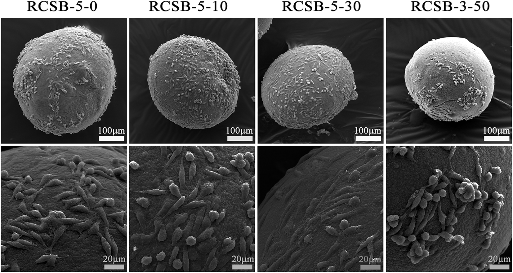
To further evaluate the potential application of RCSB-v-n as cell culture scaffold or biomaterials, L929 cells were used as seed cells to observe their adhesion, proliferation, and distribution. During cell culture, many factors may affect the adhesion, migration, and proliferation of the cells. For beads, these factors may include composition, roughness, pore structure, and diameter. To avoid the effects of different bead diameters, RCSB-5-0, RCSB-5-10, RCSB-5-30, RCSB-3-50 with very close bead diameters (about 400 μm) were chosen for cell culture. SEM images of L929 cells cultured with RCSB-v-n for 72 h are shown in Fig. 10. Cells adhered well on the surface of RCSB-v-n, and the interaction between cells and RCSB-v-n could be clearly observed from the SEM images. The L929 cells adhered through protrusions and grew well on the surface of the composite beads. In some cases, cells layered on bead surfaces and exhibited good proliferation, especially on the RCSB-5-10 and RCSB-5-30 composite beads. Moreover, as shown in Fig. 10, the cells adhered to almost the entire surface of RCSB-5-10 and RCSB-5-30 and grew uniformly on the surface of these beads. The cell density on the surface of RCSB-5-10 and RCSB-5-30 were higher than that of RCSB-5-0. Cellular pseudopodium extended and adhered to the surface of the composite beads with most cells being shuttle-shaped. The incorporation of SPI into cellulose played an important role in promoting cell adhesion and proliferation, which is very important for cell culture scaffold. SPI incorporated into cellulose may modify the chemical composition and microstructure of the beads to improve cytocompatibility and cell adhesion. The surfaces of RCSB-5-10, 30, and 50 were homogeneous and relatively rough (Fig. 2), which was also beneficial for cell adherence; however, there were fewer cells on the surface of RCSB-3-50, with aggregated cells being observed. The appropriate amount of SPI hydrolysate dissolved in the culture medium could promote cell growth and proliferation, whereas a high concentration of SPI hydrolysate has slight negative effect on cell viability.55 This was consistent with our previous work.36 Moreover, alkali modification caused an increase of exposed hydrophobic regions on the surface of SPI. Measurement of the water contact indicated that RCSB-3-50 exhibited the highest water contact angle (97.9°). As a result, cell adhesion on the surface of RSCB-3-50 was the least because of its highest surface hydrophobicity. The above results indicated that RCSB-v-n, especially RCSB-5-10 and RCSB-5-30, could support cell growth well with a high adhesion efficiency and biocompatibility, showing potential for application as biomaterials.
 |
| | Fig. 10 SEM images of the L929 cells cultured on the surfaces of the RCSB-v-n (n = 0, 10, 30, and 50) beads for 72 h. | |
4. Conclusions
Herein, a facile, environmentally friendly and convenient high-voltage electrostatic technique for the fabrication of nano-porous cellulose/SPI composite beads was applied. These composite beads exhibited a 3D-structure with a particle size ranging from 300 to 1500 μm in the dry state. The size of the beads could be controlled by changing fabrication parameters such as high voltage and SPI content. In addition, the hydrophilicity/hydrophobicity of RCSB-3-n could be altered by controlling the SPI content of the beads. RCSB-v-n exhibited good thermal stability and could be sterilized by autoclave at 121 °C. Moreover, the incorporation of SPI into cellulose improves its cytocompatibility with L929 cells, particularly the RCSB-5-10 and RCSB-5-30, which exhibited a high efficiency in cell adhesion and cell proliferation. These features are important particularly for application in various biomedical fields. Therefore, these highly biocompatible beads, fabricated by simple and controllable high-voltage electrostatic technique, may be good candidates for cell culture, showing potential for application as biomaterials.
Acknowledgements
This work was supported by the National Natural Science Foundation of China (No. NSFC 81471789, NSFC 81171480, and NSFC 81270411) and “Program Cai Yuanpei” (CSC No. 201304490192 and 201304490191) from China Scholarship Council.
References
- S. Peng, H. C. Meng, Y. Ouyang and J. Chang, Ind. Eng. Chem. Res., 2014, 53, 2106–2113 CrossRef CAS.
- J. Trygg, P. Fardim, M. Gericke, E. Makila and J. Salonen, Carbohydr. Polym., 2013, 93, 291–299 CrossRef CAS PubMed.
- X. G. Luo and L. N. Zhang, J. Chromatogr. A, 2010, 1217, 5922–5929 CrossRef CAS PubMed.
- H. L. Cai, S. Sharma, W. Y. Liu, W. Mu, X. D. Zhang and Y. L. Deng, Biomacromolecules, 2014, 15, 2540–2547 CrossRef CAS PubMed.
- R. Gavillon and T. Budtova, Biomacromolecules, 2008, 9, 269–277 CrossRef CAS PubMed.
- X. G. Luo, J. Zeng, S. L. Liu and L. N. Zhang, Bioresour. Technol., 2015, 194, 403–406 CrossRef CAS PubMed.
- R. Sescousse, R. Gavillon and T. Budtova, J. Mater. Sci., 2011, 46, 759–765 CrossRef CAS.
- M. Gericke, J. Trygg and P. Fardim, Chem. Rev., 2013, 113, 4812–4836 CrossRef CAS PubMed.
- K. F. Du, M. Yan, Q. Y. Wang and H. Song, J. Chromatogr. A, 2010, 1217, 1298–1304 CrossRef CAS PubMed.
- Y. Yoshida, M. Yanagisawa, A. Isogai, N. Suguri and N. Sumikawa, Polymer, 2005, 46, 2548–2557 CrossRef CAS.
- S. Fischer, H. Leipner, K. Thummler, E. Brendler and J. Peters, Cellulose, 2003, 10, 227–236 CrossRef CAS.
- M. Egal, T. Budtova and P. Navard, Cellulose, 2008, 15, 361–370 CrossRef CAS.
- H. J. Jin, C. X. Zha and L. X. Gu, Carbohydr. Res., 2007, 342, 851–858 CrossRef CAS PubMed.
- W. Q. Liu, T. Budtova and P. Navard, Cellulose, 2011, 18, 911–920 CrossRef CAS.
- L. N. Zhang, D. Ruan and S. J. Gao, J. Polym. Sci., Part B: Polym. Phys., 2002, 40, 1521–1529 CrossRef CAS.
- B. Duan, X. Zheng, Z. X. Xia, X. L. Fan, L. Guo, J. F. Liu, Y. F. Wang, Q. F. Ye and L. N. Zhang, Angew. Chem., Int. Ed., 2015, 54, 5152–5156 CrossRef CAS PubMed.
- B. Peng, E. Wee, A. Imhof and A. Blaaderen, Langmuir, 2012, 28, 6776–6785 CrossRef CAS PubMed.
- A. Gharsallaoui, G. Roudaut, O. Chambin, A. Voilley and R. Saurel, Food Res. Int., 2007, 40, 1107–1121 CrossRef CAS.
- W. B. Wu, J. Gu, G. C. Zhou, L. Zhang, M. R. Gong and H. Q. Dai, J. Appl. Polym. Sci., 2014, 131, 40656 Search PubMed.
- R. Stojanovic, A. B. Cvitanovic, V. Manojlovic, D. Komes, V. Nedovic and B. Bugarski, J. Sci. Food Agric., 2012, 92, 685–696 CrossRef CAS PubMed.
- Y. Shang, F. Y. Ding, L. Xiao, H. B. Deng, Y. M. Du and X. W. Shi, Carbohydr. Polym., 2014, 102, 413–418 CrossRef CAS PubMed.
- S. K. Papadopoulou, C. Tsioptsias, A. Pavlou, K. Kaderides, S. Sotiriou and C. Panayiotou, Colloids Surf., A, 2011, 387, 71–78 CrossRef CAS.
- L. Sun, X. F. Yu, M. D. Sun, H. G. Wang, S. F. Xu, J. D. Dixon, Y. A. Wang, Y. X. Li, Q. B. Yang and X. Y. Xu, J. Colloid Interface Sci., 2011, 358, 73–80 CrossRef CAS PubMed.
- D. G. Yu, J. H. Yang, X. Wang and F. Tian, Nanotechnology, 2012, 23, 105606 CrossRef PubMed.
- V. L. Workman, L. B. Tezera, P. T. Elkington and S. N. Jayasinghe, Adv. Funct. Mater., 2014, 24, 2648–2657 CrossRef CAS PubMed.
- M. Hirota, N. Tamura, T. Saito and A. Isogai, Cellulose, 2009, 16, 841–851 CrossRef CAS.
- W. D. Oliveira and W. G. Glasser, J. Appl. Polym. Sci., 1996, 60, 63–73 CrossRef.
- H. F. Xia, D. Q. Lin, L. P. Wang, Z. J. Chen and S. J. Yao, Ind. Eng. Chem. Res., 2008, 47, 9566–9572 CrossRef CAS.
- B. Volkert, B. Wolf, S. Fischer, N. Li and C. H. Lou, Macromol. Symp., 2009, 280, 130–135 CrossRef CAS.
- J. L. Ye, J. X. Yun, D. Q. Lin, L. H. Xu, H. Kirsebom, S. C. Shen, G. S. Yang, K. J. Yao, Y. X. Guan and S. J. Yao, J. Sep. Sci., 2013, 36, 3813–3820 CrossRef CAS PubMed.
- F. Shi, D. Q. Lin, W. Phottraithip and S. J. Yao, J. Appl. Polym. Sci., 2011, 119, 3453–3461 CrossRef CAS.
- J. L. Wang, L. G. Wei, Y. C. Ma, K. L. Li, M. H. Li, Y. C. Yu, L. Wang and H. H. Qiu, Carbohydr. Polym., 2013, 98, 736–743 CrossRef CAS PubMed.
- T. Cserhati and E. Forgacs, Amino Acids, 2005, 28, 99–103 CrossRef CAS PubMed.
- C. M. Vaz, L. A. Graaf, R. L. Reis and A. M. Cunha, NATO Sci. Ser., 1, 2002, 86, 93–110 CAS.
- G. A. Silva, C. M. Vaz, O. P. Coutinho, A. M. Cunha and R. L. Reis, J. Mater. Sci.: Mater. Med., 2003, 14, 1055–1066 CrossRef CAS PubMed.
- L. H. Luo, X. M. Wang, Y. F. Zhang, Y. M. Liu, P. R. Chang, Y. Wang and Y. Chen, J. Biomater. Sci., Polym. Ed., 2008, 19, 479–496 CrossRef CAS PubMed.
- J. Cai and L. N. Zhang, Biomacromolecules, 2006, 7, 183–189 CrossRef CAS PubMed.
- F. Y. Du, H. Wang, W. Zhao, D. Li, D. L. Kong, J. Yang and Y. Y. Zhang, Biomaterials, 2012, 33, 762–770 CrossRef CAS PubMed.
- S. Levic, I. P. Lijakovic, V. Đordevic, V. Rac, V. Rakic and T. S. Knudsen, et al., Food Hydrocolloids, 2015, 45, 111–123 CrossRef CAS.
- M. Devaraj, R. K. Deivasigamani and S. Jeyadevan, Colloids Surf., B, 2013, 102, 554–561 CrossRef CAS PubMed.
- S. Yun, H. J. Luo and Y. F. Gao, RSC Adv., 2014, 4, 4535–4542 RSC.
- X. S. Du, Y. P. Li, X. Liu, X. Wang, C. Huselstein, Y. T. Zhao, P. R. Chang and Y. Chen, J. Mater. Sci.: Mater. Med., 2014, 25, 823–833 CrossRef CAS PubMed.
- S. M. K. Mohamed, K. Ganesan, B. Milow and L. Ratke, RSC Adv., 2015, 5, 90193–90201 RSC.
- M. Subirade, I. Kelly, J. Gueguen and M. Pezolet, Int. J. Biol. Macromol., 1998, 23, 241–249 CrossRef CAS PubMed.
- V. Schmidt, C. Giacomelli and V. Soldi, Polym. Degrad. Stab., 2005, 87, 25–31 CrossRef CAS.
- L. H. Luo, Y. F. Zhang, X. M. Wang, Y. Wan, P. R. Chang, D. P. Anderson and Y. Chen, J. Biomater. Appl., 2010, 24, 503–526 CrossRef CAS PubMed.
- I. Keizo and K. Shiro, Agric. Biol. Chem., 1980, 44, 537–543 Search PubMed.
- H. Weining and S. Xiuzhi, J. Am. Oil Chem. Soc., 2000, 77, 101–104 CrossRef.
- D. F. Xu, X. Xiao, J. Cai, J. Zhou and L. N. Zhang, J. Mater. Chem. A, 2015, 3, 16424–16429 CAS.
- N. Shuryo, J. Agric. Food Chem., 1983, 31, 676–683 CrossRef.
- D. Srinivasan, J. Agric. Food Chem., 1988, 36, 262–269 CrossRef.
- C. H. Lin, W. C. Jao, Y. H. Yeh, W. C. Lin and M. C. Yang, Colloids Surf., B, 2009, 70, 132–141 CrossRef CAS PubMed.
- X. M. Zhang, H. W. Yu, H. J. Yang, Y. C. Wan, H. Hu, Z. Zhai and J. M. Qin, J. Colloid Interface Sci., 2015, 437, 277–282 CrossRef CAS PubMed.
- F. Franek, O. Hohenwarter and H. Katinger, Biotechnol. Prog., 2008, 16, 688–692 CrossRef PubMed.
- B. H. Chun, J. H. Kim, H. J. Lee and N. Chung, Bioresour. Technol., 2007, 98, 1000–1005 CrossRef CAS PubMed.
Footnote |
| † Electronic supplementary information (ESI) available. See DOI: 10.1039/c6ra02364a |
|
| This journal is © The Royal Society of Chemistry 2016 |
Click here to see how this site uses Cookies. View our privacy policy here.