
Mass spectrometry based chemical imaging of foods
Shekhar U. Kadam
a,
N. N. Misra
 *b and
Nobuhiro Zaima
c
*b and
Nobuhiro Zaima
c
aResearch, BioAtlantis Ltd., Tralee, Co., Kerry, Ireland
bGTECH, Research & Development, General Mills India Pvt. Ltd., Mumbai, India. E-mail: misra.cftri@gmail.com; Tel: +91 22 420 31566
cDepartment of Applied Biological Chemistry, Graduate School of Agricultural Science, Kindai University, 204-3327 Nakamachi, Nara City, Nara 631-8505, Japan
First published on 11th March 2016
Abstract
Chemical imaging based on mass spectrometry is an emerging technology which has opened opportunities for fundamental research in food science. The ability to quantitatively determine the spatial distribution of several chemical entities simultaneously makes it a method of choice for chemical characterization of plant materials and foods. In this review, an overview of the ionization methods is presented, followed by a discussion of the current state of the art in mass spectrometry imaging (MSI) of relevance to practices in food research. The known applications, analytical challenges, and future research potential are highlighted. MSI has been successfully utilized to a variable degree for obtaining information about spatial distribution of a variety of molecules and metabolites in foods. It allows visualizing the spatial molecular maps without extraction, purification, separation, or labelling of foods. The method is likely to find wider adoption with developments in ionization sources and inter-laboratory collaborations.
 Shekhar U. Kadam | Shekhar U. Kadam is a researcher in chemistry at Bioatlantis Ltd., Ireland. He obtained his PhD in biosystems and food engineering from University College Dublin, Ireland in 2015 and masters in food technology from CSIR-Central Food Technological Research Institute, India in 2010. His research interests include marine foods, seaweeds, bioactive compounds and power ultrasound assisted extraction. Shekhar has authored 15 publications in international peer-reviewed journals. |
 N. N. Misra | N. N. Misra is a senior scientist with the technology development team of R&D, General Mills. He obtained his PhD in food engineering from Dublin Institute of Technology, Ireland in 2014, and his M.Sc. in food technology from CSIR-Central Food Technological Research Institute, India in 2010. His research interests include food physics, cold plasma, chemometrics and mathematical modelling. |
 Nobuhiro Zaima | N. Zaima is an associate professor with the department of applied biological chemistry, Kinki University. He obtained his PhD in agriculture from Kyoto University, Japan in 2007. His research interests are in the areas of food chemistry, lipid biology, vascular diseases such as abdominal aortic aneurysm, atherosclerosis, or triglyceride deposit cardiomyovasculopathy. His research aims to clarify the biological meaning of characteristic distribution of metabolites found by MALDI mass spectrometric imaging analysis. |
1 Introduction
Mass spectrometry is an important analytical tool which offers high sensitivity and molecular specificity for a wide range of molecules, and the flexibility to address many varied analytes on a single platform. These advantages have led to the growth of mass spectrometry based ‘omics’ technologies for rapid analysis of a large class of metabolites in food systems. In fact, metabolomics based on mass spectrometry is now a mature technology in food science and several detailed reviews are available.1–4 Mass spectrometry techniques are conventionally used in tandem with separation techniques such as liquid chromatography (LC) and gas chromatography (GC) to form LC-MS and GC-MS, which can achieve higher efficiency in characterization of molecules. However, extraction methods involved in LC-MS and GC-MS result in loss of all the information about its spatial distribution in the sample. This information can be retained if extraction is carried out locally in the sample or best avoided.5Chemical imaging methods allow identification of differences in chemical composition and structure of biological tissues.6 Spectroscopic techniques such as Fourier-transform infrared (FTIR),7 NIR8 and Raman9 have been frequently used to identify the spatial distribution of bioactive compounds and chemical markers in food matrices. In recent times, however, the interest in use of ambient ionization based mass spectrometry as a tool for chemical analysis and mass spectral imaging of biological matter is steadily growing. This trend is driven by the success of MSI in several other areas of science and engineering, including pharmaceuticals, medicine10 and polymers.11,12 While the use of MS based food-omics is well established, the use of mass spectrometry imaging (hereafter abbreviated as MSI) in food research is sparse. Low spatial resolution, limitations in molecular identification capabilities, low throughput and requirement of very specific instrumentation for desired results render MSI into a perfect tool for direct analysis of biological tissue.
Mass spectrometry based chemical imaging involves combining on a common platform, the elemental or molecular mass measurement obtained via mass spectrometry and spatial information obtained via imaging. This enables visualizing the spatial distribution of elements/molecules of interest on complex surfaces. The overall framework of MSI is similar to near-infrared chemical imaging,13 which has earned recognition for food quality monitoring within the recent past.14 Mass analysis of molecules/isotopes are to MSI, what molecular vibrations and infrared spectrum analysis are to near-infrared chemical imaging. However, MSI certainly offers a very high chemical specificity compared to infrared imaging, especially for complex biological matrices such as foods and plant materials.
MSI involves the general steps of sample preparation, desorption/ionisation of the sample, followed by analysis of ion masses using a mass spectrometer, the image registration and subsequent multivariate analyses. MSI ionization methods allow desorbing analytes directly from the food surface, thereby avoiding traditional liquid extractions. What differs essentially between different MSI techniques is the way ions are generated. The ionization of the sample is an important aspect to be considered for analysis of a given food sample, and is discussed later in Section 2. The mass analysis is carried out using mass spectrometers, which depending on the instrument, could either be a time of flight (TOF), orbitrap, or Fourier transform ion cyclotron resonance (FTICR). The topic of mass analysis has already rendered itself into several volumes of literature.15,16 For ease of presentation, the sample preparation involved is relegated to Section 3 – after describing the ionization methods. In practice, the mass spectrum is recorded as the ionization spot moves spatially, generating data which is automatically mapped to spatial coordinates; this is known as ‘image registration’. The concept of image registration is explained within the Data analysis section.
MSI is an exceedingly multidisciplinary field and its application to biological systems, including food and plant material, often involves the joint efforts of chemists, food scientists, physicists and statisticians.17 There has been a surge of interest in use of MSI as a tool for pathological analysis,18 understanding pharmaceutical mechanisms, drug metabolism19 and biomarker discovery. This is evident from the growing number of publications in this area (see Fig. 1). However, these tools are relatively less known and yet to gain popularity among food scientists. MSI offers several fundamental research opportunities within food science, as will be discussed later in the review. Within its true spirit, MSI has a huge potential to enable a detailed understanding of the biological processes in foods over a range of different length scales, ranging from sub-cellular to tissue, all the way up to whole foods.
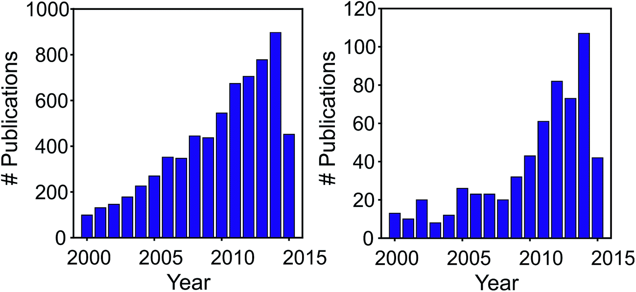 | ||
| Fig. 1 Number of publications retrieved from Web of Science, for ‘mass spectrometry imaging’ (left) and ‘food mass spectrometry imaging’; accessed on 27 June, 2015. | ||
In this review, the ionization methods, sample preparation, and data analysis in MSI are briefly introduced, followed by a discussion of the progress made until date in its application to probe food systems. The potential opportunities and challenges in chemical imaging of complex food matrices using MSI have been factually and also speculatively highlighted. We target this review at food scientists and mass spectroscopists, who are unaware of the work from the other field. We hope that this review will attract their interest to adopt this tool in advancing food science.
2 Ionisation methods
In the following section, a summary of the desorption and ionisation methods pertinent to MSI is presented. These aspects have been reviewed earlier by Pacholski and Winograd,20 Rubakhin and Sweedler.212.1 Secondary ion mass spectrometry (SIMS)
SIMS imaging involves the precise focusing of a primary ion beam on to the surface of the sample. This causes sputtering of atoms and molecules to yield the secondary ions, which are analysed using a mass spectrometer. With SIMS, the images could either be developed for specific molecules or for molecular fragments of interest, which directly scales with the relative abundance of the compound over the imaged surface. Recently, a high lateral resolution imaging (∼40 nm) with high-sensitivity SIMS analysis, the “nanoSIMS” has been developed by Cameca.22 The nanoSIMS is particularly advantageous for research in biological, materials and geological science. The nanoSIMS achieves its high lateral resolution by means of the reactive primary ions, which enhances sensitivity, without compromising the mass resolving power, and therefore, specificity.232.2 Matrix assisted laser desorption ionization (MALDI)
MALDI imaging analyses and detects a wide range of biomolecules directly from tissue sections. The tissue sections are covered with a matrix which is generally a UV-absorbing weak organic acid such as sinapinic acid or α-cyano-4-hydroxycinnamic acid (α-CHC). This matrix helps in extracting target molecules from the tissue sample, formation of analyte-doped crystals and absorption of laser energy for soft-ionization of target molecules. In MALDI, a laser is fired on the matrix layer, which absorbs energy and transfers analytes to gas phase and then finally the MALDI stage is moved on to the x/y axis to assay each sample position. Additionally, the matrix plays the role of both proton donor and receptor to ionise the analyte in positive as well as negative modes. In MALDI imaging, histological tissue examination is feasible as the laser is fired only at the matrix layer, keeping tissue intact in its form.18,242.3 Desorption electrospray ionization (DESI)
Unlike SIMS and MALDI, DESI operates under ambient pressure conditions. It utilizes both electrospray ionization and desorption ionization methods. In DESI, electrospray is generated by a flow of solvent and the application of a nebulizer gas and a high voltage potential resulting in a beam of charged microdroplets. These microdroplets are directed towards the surface where simultaneous extraction, desorption and ionization is carried out from the sample spot below the spray.5 The ion formation occurs by ion emission or by evaporation of neutral solvent molecules.2.4 Novel ionisation methods
Considering that ionisation under atmospheric pressure conditions is more suited for routine analysis and easy to maintain, research efforts to develop atmospheric pressure plasma ionisation sources have increased. Among these, dielectric barrier discharges,25 and micro-plasma jets26 have been investigated with promising results. The ease of ionisation using portable plasma sources (typically operating in helium) could complement well with portable mass spectrometers.For the sake of brevity, a discussion of other ionization methods, such as Laser Ablation Electrospray Ionization (LAESI), Probe Electrospray Ionization (PESI), and Laser Electrospray Mass Spectrometry (LEMS) has been omitted here. These are also popular for MSI and have been successfully applied to biological samples.27–31
2.5 Selection criteria
Where options are available, the choice of a MSI technique will depend on several factors such as the spatial and mass resolution desired, the operating pressure, the ease of ionisation, etc. SIMS imaging utilizes a primary ion beam and is preferable when a higher spatial resolution of images with a low mass range (<1000 Da) is desired. MALDI-MSI relies on exposure of the sample surface to a laser beam, and is limited by its lower spatial resolution, but turns out advantageous in situations when a much wider mass range is to be covered (≥100![[thin space (1/6-em)]](https://www.rsc.org/images/entities/char_2009.gif) 000 Da). The fragmentation in SIMS and matrix interference in MALDI restrict the analysis of large and small molecules, respectively.
000 Da). The fragmentation in SIMS and matrix interference in MALDI restrict the analysis of large and small molecules, respectively.
DESI is based on use of charged droplet flux for ionization and desorption. Similar to MALDI-MSI, DESI is characterized by a lower spatial resolution, but equips an analyst with a tool to analyze small molecules at ambient pressure. On the contrary, SIMS is performed under vacuum conditions. Until recently, MALDI-MSI was ordinarily performed under vacuum; however, Harada et al.32 successfully demonstrated the development and application of an atmospheric pressure laser desorption/ionization microscope (AP-LDI) imager.
Spatial resolutions that can be routinely obtained with MALDI, DESI and SIMS are in the order of 15 μm, 250 μm, and ca. 1 μm respectively. However, with nanoSIMS a lateral resolution of ∼30–50 nm can be achieved.
3 Sample preparation
The preservation of structural organization of the biological or food samples to be imaged and facilitation of the mass analysis are the primary objectives of sample preparation in MSI. As with any analytical method, sample preparation techniques for MSI should embrace sensitivity, reproducibility and accuracy, and therefore exposure of the samples to unfavourable environments (high moisture, temperature) should be avoided. When developing methods for preparation of food samples for MSI, one should note that each treatment step adds a risk of chemical contamination and molecular diffusion.33 Therefore, the number of steps should be minimised.For MALDI, the sample is typically flash frozen using liquid nitrogen and cryosectioned, following which the chemical matrix is applied. When obtaining reasonable cryosections turns out difficult, the freshly cut surface can be blotted on to pre-coated cellulose membranes for direct loading. This method, known as imprinting or blotting, has been successfully employed for MSI of potato tuber.34 Sometimes, an adhesive tape is useful for mounting samples where the thaw-mounting preparation of the sample section is difficult (e.g. plant seeds or whole-body sections).35,36 When preparing samples for MALDI, inhomogeneous distributions of matrix crystals can result in position dependent ion signals. This could potentially lead to errors and should be taken care of. In recent times, the sensitivity of MSI has been enhanced by coupling it to thin layer chromatography (TLC) blotting. This overcomes the difficulty arising due to competition between numerous molecular species for ionization in non-separated matrices (which permits identification of ‘easy to ionize’ molecules only).37
For SIMS imaging, removal of water and minimisation of salts in the sample is the first requirement. Rapid freezing methods using liquid ethane or propane, which prevent water crystallisation, while keeping the sample frozen and subliming off the vitreous ice are recommended. Imprinting of samples onto hard surfaces prior to SIMS imaging experiments has also been employed to boost signal intensities compared to direct imaging; e.g. leaf samples.38
DESI being an ambient ionization technique in which desorption and ionization occur in the native environment, often minimal or no sample preparation is required. The reduced sample preparation steps compared with vacuum imaging techniques is a specific reason for the rising popularity of DESI. The positioning of the nozzle, the sample, and the inlet of the mass spectrometer is crucial for success with DESI-MSI.39 The use of solvent extraction and/or heating during imprinting for DESI-MSI has also been successfully applied for transfer of chemicals.40
4 Data analysis
The data generated from a typical MSI experiment comprises of 5000 to 50000 mass spectra acquired at individual pixels of the sample.41 The number of pixels depend on the spatial resolution chosen. The size of the mass spectrum varies depending on the ionisation method and the mass analyser. It is worthwhile noting that the mass analyser decides the mass resolution, which in turn dictates the degree of chemical specificity. These facts by themselves indicate that MSI yields ‘big’ datasets (which often occupy several gigabytes of memory). Given that each unique m/z signal corresponds to a metabolite, a spatial assembly of the intensity of m/z of interest from all mass spectra will constitute a metabolite distribution map. Such maps are pseudo-coloured to visualise the distribution as an image and all the images put together make a hyperspectral dataset over the m/z spectrum (see Fig. 2 for details). A new imaging data standard with a file extension imzML has been developed recently42 and demonstrates the global efforts towards easy data sharing.
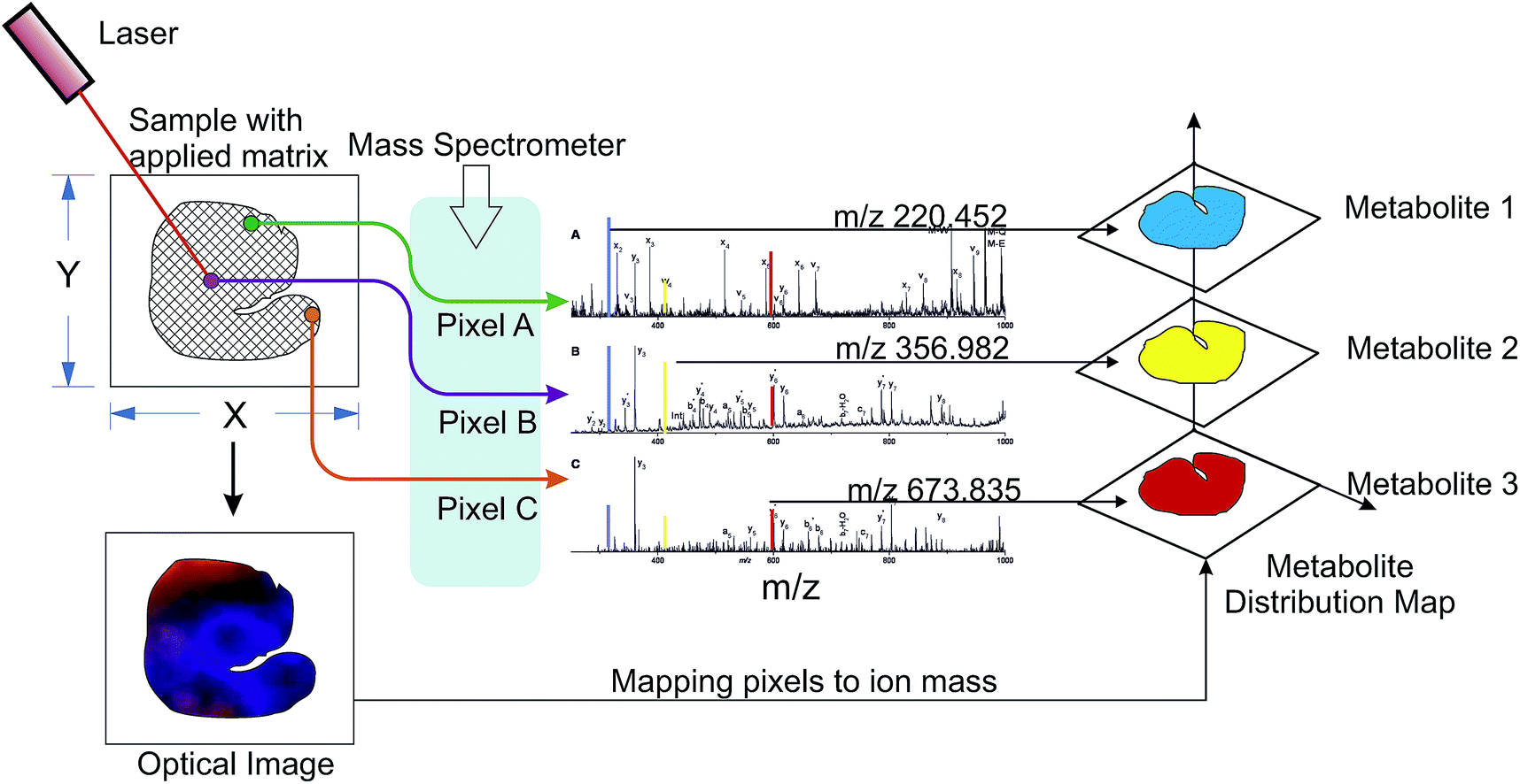 | ||
| Fig. 2 A schematic describing the structure of chemical imaging data obtained by mass spectrometry. This exemplary figure shows a MALDI ionisation method. | ||
The analysis of MSI data typically begins with the application of denoising and smoothing routines, which help to subtract the background noise from the spectrum, followed by optional normalization of the mass intensities. Methods such as nearest neighbour averaging and edge-preserving smoothing algorithms have been proposed for reducing the noise in images.43,44 Wavelets based smoothing and data reduction has also been explored45 and is likely to gain popularity in the near future considering its versatility and speed. Following the application of pre-processing routines, the next challenge involves the feature extraction. The large datasets from MSI studies demand automated methods of data analysis to find differences among spatial distribution in different samples. Many of the assumptions made in the analysis of LC-MS or GC-MS omics datasets are not suitable for imaging.17 For such complex data analysis problems, multivariate statistical methods have stood the test of time, and are also useful for MSI data. Alexandrov et al.41 developed and demonstrated the use of unsupervised cluster analysis, through which m/z images (corresponding to a metabolite) are clustered according to their spatial similarity, resulting in each cluster containing spatially similar metabolite images. Recently, Jaumot and Tauler46 proposed the use of multivariate curve resolution for MSI data, which allows identifying components in distribution maps using resolved pure high-resolution mass spectra. Other options for data analysis include chemometrical methods such as Multivariate Image Analysis (MIA) and Principal Component Analysis (PCA). The recently developed package Cardinal for open source software R allows segmenting and classifying mass spectrometry images, and is based on mixture modeling and regularization statistics.47 Detailed protocols and subtleties of MSI data analysis on commercial software, have been reviewed in a recent book by Setou.48 The general framework for MSI data analysis and necessary precautions have been reviewed by Jones et al.17
5 Current applications
When compared to optical spectrometry based imaging techniques such as Raman, FTIR and NIR, chemical imaging with mass spectrometry allows visualization of spatial distribution of chemical entities and/or target molecules with very high specificity and precision. Unlike most optical spectrometry, MSI is virtually free from the problem of overlapping/convoluted peaks. This specificity is particularly important when analyzing chemically complex biological structures, including foods. MSI has been applied for visualizing the spatial distribution of lipids, polyphenols, allergens and toxins, besides other known phytochemicals and bioactive molecules. In the following subsections an overview of the applications in food science is provided.5.1 Food lipidomics
MSI has been successfully used to visualize and analyze distribution of lipids in foods. Among all the available ionization methods, MALDI and DESI can detect most lipid classes from a variety of surfaces, though MALDI has enjoyed popularity for lipidomics. These techniques allow revealing the distinct distribution of lipid species within the sections of interest.MALDI-MSI has been used to visualize the spatial distribution of lipids in rice;49 see Fig. 3. It may be noted from Fig. 3 that the lysophosphatidylcholine (LPC) (18:2) and LPC (18:1) are distributed in the outer area of rice endosperm. On the other hand, LPC (18:0) is localized in the core of rice. The distinct distribution of LPC species has been suspected to be a possible reason for the high quality of sake (Japanese traditional alcoholic beverage) made from rice.49 The distribution of triacylglycerols (TAGs) in avocado mesocarp was studied by Horn et al.50 using MALDI-MSI. The mesocarp was found to be rich in monounsaturated fatty acids, and TAG-52:2 (TAGs with two oleic and one palmitic acid). However, Horn et al.50 for the first time found that the species with oleic acid at each position, TAG-54:3, was relatively inhomogeneous in the mesocarp. This distinct behaviour requires further analysis.
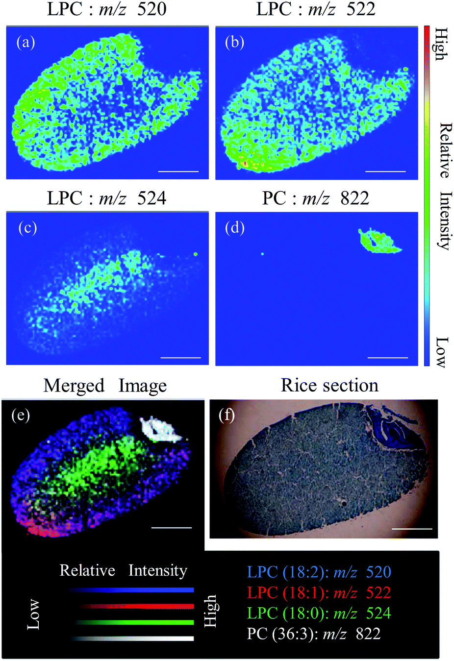 | ||
| Fig. 3 Visualization of lipid species in rice grain. Distribution of (a) m/z 520; lysophosphatidylcholine (LPC) (18:2), (b) m/z 522; LPC (18:1), (c) m/z 524; LPC (18:0), and (d) m/z 822; PC (36:3). (e) Merged image of LPC species and PC. (f) Toluidine blue staining of rice section.49 Scale bar: 1.0 mm. | ||
To avoid matrix-related ions in the spectrum and additional steps in the sample preparation procedure, matrix-free methods such as nano-structure-initiator mass spectrometry (NIMS) and nanowire-assisted laser desorption ionization (NALDI) have also emerged. These methods use an active nanostructured surface to couple the laser energy for the desorption/ionization of analytes present on the surface.51 Essentially NIMS uses a porous, nanostructured silicon substrate doped typically with siloxanes or silanes. NIMS and NALDI methods are emerging techniques in lipid surface analysis by virtue of enhanced sensitivity. SIMS techniques also have the ability to produce higher spatial resolution.
MSI methods can be coupled with additional analytical methods such as thin layer chromatography (TLC) to gain more information at the molecular level. Goto-Inoue et al.52 developed the TLC-Blot-MALDI-MSI technology, discussed earlier (Sample preparation section). TLC-Blot involves transferring separated lipids from TLC plates onto polyvinylidene fluoride membranes to achieve better sensitivity, mass resolution, and minimal background interference in the analysis.53,54 Zaima et al.53 employed this method to visualize and identify major phospholipids, including phosphatidylethanolamine, phosphatidylinositol, phosphatidylserine, phosphatidylcholine and sphingomyelin in tuna fish (Thunnus thynnus).
When food products are tested for their shelf-life, spatial heterogeneity in degradation is often overlooked. Consequently, localised degradation could severely influence end-user perception, while it could remain undetected during holistic evaluations. Within this context, advances in imaging of lipids have enabled researchers to track spatial degradation of lipids directly from meat tissue without extraction.55 Identification of lipid degradation mechanisms directly from muscle tissues will empower food scientists to better understand safety and shelf-life issues related to meat products without any chemical extraction or modification of lipids.
5.2 Spatial distribution of bioactives
MSI facilitates mapping at molecular level, and detection and identification of bioactive components, which includes the parent molecule and its metabolites. Thus, MSI is important for finding the exact role of bioactives through pathological analysis and bioactive mechanism. While information about biosynthetic pathways, in vivo mechanism, and variation in bioactive content can be depicted by MSI,56 mass spectrometric or other optical measures can analyse bioactive compounds only quantitatively or qualitatively.57,58Recently, MSI has been used to find spatial distribution of nutrients and bioactive compounds in foods such as strawberries, celery, sage leaf, potato, cooked chicken egg and turkey.59 Kim et al.60 used MALDI-MSI for visualizing the bioactive strictinin (an ellagitannin found in green tea cultivars), on orally dosed mouse kidney tissue. Authors found intact strictinin within kidney after 1 h of oral dosing and suggested that 1,5-diaminonaphthalene is the best matrix for high sensitivity in the negative ionization mode. Flavonoids in foods have also been subject of MSI studies. Seyer et al.61 used SIMS to localize the flavonoids in fresh pea and Arabidopsis thaliana seeds. Subsequently, Li et al.62 detected and visualized free flavonoids, flavonoid glycosides and saponins with mass difference of 0.02 Da by using tandem mass spectrometry imaging.
Polyphenols, particularly anthocyanins, are known for many beneficial effects to the human health. Using MSI, these compounds have been imaged by Yoshimura et al.63,64 in blueberry and black rice. The authors found that black rice anthocyanins with a pentose moiety were localized in the entire pericarp, whereas anthocyanin species with hexose moiety were focally localized in the dorsal pericarp. On the contrary, in case of blueberry, the anthocyanins delphinidin and petunidin were dominantly distributed in the exocarp, while cyanidin, peonidin and malvidin were found in exocarp as well as the endocarp.
Cabral et al.40 successfully imaged strawberry fruit (Fragaria × ananassa Duch.) and ginkgo leaf (Ginkgo biloba L.) sections by imprinting the samples on TLC plates, followed by DESI-MSI. In the strawberry analysis, authors found that sugar ions were distributed throughout the fruit whereas the anthocyanin pelargonidin-3-glucoside was highly concentrated in the margin region of strawberry. However, the exact location of anthocyanin pelargonidin-3-glucoside should be further investigated as it could be the result of pressure induced during imprinting.
The knowledge of anthocyanins (or other bioactive or macromolecule) distribution patterns in grains could serve to guide and accelerate research in the area of grain dry-fractionation.65,66 One could, in principle, design processes for separation of these regions through dry milling and classification to obtain natural ingredients for food use. Furthermore, extending this concept to the case of plant materials, extraction efficiencies for bioactives can be increased by using selected material fractions rich in the component, against the current practice of whole plant material usage in most cases.
5.3 Allergen and chemical hazards
Considering the potential threats to food safety and quality issues posed by chemical toxins such as herbicides, fungicides, pesticides, adulterants and allergens, these should be analyzed with high sensitivity and accuracy.67 In this regard, MSI techniques can be useful due to the added advantage of spatial distribution over mass spectrometric methods. MALDI-MSI has been used for detection and imaging of the mesotrione and azoxystrobin, a herbicide and a fungicide respectively, present on the surface of the soya plant structures.68 Imaging of 2,4-dichlorophenoxyacetic acid (herbicide) distribution using laser-ablation electrospray ionization and imaging on cabbage leaves has been reported.69 MALDI-MSI has also been used to image the distribution of the pesticide nicosulfuron (parent compound and a phase 1 metabolite) in plant tissue following root and foliar uptake.70 Aflatoxin and phytoalexin identification and detection on peanut surface has been carried out directly using MALDI-MSI. This method is characterized by solvent-free and matrix-free analysis without any sample preparation or separation steps.71 Nielen and van Beek72 have used this method for macroscopic and microscopic imaging of pesticides, mycotoxins and plant metabolites on rose leaves, orange and lemon fruit peel, ergot bodies, cherry tomatoes (Fig. 4), and maize kernels.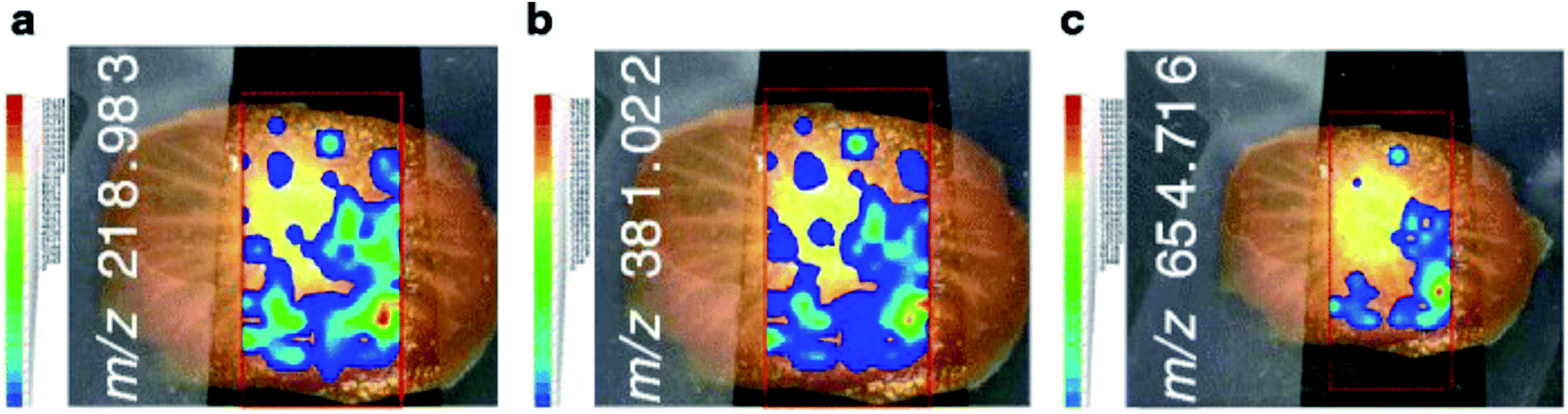 | ||
| Fig. 4 Spatial distribution of the natural carbohydrates and lycoperosides F and/or G and/or esculeoside A on cherry tomato slice through ion maps of (a) m/z 218.983, (b) m/z 381.022 and (c) m/z 654.716. Adapted from Nielen and van Beek72 by courtesy of Springer. | ||
Arsenic (As) is a toxic element entering the food chain through arsenic contaminated water used for irrigation. Rice (Oryza sativa) is a staple food in As-epidemic zones of world. Moore et al.73 employed high-resolution SIMS (nano-SIMS) to probe the distribution of arsenic in rice grains. They observed that arsenic was confined to the sub-aleurone endosperm cells in association with the protein matrix rather than in the aleurone cells.
The adulteration of food products is of primary concern to consumers and regulatory bodies. Melamine is well known adulterant in protein and infant food products. DESI-MSI has been used to detect the adulterant melamine in edible eggs. According to Yang et al.74 significant proportions of melamine are confined to the egg white and only little amount is present in the egg yolk region. On a similar subject, lipid transfer proteins (LTPs) are pan-allergens found in plants, cf. Pru p-3 in peach. Bencivenni et al.75 used MALDI-MSI to confirm the localization of the tomato LTP isoforms in the seeds and its absence in peel and pulp tissues. Thus, separation of seed fraction from tomato would eliminate the risk of having LTP in the diet. On the contrary, peach peel is the most dangerous for allergic people since MALDI-MSI shows Pru p-3 LTP distribution in peel.76
Cabral et al.40 successfully imaged potato sprout (Solanum tuberosum L.) to identify the distribution of glycoalkaloid by imprinting the samples on to TLC plates, followed by DESI MSI. It may be recalled that glycoalkaloids are natural toxins occurring in potato, which impart antimicrobial, fungicidal and insecticidal properties to defend against invading pathogens. This group visualised higher intensities for α-chaconine at m/z 852 and α-solanine at m/z 868 at the sprout tip relative to the remainder of the potato. In addition, all the imprinting conditions showed similar intensity and image quality (see Fig. 5). Based on the aforementioned studies, it may be concluded that MSI is a very useful tool in food research for identifying and localizing allergens, pesticides, toxins, adulterants. This could occasionally help in selective and safe utilization of foods, otherwise known to be harmful and completely rejected.
 | ||
| Fig. 5 (a) Spatial distribution of potato sprout imprinted directly onto the TLC plate with solvent mixture deposited and heat applied. (a1) The distribution of α-chaconine (m/z 852) and (a2) α-solanine (m/z 868). (b) The mass spectrum was acquired in positive ion mode. MS/MS analysis was also carried out for confirmation. Adapted from Cabral et al.,40 by courtesy of Springer-Verlag. | ||
5.4 Other plant metabolites
Applications of MSI for identifying the spatial distribution of plant metabolites were recently reviewed by Bjarnholt et al.5 Therefore, discussions hereafter are confined to MSI applications of significance to food science. GABA (γ-aminobutyric acid) is an important metabolite involved in regulating plant development. Using MALDI-MSI, Goto-Inoue et al.77 detected GABA in eggplant (Solanum melongena) seeds inner area and verified its distribution by using higher-resolution imaging at spatial resolution of 25 μm in positive ion mode.Taira et al.78 used MALDI-MSI to identify spatial distribution of metabolites from Panax ginseng and from Capsicum fruits. Ginsenosides are bioactive constituents of Panax ginseng, known for health effects on the immune system, cardiovascular system and central nervous system. MALDI-MSI revealed that the ginsenosides were located in the pericarp and a part of the cortex in the center of the lateral root and the entire fibril of Panax ginseng. The other metabolite, analyzed by Taira et al.78 was capsaicin from Capsicum fruits. It is an alkaloid which was shown to be present at the placenta, pericarp and seed regions of Capsicum fruits. In another distinct study, monoterpenes and 6-gingerol, compounds responsible for pungency and flavor in fresh ginger, were visualized in oil drop-containing organelles of fresh rhizome using atmospheric pressure desorption/ionization based mass-micrometry.32
Burrell et al.34 studied the spatial distribution of metabolites in wheat grain by MALDI-MSI using the matrices such as α-cyano-4-hydroxycinnamic acid (α-CHCA) and 9-aminoacridine. They successfully mapped the grain anatomy to arginine, sucrose and glucose-6-phosphate distributions in the grain. Burrell et al.34 also recorded differences in sucrose distribution amongst wheat grown at different temperatures, with higher temperatures leading to lower yield. This study is of significance considering its broader implications regarding obtaining targeted functionality or selective nutrient enrichment through dry fractionation (as mentioned earlier) or wet extraction routes.
Catabolic processes of leaf senescence and fruit ripening have remained quite under-researched. Müller et al.79 used DESI-MSI to study chlorophyll degradation products (e.g. non-fluorescent chlorophyll catabolites), both directly and after imprinting on PTFE from plant leaf tissues. Imprinting on PTFE enhanced the sensitivity and identification of chlorophyll catabolites concentrated in minute quantities.
Selenium is an important trace element in human nutrition. The distribution of selenium in wheat (Triticum aestivum cv. Hereward) was recently investigated by Moore et al.73 using high-resolution nanoSIMS. The study found that majority of the Se was associated with the protein circumscribing the starch granules of the endosperm and more uniformly present in the aleurone, with some hotspots rich in selenium. Accumulation of Pb, Mg and Cu in sunflower (Helianthus annuus L.) leaves was localized by using LIBS (Laser-Induced Breakdown Spectroscopy) and LA-ICP-MS (Laser Ablation Inductively Coupled Plasma Mass Spectrometry) around close positions of the central vein with resolution of 200 μm.80 Kötschau et al.81 carried out a more detailed study on sunflower leaves where each part of leaf has shown to possess diverse distribution of macro- and microelements. Veins of leaves accumulate most of elements such as Cd, Ce, Cu, La, Mn, while Zn, Fe and S were found to accumulate at the tip of the leaf and K and Ni were enriched in the mesophyll area. Ca, Cr and P were found to be homogeneously distributed over the whole sunflower leaf. Overall, one could appreciate the applications of MSI to not only map organic metabolites, but also mineral constituents.
6 Future trends
The kinetics of chemical changes in foods has remained a topic of wide interest due to its practical applications in shelf-life monitoring. In this context, time-series MSI data would allow to resolve the chemical make-up of the plant and food materials, not only spatially, but also temporally. Furthermore, identification of changes at different time-scales, in addition to the inherent capability of MSI to analyze over different length-scales, offers huge potential for improving the current practices in packaging, processing and shelf-life monitoring; cf. Dyer et al.55 Nevertheless, greater challenges in relation to data analysis can be expected when handling time-series MSI data. Drawing meaningful conclusions out of time-series MSI data would require active collaboration between mass spectroscopists’, ‘big data’ statisticians and computer programmers.We opine that in near future ambient pressure MSI will serve as a discovery tool in food research, whereafter it will eventually mature into an in-line/real-time process analytical tool for the food industry.13 This is justifiable considering the rapid advances in MSI instrumentation and the efforts of physicists and chemists to make instruments inexpensive and simpler. In a related context, it may be noted that the diameter of the laser focus (optical resolution) in most MSI instruments is in the order of 100 μm or less, which restricts the spatial resolution of chemical images to ca. 100 μm. However, considerable research efforts have been put to enhance the resolution in space of MALDI-MSI by an order of magnitude (say, 10 μm).82,83 Thanks to the developments in laser physics, high-resolution mass spectrometers with coaxial laser illumination ion sources capable of irradiating areas as small as 40 μm2 (∼7 μm diameter) now exist.84 Moreover, the cost of lasers is also steadily declining.
McDonnell et al.85 noted that MSI studies, at present, are largely individual endeavours, based on their expertise and laboratory infrastructure. In addition, MSI studies are largely focused towards bio-medical applications and consequently often confined to laboratories involved in medical research. Therefore, inter-laboratory collaborations at initial stages could prove useful to food scientists. That said, MSI tools are becoming easily available, though they still tend to be expensive. Development of cheaper ionization sources is expected to partially reduce the cost burden.
7 Conclusions
In this review, an overview of the mass spectrometry based imaging methods was provided and their demonstrated as well as potential applications in food science were discussed. There exist few choices for desorption and ionisation methods, and the decision to opt for a method is to be made by carefully identifying the nature of the food matrix to be analysed. The spatial distribution of plant metabolites, nutraceuticals, toxins and macromolecules, including proteins, peptides and lipids are some of the topics that have received due attention. While the limited research body available shows promising options for food scientists, there are certainly many applications and advancements to envision for the future. With significant developments on their way to improve the MSI instrumentation, the use of MSI is sure to find wider adoption among food researchers for increased understanding of the molecular basis of spatial and spatio-temporal differences in foods. The new knowledge that will be generated from the application of MSI can drive innovation, particularly for precision extraction and efficient fractionation of plant materials and grains respectively.References
- C. Simmler, J. G. Napolitano, J. B. McAlpine, S.-N. Chen and G. F. Pauli, Curr. Opin. Biotechnol., 2014, 25, 51–59 CrossRef CAS PubMed.
- R. C. De Vos, S. Moco, A. Lommen, J. J. Keurentjes, R. J. Bino and R. D. Hall, Nat. Protoc., 2007, 2, 778–791 CrossRef CAS PubMed.
- J. M. Cevallos-Cevallos, J. I. Reyes-De-Corcuera, E. Etxeberria, M. D. Danyluk and G. E. Rodrick, Trends Food Sci. Technol., 2009, 20, 557–566 CrossRef CAS.
- J.-P. Antignac, F. Courant, G. Pinel, E. Bichon, F. Monteau, C. Elliott and B. L. Bizec, TrAC, Trends Anal. Chem., 2011, 30, 292–301 CrossRef CAS.
- N. Bjarnholt, B. Li, J. D’Alvise and C. Janfelt, Nat. Prod. Rep., 2014, 31, 818–837 RSC.
- S. A. Patel, A. Barnes, N. Loftus, R. Martin, P. Sloan, N. Thakker and R. Goodacre, Analyst, 2009, 134, 301–307 RSC.
- L. G. Thygesen, M. M. Løkke, E. Micklander and S. Engelsen, Trends Food Sci. Technol., 2003, 14, 50–57 CrossRef CAS.
- A. Gowen, C. O’Donnell, P. Cullen, G. Downey and J. Frias, Trends Food Sci. Technol., 2007, 18, 590–598 CrossRef CAS.
- A. Gonzalvez, D. Martin, K. Slowing and A. G. U. Na, Food Structure, 2014, 2, 61–65 CrossRef.
- B. Prideaux and M. Stoeckli, J. Proteomics, 2012, 75, 4999–5013 CrossRef CAS PubMed.
- C. Barner-Kowollik, T. Gruendling, J. Falkenhagen and S. Weidner, Mass spectrometry in polymer chemistry, John Wiley & Sons, 2012 Search PubMed.
- N. Takai, Y. Tanaka, K. Inazawa and H. Saji, Rapid Commun. Mass Spectrom., 2012, 26, 1549–1556 CrossRef CAS PubMed.
- N. Misra, C. Sullivan and P. Cullen, Current Biochemical Engineering, 2015, 2, 4–16 CrossRef CAS.
- R. Karoui, G. Downey and C. Blecker, Chem. Rev., 2010, 110, 6144–6168 CrossRef CAS PubMed.
- E. W. Schlag, Time-of-flight mass spectrometry and its applications, Newnes, 2012 Search PubMed.
- P. H. Dawson, Quadrupole mass spectrometry and its applications, Elsevier, 2013 Search PubMed.
- E. A. Jones, S. O. Deininger, P. C. Hogendoorn, A. M. Deelder and L. A. McDonnell, J. Proteomics, 2012, 75, 4962–4989 CrossRef CAS PubMed.
- M. Aichler and A. Walch, Lab. Invest., 2015, 95, 422–431 CrossRef CAS PubMed.
- D. S. Cornett, M. L. Reyzer, P. Chaurand and R. Caprioli, Nat. Methods, 2007, 4, 828–833 CrossRef CAS PubMed.
- M. Pacholski and N. Winograd, Chem. Rev., 1999, 99, 2977–3006 CrossRef CAS PubMed.
- S. S. Rubakhin and J. V. Sweedler, Mass spectrometry imaging: principles and protocols, Humana Press, New York, 2010 Search PubMed.
- Cameca, http://www.cameca.fr/, accessed: 25 June 2015.
- S. G. Boxer, M. L. Kraft and P. K. Weber, Annu. Rev. Biophys., 2009, 38, 53–74 CrossRef CAS PubMed.
- K. Chughtai and R. M. A. Heeren, Chem. Rev., 2010, 110, 3237–3277 CrossRef CAS PubMed.
- A. Bowfield, D. A. Barrett, M. R. Alexander, C. A. Ortori, F. M. Rutten, T. L. Salter, I. S. Gilmore and J. W. Bradley, Rev. Sci. Instrum., 2012, 83, 063503 CrossRef CAS PubMed.
- A. Bowfield, J. Bunch, T. L. Salter, R. T. Steven, I. S. Gilmore, D. A. Barrett, M. R. Alexander, K. McKay and J. W. Bradley, Analyst, 2014, 139, 5430–5438 RSC.
- B. Shrestha and A. Vertes, Anal. Chem., 2009, 81, 8265–8271 CrossRef CAS PubMed.
- A. Vaikkinen, B. Shrestha, J. Koivisto, R. Kostiainen, A. Vertes and T. J. Kauppila, Rapid Commun. Mass Spectrom., 2014, 28, 2490–2496 CrossRef CAS PubMed.
- L. C. Chen, K. Yoshimura, Z. Yu, R. Iwata, H. Ito, H. Suzuki, K. Mori, O. Ariyada, S. Takeda and T. Kubota, et al., J. Mass Spectrom., 2009, 44, 1469–1477 CrossRef CAS PubMed.
- E. J. Judge, J. J. Brady, D. Dalton and R. J. Levis, Anal. Chem., 2010, 82, 3231–3238 CrossRef CAS PubMed.
- Y. Cui, C. Bhardwaj, S. Milasinovic, R. P. Carlson, R. J. Gordon and L. Hanley, ACS Appl. Mater. Interfaces, 2013, 5, 9269–9275 CAS.
- T. Harada, A. Yuba-Kubo, Y. Sugiura, N. Zaima, T. Hayasaka, N. Goto-Inoue, M. Wakui, M. Suematsu, K. Takeshita, K. Ogawa, Y. Yoshida and M. Setou, Anal. Chem., 2009, 81, 9153–9157 CrossRef CAS PubMed.
- R. J. Goodwin, J. Proteomics, 2012, 75, 4893–4911 CrossRef CAS PubMed.
- M. Burrell, C. Earnshaw and M. Clench, J. Exp. Bot., 2006, 58, 757–763 CrossRef PubMed.
- N. Zaima, N. Goto-Inoue, T. Hayasaka and M. Setou, Rapid Commun. Mass Spectrom., 2010, 24, 2723–2729 CrossRef CAS PubMed.
- M. Stoeckli, D. Staab and A. Schweitzer, Int. J. Mass Spectrom., 2007, 260, 195–202 CrossRef CAS.
- N. Goto-Inoue, T. Hayasaka, N. Zaima and M. Setou, Biochim. Biophys. Acta, Mol. Cell Biol. Lipids, 2011, 1811, 961–969 CrossRef CAS PubMed.
- P. Sjövall, J. Lausmaa, H. Nygren, L. Carlsson and P. Malmberg, Anal. Chem., 2003, 75, 3429–3434 CrossRef.
- V. Kertesz and G. J. Van Berkel, Anal. Chem., 2008, 80, 1027–1032 CrossRef CAS PubMed.
- E. C. Cabral, M. F. Mirabelli, C. J. Perez and D. t. Ifa, J. Am. Soc. Mass Spectrom., 2013, 24, 956–965 CrossRef CAS PubMed.
- T. Alexandrov, I. Chernyavsky, M. Becker, F. von Eggeling and S. Nikolenko, Anal. Chem., 2013, 85, 11189–11195 CrossRef CAS PubMed.
- T. Schramm, A. Hester, I. Klinkert, J.-P. Both, R. M. Heeren, A. Brunelle, O. Laprévote, N. Desbenoit, M.-F. Robbe, M. Stoeckli, B. Spengler and A. Römpp, J. Proteomics, 2012, 75, 5106–5110 CrossRef CAS PubMed.
- L. A. McDonnell, A. van Remoortere, R. J. M. van Zeijl and A. M. Deelder, J. Proteome Res., 2008, 7, 3619–3627 CrossRef CAS PubMed.
- T. Alexandrov, M. Becker, S.-O. Deininger, G. Ernst, L. Wehder, M. Grasmair, F. von Eggeling, H. Thiele and P. Maass, J. Proteome Res., 2010, 9, 6535–6546 CrossRef CAS PubMed.
- R. Van de Plas, B. De Moor and E. Waelkens, Proceedings of the 2008 ACM Symposium on Applied Computing, New York, NY, USA, 2008, pp. 1307–1308 Search PubMed.
- J. Jaumot and R. Tauler, Analyst, 2015, 140, 837–846 RSC.
- K. D. Bemis, A. Harry, L. S. Eberlin, C. Ferreira, S. M. van de Ven, P. Mallick, M. Stolowitz and O. Vitek, Bioinformatics, 2015, 31, 2418–2420 CrossRef PubMed.
- M. Setou, Imaging mass spectrometry: protocols for mass microscopy, Springer Science & Business Media, 2010 Search PubMed.
- N. Zaima, Y. Yoshimura, Y. Kawamura and T. Moriyama, Rapid Commun. Mass Spectrom., 2014, 28, 1515–1520 CrossRef CAS PubMed.
- P. J. Horn, C. N. James, S. K. Gidda, A. Kilaru, J. M. Dyer, R. T. Mullen, J. B. Ohlrogge and K. D. Chapman, Plant Physiol., 2013, 162, 1926–1936 CrossRef CAS PubMed.
- S. R. Ellis, S. H. Brown, M. In Het Panhuis, S. J. Blanksby and T. W. Mitchell, Prog. Lipid Res., 2013, 52, 329–353 CrossRef CAS PubMed.
- N. Goto-Inoue, T. Hayasaka, T. Taki, T. V. Gonzalez and M. Setou, J. Chromatogr. A, 2009, 1216, 7096–7101 CrossRef CAS PubMed.
- N. Zaima, N. Goto-Inoue, K. Adachi and M. Setou, J. Oleo Sci., 2011, 60, 93–98 CrossRef CAS PubMed.
- T. Taki, Methods Mol. Biol., 2015, 1314, 219–241 Search PubMed.
- J. M. Dyer, S. Deb-Choudhury, C. D. Cornellison, G. Krsinic, P. Dobbie, K. Rosenvold and S. Clerens, J. Food Compos. Anal., 2014, 33, 203–209 CrossRef CAS.
- Y. Fujimura and D. Miura, Metabolites, 2014, 4, 319–346 CrossRef PubMed.
- S. U. Kadam, C. P. O’Donnell, D. K. Rai, M. B. Hossain, C. M. Burgess, D. Walsh and B. K. Tiwari, Mar. Drugs, 2015, 13, 4270 CrossRef PubMed.
- S. U. Kadam, B. K. Tiwari, T. J. Smyth and C. P. O’Donnell, Ultrason. Sonochem., 2015, 23, 308–316 CrossRef CAS PubMed.
- E. Handberg, K. Chingin, N. Wang, X. Dai and H. Chen, Mass Spectrom. Rev., 2015, 34, 641–658 CrossRef CAS PubMed.
- Y. H. Kim, Y. Fujimura, M. Sasaki, X. Yang, D. Yukihira, D. Miura, Y. Unno, K. Ogata, H. Nakajima, S. Yamashita, K. Nakahara, M. Murata, I. C. Lin, H. Wariishi, K. Yamada and H. Tachibana, J. Agric. Food Chem., 2014, 62, 9279–9285 CrossRef CAS PubMed.
- A. Seyer, J. Einhorn, A. Brunelle and O. Laprévote, Anal. Chem., 2010, 82, 2326–2333 CrossRef CAS PubMed.
- B. Li, D. R. Bhandari, C. Janfelt, A. Rompp and B. Spengler, Plant J., 2014, 80, 161–171 CrossRef CAS PubMed.
- Y. Yoshimura, H. Enomoto, T. Moriyama, Y. Kawamura, M. Setou and N. Zaima, Anal. Bioanal. Chem., 2012, 403, 1885–1895 CrossRef CAS PubMed.
- Y. Yoshimura, N. Zaima, T. Moriyama and Y. Kawamura, PLoS One, 2012, 7, e31285 CAS.
- M. Schutyser, P. Pelgrom, A. van der Goot and R. Boom, Trends Food Sci. Technol., 2015, 45, 327–335 CrossRef CAS.
- D. Gray, R. Auerbach, S. Hill, R. Wang, G. Campbell, C. Webb and J. South, J. Cereal Sci., 2000, 32, 89–98 CrossRef CAS.
- N. Misra, Trends Food Sci. Technol., 2015, 45, 229–244 CrossRef CAS.
- A. K. Mullen, M. R. Clench, S. Crosland and K. R. Sharples, Rapid Commun. Mass Spectrom., 2005, 19, 2507–2516 CrossRef CAS PubMed.
- S. J. Rumbelow, G. Lindner, H. Henderson, M. Moury and H. Goodman, 61st American Society for Mass Spectrometry annual conference, Minneapolis, 2013 Search PubMed.
- D. M. G. Anderson, V. A. Carolan, S. Crosland, K. R. Sharples and M. R. Clench, Rapid Commun. Mass Spectrom., 2009, 23, 1321–1327 CrossRef CAS PubMed.
- D. N. de Oliveira, M. S. Ferreira and R. R. Catharino, PLoS One, 2014, 9, e90901 Search PubMed.
- M. W. Nielen and T. A. van Beek, Anal. Bioanal. Chem., 2014, 406, 6805–6815 CrossRef CAS PubMed.
- K. L. Moore, M. Schröder, E. Lombi, F.-J. Zhao, S. P. McGrath, M. J. Hawkesford, P. R. Shewry and C. R. M. Grovenor, New Phytol., 2010, 185, 434–445 CrossRef CAS PubMed.
- S.-P. Yang, H.-W. Chen, Y.-L. Yang, B. Hu, X. Zhang, Y.-F. Zhou, L.-L. Zhang and H.-W. Gu, Chin. J. Anal. Chem., 2009, 37, 315–318 CAS.
- M. Bencivenni, A. Faccini, R. Zecchi, F. Boscaro, G. Moneti, A. Dossena and S. Sforza, J. Mass Spectrom., 2014, 49, 1264–1271 CrossRef CAS PubMed.
- V. Cavatorta, S. Sforza, G. Mastrobuoni, G. Pieraccini, S. Francese, G. Moneti, A. Dossena, E. A. Pastorello and R. Marchelli, J. Mass Spectrom., 2009, 44, 891–897 CrossRef CAS PubMed.
- N. Goto-Inoue, M. Setou and N. Zaima, Anal. Sci., 2010, 26, 821–826 CrossRef CAS PubMed.
- S. Taira, K. Uematsu, D. Kaneko and H. Katano, Anal. Sci., 2014, 30, 197–203 CrossRef CAS PubMed.
- T. Müller, S. Oradu, D. R. Ifa, R. G. Cooks and B. Kräutler, Anal. Chem., 2011, 83, 5754–5761 CrossRef PubMed.
- J. Kaiser, M. Galiová, K. Novotný, R. Červenka, L. Reale, J. Novotný, M. Liška, O. Samek, V. Kanický, A. Hrdlička, K. Stejskal, V. Adam and R. Kizek, Spectrochim. Acta, Part B, 2009, 64, 67–73 CrossRef.
- A. Kötschau, G. Büchel, J. W. Einax, C. Fischer, W. von Tümpling and D. Merten, Microchem. J., 2013, 110, 783–789 CrossRef.
- S. Guenther, M. Koestler, O. Schulz and B. Spengler, Int. J. Mass Spectrom., 2010, 294, 7–15 CrossRef CAS.
- J. C. Jurchen, S. S. Rubakhin and J. V. Sweedler, J. Am. Soc. Mass Spectrom., 2005, 16, 1654–1659 CrossRef CAS PubMed.
- P. Chaurand, K. E. Schriver and R. M. Caprioli, J. Mass Spectrom., 2007, 42, 476–489 CrossRef CAS PubMed.
- L. A. McDonnell, R. M. Heeren, P. E. Andren, M. Stoeckli and G. L. Corthals, J. Proteomics, 2012, 75, 5113–5121 CrossRef CAS PubMed.
| This journal is © The Royal Society of Chemistry 2016 |