Reduced graphene oxide/ZnO nanocomposite for application in chemical gas sensors

Vardan Galstyanab, Elisabetta Cominiab, Iskandar Kholmanovac, Guido Fagliaab and Giorgio Sberveglieri*ab
aSensor Lab, CNR, National Institute of Optics (INO), Via Valotti 9, 25133 Brescia, Italy. E-mail: giorgio.sberveglieri@unibs.it
bSensor Lab, Department of Information Engineering, University of Brescia, Via Valotti 9, 25133 Brescia, Italy
cDepartment of Mechanical Engineering, The University of Texas at Austin, Austin, TX 78712, USA

Received 21st January 2016 , Accepted 26th March 2016

First published on 1st April 2016


Abstract

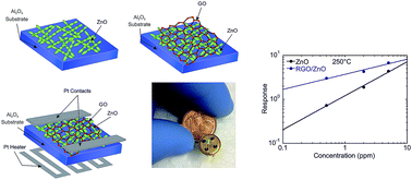
Coupling of graphene-based materials with metal oxide nanostructures is an effective way to obtain composites with improved gas sensing properties. In this work, we prepared a hybrid structure based on graphene oxide (GO) and ZnO nanostructures. The morphological, compositional and structural analyses of the composite material have been investigated using scanning electron microscopy, X-ray diffraction spectroscopy, energy dispersive X-ray analysis and Raman spectroscopy. The gas sensing properties of the obtained structure have been studied towards nitrogen dioxide, hydrogen and methane at relatively low (about 200 °C) operating temperatures. It has been demonstrated that the reduced graphene oxide (RGO)/ZnO composites exhibit 40–50% better response to NO2 and H2 compared to pure ZnO sensors. The obtained results show that the functionalization of the nanostructured ZnO with the RGO sheets is a promising strategy to develop chemical gas sensors with improved gas sensing properties.


1. Introduction

The importance of gas sensors is rapidly increasing for a wide range of applications, including detection of harmful chemical vapors and explosives in public spaces, government and military facilities, and chemical processing plants. Consequently, the development of chemical gas sensors is a highly critical research area that involves health, safety and environmental risks.

One-dimensional (1D) transition metal oxide nanostructures have been extensively investigated for application in gas sensing devices because of their advantages, such as good chemical stability, low cost, easy production, simple measuring electronics, etc.1–5 Among the transition metal semiconducting materials ZnO is one of the most widely investigated structures for chemical sensing due to its high thermal/chemical stability and good oxidation resistance.2,6 Zinc oxide is an n-type semiconductor with a wide band gap energy of 3.37 eV and a large exciton binding energy of 60 meV at room temperature.2,7 The sensing mechanism of ZnO conductometric gas sensor is based on the variation of its electrical conductivity in the presence of reducing and oxidizing gases. Under the exposure to air, oxygen is adsorbed on the surface of the metal oxide, thereby extracting electrons from the conduction band and forming a depletion layer. Oxygen adsorbs as O2, O2 and O2− species depending on surrounding atmosphere, temperature and material. When exposed to reducing gases, the negatively charged oxygen species adsorbed on the surface interact with the reducing gas – oxidizing it exchanging charge. Oxygen ions re-injects the electrons back into the conduction band of the semiconductor, resulting in an increase in electrical resistance. The reaction between the surface adsorbed oxygen species and the target gas may result in a significant change in the electrical resistance of ZnO.8,9 ZnO-based gas sensors usually operate at typical working temperatures of about 200–450 °C.10–12 The high temperature promotes the adsorption/desorption process of oxygen and increase the material response in presence of reducing and oxidizing gases.8 However, the decrease of the material working temperature is an important issue for fabrication of low power consumption and small size sensing devices.

It was reported that ZnO has very high sensitivity mainly towards ethanol and acetone.11,13–17 Detection of other technologically important gases, such as methane (CH4), nitrogen dioxide (NO2), and hydrogen (H2), is of vital interest for wide range of practical applications. CH4 is a highly flammable gas and small spark can cause a violent explosion if the concentration exceeds the threshold limit. It is the major part of natural gas with wide applications in cars, home and industry.18,19 Therefore, the development of CH4 sensors is very important to allow its safe and controlled use. NO2 is a toxic and colorful gas, and it is generated during different combustion processes.20 Detecting and controlling of NO2 in power plants, factories and combustion engines has become an important issue for the environmental monitoring.21,22 H2 is an invisible and odorless gas promising as an alternative fuel for our future energy needs.23 However, because of its high flammability we have to be able to detect any H2 leakage and precisely measure the H2 concentration in fuel cell systems at all stages of H2 production, transportation and storage.24,25 Therefore, the improvement of sensing performance of ZnO for the detection of these toxic, explosive and hazardous gases is of vital importance.

Nanostructured ZnO with different morphologies has attracted an enhanced interest because it can provide large surface area that is advantageous for the enhancement of the sensing performance.2,12,13,26,27 In addition, different methods, such as surface modification, addition of noble catalytic metal, doping and use of multi-compositional structures, have been applied to increase the sensitivity, response and recovery times of ZnO nanostructures.28–32 In this regard, fabrication of composite structures using different materials in single or multiple layers could prove to be an effective way to enhance the sensing properties of ZnO based materials.

Graphene and modified graphene nanomaterials have been studied for applications in gas sensing devices.11,33–35 In addition, graphene and its oxides have been applied as new additives in composite materials for conductometric gas sensors.32,36–39 It was shown, that the combination of p-type reduced graphene oxide (RGO) with the n-type ZnO nanofibers greatly enhanced the response of the ZnO at an operating temperature of 400 °C by creating local heterointerfaces with the n-type ZnO nanograins and acted as the electron acceptors.40 A few recent studies show that the graphene oxide (GO) improves the sensitivity of ZnO to H2, ammonia and oxygen gases indicating that GO is a promising material for the enhancement of the ZnO sensing performance.36–38

In this manuscript, we report the fabrication of RGO/ZnO composites by incorporating the GO sheets to the ZnO nanostructures with subsequent thermal reduction of GO at 250 °C under argon gas atmosphere. The fabricated nanocomposites were tested as a sensing material for detection of NO2, H2 and CH4 gases, in comparison with pure ZnO nanostructures. We found out that the presence of reduced graphene oxide (RGO) enhanced the nanocomposite conductance and improved the response of the composite to all measured gases except CH4. The best improvement (50%) was obtained for H2 at 250 °C. The obtained results showed that the fabricated nanocomposite material is promising for applications in gas sensing devices.

2. Experimental

2.1 Synthesis of the ZnO nanostructures

ZnO nanostructures were synthesized based on the procedure reported in our previous work.41 Briefly, thin films of metallic Zn were deposited on 2 mm square alumina and SiO2 substrates using radio frequency (RF) magnetron sputtering. The average thickness of the obtained Zn thin films was 500 nm. Afterwards, thin films of Zn were anodized in Teflon electrochemical cell using two-electrode system. A platinum foil was used as a counter electrode. Anodization was carried out at room temperature in the solution of 2 M oxalic acid dihydrate (C2H2O4·2H2O) containing ethanol. The applied voltage was 75 V and the anodization time was 15 min. As-anodized structures were zinc oxalate dihydrate (ZnC2O4·2H2O). ZnC2O4·2H2O-to-ZnO transformation of the samples was performed by thermal annealing in the furnace at 400 °C under the atmosphere of 50% O2 + 50% Ar for 4 h.

2.2 Fabrication of RGO/ZnO nanocomposites

Graphite oxide was produced from natural graphite (SP-1, Bay Carbon) using a modified Hummers method, as described elsewhere.42,43 Aqueous dispersions of GO at various concentrations were prepared by stirring graphite oxide solids in pure water (18.0 MΩ cm resistivity, purchased from Barnstead) for 3 hours, and then sonicating the resulting mixture (VWR B2500A-MT bath sonicator) for 45 minutes. For the fabrication of the composite structure, we drop casted 5 μl homogeneous dispersion of the GO with concentration of 0.05 mg ml−1 onto ZnO nanostructures prepared on 2 mm square alumina substrates. Afterwards, the obtained nanocomposite was annealed in the furnace at 250 °C in the atmosphere made of 20% O2 and 80% Ar for 1 h that results in partial reduction of GO in the composite structure.

2.3 Morphological and structural analyses of the obtained structures

The morphological analyses of GO and ZnO structures were carried out using a LEO 1525 scanning electron microscope (SEM) equipped with field emission gun. Elemental composition of the obtained samples was studied by means of energy dispersive X-ray analysis (EDX). Crystal structure of the prepared ZnO was investigated trough the X-ray diffraction (XRD) spectroscopy. XRD was performed using an Empyrean diffractometer (PANalytical, Almelo, The Netherlands) mounting a Cu-LFF (λ = 1.5406 Å) tube operated at 40 kV to 40 mA. XRD spectra were recorded by a parallel-plate collimated proportional Xe detector with a nickel large-β filter, in glancing-angle mode (ω = 1°). An acquisition time of 20 s, a divergence slit of 1/8°, an antiscatter slit of 1/4° and a soller slit of 0.04 rad were used. The diffraction angle was scanned between 30° and 80°. Raman spectroscopy analyses of GO samples were performed using a WITec Micro-Raman spectrometer Alpha 300 with a laser beam wavelength of λ = 488 nm and a 50× optical microscope objective.

2.4 Fabrication and functional characterization of the gas sensors

For gas sensing measurements, platinum electrodes were deposited on the surface of the obtained samples using RF magnetron sputtering. Then a platinum heater was deposited on the backside of the alumina substrate. The schematic and the digital image of the sample and device fabricated for the gas sensing measurements are shown in Fig. 1.
image file: c6ra01913g-f1.tif
Fig. 1 The schematic of the sample and device fabrication steps for gas sensing measurements. (a) Synthesis of ZnO nanostructures on an alumina substrate, (b) preparation of RGO/ZnO nanocomposite and (c) deposition of Pt electrodes and the heater for gas sensing measurements. (d) Digital photograph of the gas sensing device.

Gas sensing properties were tested by means of the flow-through technique at atmospheric pressure, using a constant synthetic airflow (0.3 l min−1) as carrier gas for the analyte dispersion. The desired gas mixtures were obtained by means of a computer controlled gas flow system and 40% relative humidity at 20 °C by mixing dry air with saturated humid air (produced by bubbling synthetic air through a column of water kept at 25 °C and then condensed at 20 °C) in the desired proportions. Before each measurement, the sensors were stabilized at the selected working temperature for 8 h. The sensors were purged with humid synthetic air after every exposure to test gases for at least 30–60 min to allow a full recovery. Sensor conductance was monitored by means of the volt-amperometric technique at constant bias voltage. The working temperature of the sensors was controlled from room temperature to 250 °C, and the resistance of the films was recorded every 30 s.

Gas response (R) to reducing gases was defined as [R = (GfG0)/G0], where G0 is the sample conductance in air, and Gf is the sample conductance in presence of the target gas. The response to oxidizing gases was defined as [R = (RfR0)/R0], where R0 is the sample resistance in air, while Rf is the sample resistance in presence of oxidizing gas. Preliminary measurements were performed to ensure repeatability and reproducibility of the nanocomposite material with the tested gases. The results were reproducible.

3. Results and discussions

3.1 Morphological and structural analysis

Fig. 2(a) shows typical SEM image of the obtained ZnO nanostructures. The ZnO nanoparticles with average diameter of 20 ± 2 nm (standard deviation (SD) is ∼7%) agglomerated into chain-like shaped structures forming ZnO nanowires, as shown in Fig. 2(b). The XRD pattern of annealed ZnO samples shown in Fig. 2(c) indicates the nanocrystalline nature of the oxide film. They can be indexed as a zinc oxide hexagonal structure (ZnO) with unit cell parameter a = b = 3.25 Å and c = 5.2 Å (JCPDS files no. 36-1451). We acquired the results of the EDX analysis from the different regions of the samples using SEM imaging with the high and low magnifications. The EDX spectrum (Fig. 2(d)) and the table of the quantitative analysis (Fig. 2(e)) show stoichiometric ZnO with Zn[thin space (1/6-em)]:[thin space (1/6-em)]O atomic ratio of ∼1[thin space (1/6-em)]:[thin space (1/6-em)]1.
image file: c6ra01913g-f2.tif
Fig. 2 (a) and (b) SEM images of the obtained ZnO nanostructures with the different resolutions. (c) XRD measurements carried out for samples annealed at 400 °C. The layers were prepared on SiO2/Si substrates. The XRD measurements collected at 1° of incidence show the clear presence of ZnO hexagonal structure (° peaks JCPDS files no. 36-1451), * few smaller peaks correspond to the substrate. (d) EDX spectra of the ZnO nanostructure and (e) the table of the quantitative analysis of the obtained structure.

Fig. 3(a) is an optical image of the aqueous solution of GO with a concentration of 0.05 mg ml−1, and the homogeneous color of the solution indicates the highly oxidized and dispersed GO sheets. A SEM image of GO sheets deposited onto a SiO2/Si substrate is shown in Fig. 3(b). The fabrication process yields GO sheets with a wide range of individual sheet dimensions. The lateral size of sheets can range from few nanometers up to some tens of micrometers. Raman spectroscopy characterization of obtained GO sheets deposited onto SiO2/Si wafer is shown in Fig. 3(c). The spectrum shows the presence of two high intensity D and G Raman modes, typical for GO sheets. The Raman D band (∼1365 cm−1) of graphene is activated by the defects that cause an intervalley double resonance involving transitions near two inequivalent K points at neighboring corners of the first Brillouin zone of graphene, and G band (∼1580 cm−1) is the first order Raman peak representing sp2 atomic arrangement of carbon atoms.44 Due to the decrease in size of the in-plane sp2 domains after extensive oxidation and ultrasonic exfoliation,45 the GO sheets exhibit a broad and intense D band.


image file: c6ra01913g-f3.tif
Fig. 3 (a) Image of the aqueous dispersion of GO, (b) SEM image of the GO sheets on SiO2/Si wafer and (c) Raman spectrum of the GO sheets deposited onto SiO2/Si wafer.

Fig. 4 reports the morphology and the compositional analysis of the RGO/ZnO nanocomposite structure. Fig. 4(a) and its magnified image ((b)) clearly show the RGO-decorated ZnO nanoparticles. The EDX spectrum (Fig. 4(c)) and the quantitative analyses of the nanocomposite structure (Fig. 4(d)) confirm the presence of RGO in the samples.


image file: c6ra01913g-f4.tif
Fig. 4 (a) SEM micrograph of the prepared RGO/ZnO nanocomposite, (b) magnified images of (a), (c) EDX spectra of the RGO/ZnO nanocomposite and (d) the table of the quantitative analysis of the obtained RGO/ZnO nanocomposite.

Gas sensing properties of the composite were tested at 200 °C and 250 °C, as described below. It is known that annealing of GO sheets changes the chemical structure of the sheets. In order to obtain reproducible results, the nanocomposites were annealed at 250 °C under the atmosphere of a gas mixture of 20% O2 + 80% Ar for 1 h. In addition, to study the effect of annealing on chemical structure of GO, we annealed the pure GO sheets under the same conditions as we used for the preparation of RGO/ZnO nanocomposites. The variation of C[thin space (1/6-em)]:[thin space (1/6-em)]O ratio in the sheets was checked by EDX after each annealing step in the temperature range of 50–250 °C with a step of 50 °C. The EDX observations were carried out on five different regions of the samples using SEM imaging with different magnifications. The results of the compositional analysis of GO after the each annealing temperature are reported in Table 1. The C[thin space (1/6-em)]:[thin space (1/6-em)]O ratio increased with increasing the annealing temperature (Table 1). Based on these data we argue that the GO sheets in GO/ZnO nanocomposites after annealing at 250 °C are partially reduced resulting in C[thin space (1/6-em)]:[thin space (1/6-em)]O ratio of 82[thin space (1/6-em)]:[thin space (1/6-em)]18, therefore, the annealed nanocomposites were denoted as RGO/ZnO.

Table 1 The results of the compositional analysis of GO annealed at 50–250 °C after each thermal treatment step
Annealing temperature (°C) C (atomic%, ±3%) O (atomic%, ±10%)
As-prepared sample 66 34
50 71 29
100 76 24
150 79 21
200 80 20
250 82 18


3.2 Gas sensing properties of the obtained structures

It was previously demonstrated that graphene in graphene/ZnO composite structures can significantly improve the sensitivity of ZnO to hydrogen, ammonia and oxygen,36–38 and increase the response to ethanol.32,39 Here, in order to check the effect of RGO on gas sensing properties of ZnO we have measured the sensing performances of both the materials (ZnO and RGO/ZnO) for nitrogen dioxide, methane and hydrogen gases. The measurements were carried out at different temperatures: from room temperature to 250 °C (50 °C step). At temperatures lower than 100 °C no significant response of the structures to the different gases was observed. The response and the kinetics increased slowly as a function of the operating temperature at 150 °C. The best results in terms of sensing performances were obtained in the temperature range of 200–250 °C. It should be noted that the RGO in the RGO/ZnO composite structures has stable compositional and electrical properties in the temperature range of the measurements. This is due to the thermal annealing of the composite structures during their preparation, as described the Section 2.2.

Fig. 5 shows the isothermal conductance variation of pure ZnO nanostructures and RGO/ZnO nanocomposite upon exposure to NO2 (1, 2 and 5 ppm) (Fig. 5(a)) and CH4 (100, 250 and 500 ppm) (Fig. 5(b)) at the operating temperature of 200 °C. The relative humidity in the test chamber during functional test was 40% at 20 °C. As one can observe, the presence of RGO increases the conductance value of the composite in air likely because the RGO sheets can serve as a two dimensional conductive platform to electrically interconnect the ZnO nanostructures. Thus, the functionalization of ZnO nanostructures with RGO yielded a highly conductive composite material.


image file: c6ra01913g-f5.tif
Fig. 5 Dynamical response of nanostructured ZnO and RGO/ZnO nanocomposite towards 1, 2 and 5 ppm of NO2 (a) and 100, 250 and 500 ppm of CH4 (b) at a working temperature of 200 °C. The relative humidity in the test chamber was 40% at 20 °C.

Upon gas exposure, the conductance of the layers increased/decreased, in agreement with the behavior of n-type semiconducting materials and reducing/oxidizing gases. Furthermore, with the tested gases, the conductance variation with the species is proportional to the analyte concentration, without displaying saturation effects. The recovery of the air conductance value after sensing tests is complete for methane proving reversible interactions between the analytes and the active materials. For nitrogen dioxide the recovery is not complete that shows a partially irreversible interaction.

To obtain a deeper insight into the system behavior, the response of RGO/ZnO composites to the different analytes has been studied in comparison with the response of pure ZnO samples at 200 and 250 °C temperatures, as shown in Fig. 6(a) and (b). The response spectra are different for the two materials. RGO/ZnO has a higher response to NO2 (6.8@250 °C), and a lower response to CH4 (0.5@250 °C) compared to pure ZnO at both 200 and 250 °C temperatures. The response of both samples (ZnO and RGO/ZnO) is higher at 250 °C compared to the responses at 200 °C. The highest response for both samples is obtained for NO2. The sensor response of RGO/ZnO to 5 ppm NO2 is 5.4 that is about 1.4 times higher than that of pure ZnO nanostructure sensors obtained under the same operating conditions. These experimental data demonstrate that the presence of RGO in the nanocomposites results in almost 40% improvement in NO2 sensing response of pure ZnO nanostructures. Concerning H2 gas detection, the effect of RGO in the nanocomposite sample exhibits a strong dependence on the working temperature: the response of the RGO/ZnO is lower at 200 °C and higher at 250 °C compared to that of the pure ZnO sample (Fig. 6(a) and (b)). The different sensing behavior of the RGO/ZnO samples for different gases may have an impact in development of nanocomposites with an enhanced selectivity for gas detection.


image file: c6ra01913g-f6.tif
Fig. 6 (a) and (b) are the responses towards NO2, H2 and CH4 at 5, 500 and 500 ppm respectively, and working temperatures of 200 °C and 250 °C. (c) and (d) are the calibration curves for nitrogen dioxide of ZnO and RGO/ZnO nanocomposite at an operating temperature of 200 °C and 250 °C. The relative humidity in the test chamber was 40% at 20 °C.

The sensing performances of these materials were also investigated as a function of gas concentrations. The calibration curves (response vs. concentration) obey to the typical relation for semiconductor metal oxide sensing devices:46 response = A[gas concentration]B, where B depends on many factors and changes among different analytes. As an example, the calibration curves for nitrogen dioxide at 200 and 250 °C are reported in Fig. 6(c) and (d), respectively. The responses at both temperatures showed no evidence of saturation throughout the investigated concentration range. Assuming the validity of the previous power law even at low gas concentrations and with the experimental trends reported in Fig. 6(c) and (d), detection limits lower than 100 ppm of nitrogen dioxide may be expected for the nanocomposite.

The Table 2 compares the gas sensing performance of the nanocomposite structure proposed in this work with the recently reported results obtained for the similar composites. A significant beneficial effect of the RGO sheets has been found on the sensor performances, even compared to the recently reported results on RGO/ZnO for the same gases.47 The observed conductance variations were higher for the nanocomposites with respect to the pure ZnO nanostructures for hydrogen and nitrogen dioxide with a big influence of the operating temperatures. RGO may act itself as a catalytic promoter, favoring surface reactions between the specific gases and the oxygen species adsorbed on the sensor surface.48

Table 2 Sensing performance of the composite structure presented in this work in comparison with the previous works
Ref. Structure Shape of ZnO Operating temperature (°C) Test gas (concentration, ppm) Definition of response (S) Response
32 Graphene/ZnO Nanorods 300 Ethanol (10, 50) Ra/Rg 9, 50
36 GO/ZnO Nanorods/nanoplates 340 Ethanol (100) (RaRg)/Ra × 100% 94%
37 RGO/ZnO Nanowires 200 O2 (500) (IaIg)/Ia × 100% 25%
39 GO/ZnO Nanorods/nanoparticles 150 H2 (200) Ra/Rg 3.5
40 p-RGO/ZnO Nanofibers 300 H2 (10) Ra/Rg 1007
47 RGO/ZnO Nanorods 190 CH4 (500) (RaRg)/Ra × 100% 8%
H2 (100) 1%
49 Graphene/ZnO Thin film 200 NO2 (5) (GgGa)/Ga × 100% 36.7%
50 RGO/ZnO Nanoparticles 25 NO2 (5) (GgGa)/Ga × 100% <1%
51 RGO/ZnO Nanoparticles Room temperature NO2 (5) (RaRg)/Ra × 100% 25.6%
This paper RGO/ZnO Nanoparticles-chain-like 250 NO2 (5) (RgRa)/Ra × 100% 680%
This paper RGO/ZnO Nanoparticles-chain-like 250 H2 (500) (GgGa)/Ga × 100% 30%
This paper RGO/ZnO Nanoparticles-chain-like 250 CH4 (500) (GgGa)/Ga × 100% 40%


4. Conclusions

In summary, we fabricated RGO/ZnO nanocomposites using ZnO nanostructures obtained by electrochemical anodization of metallic zinc and GO dispersions produced by the modified Hummers method, and with subsequent annealing at 250 °C under the atmosphere of 20% O2 and 80% Ar. The gas sensing properties of the obtained nanocomposites were investigated by exposing them to NO2, H2 and CH4 gases. RGO/ZnO composites showed better gas sensing performance compared to the pure ZnO nanostructures for NO2 at relatively low working temperatures and for H2 at 250 °C. The response of the RGO/ZnO was significantly higher even compared to the recently reported results on similar composites (based on GO and ZnO) for the same gases.47 The obtained results demonstrate that the architecture of the nanostructured ZnO and its combination with the RGO are very promising for fabrication of high performance gas sensor devices.

Acknowledgements

The research leading to these results has received funding from the following projects: “New approaches and methodologies for bioremediation of water contaminated by chlorinated aliphatic solvents (SUSBIOREM)” (funded by the National Research Council (CNR) and Lombardia Region); “MSP: Multi Sensor Platform for Smart Building Management” (grant agreement no. 611887, funded by the European Commission through its 7th Framework Programme); “FIRB – Oxides at the nanoscale: multifunctionality and applications” (Protocollo: RBAP115AYN, funded by the Italian Ministry of Education); “ORAMA” (grant agreement NMP3-LA-2010-246334, funded by the European Commission through its 7th Framework Programme); “WIROX: Oxide Nanostructures for Wireless Chemical Sensing” (PEOPLE MARIE CURIE ACTIONS, International Research Staff Exchange Scheme, Call: FP7-PEOPLE-2011-IRSES, 2012–2015).

References

  1. M. Righettoni, A. Amann and S. E. Pratsinis, Mater. Today, 2015, 18, 163–171 CrossRef CAS.
  2. M. J. S. Spencer, Prog. Mater. Sci., 2012, 57, 437–486 CrossRef CAS.
  3. V. Galstyan, E. Comini, G. Faglia and G. Sberveglieri, Sensors, 2013, 13, 14813–14838 CrossRef PubMed.
  4. E. Comini, C. Baratto, G. Faglia, M. Ferroni, A. Vomiero and G. Sberveglieri, Prog. Mater. Sci., 2009, 54, 1–67 CrossRef CAS.
  5. Y.-B. Hahn, R. Ahmad and N. Tripathy, Chem. Commun., 2012, 48, 10369–10385 RSC.
  6. X. Liu, N. Chen, X. Xing, Y. Li, X. Xiao, Y. Wang and I. Djerdj, RSC Adv., 2015, 5, 54372–54378 RSC.
  7. A. B. Djurisic and Y. H. Leung, Small, 2006, 2, 944–961 CrossRef CAS PubMed.
  8. N. Yamazoe and K. Shimanoe, Sens. Actuators, B, 2008, 128, 566–573 CrossRef CAS.
  9. G. Sakai, N. Matsunaga, K. Shimanoe and N. Yamazoe, Sens. Actuators, B, 2001, 80, 125–131 CrossRef CAS.
  10. Q. Zhao, Q. Shen, F. Yang, H. Zhao, B. Liu, Q. Liang, A. Wei, H. Yang and S. Liu, Sens. Actuators, B, 2014, 195, 71–79 CrossRef CAS.
  11. N. Hu, Z. Yang, Y. Wang, L. Zhang, Y. Wang, X. Huang, H. Wei, L. Wei and Y. Zhang, Nanotechnology, 2014, 25(2), 025502,  DOI:10.1088/0957-4484/25/2/025502.
  12. W. Guo, T. Liu, H. Zhang, R. Sun, Y. Chen, W. Zeng and Z. Wang, Sens. Actuators, B, 2012, 166, 492–499 CrossRef.
  13. T.-J. Hsueh, S.-J. Chang, C.-L. Hsu, Y.-R. Lin and I. C. Chen, J. Electrochem. Soc., 2008, 155, K152–K155 CrossRef CAS.
  14. X. L. Xu, Y. Chen, S. Y. Ma, W. Q. Li and Y. Z. Mao, Sens. Actuators, B, 2015, 213, 222–233 CrossRef CAS.
  15. Q. Jia, H. Ji, Y. Zhang, Y. Chen, X. Sun and Z. Jin, J. Hazard. Mater., 2014, 276, 262–270 CrossRef CAS PubMed.
  16. M. H. Darvishnejad, A. Anaraki Firooz, J. Beheshtian and A. A. Khodadadi, RSC Adv., 2016, 6, 7838–7845 RSC.
  17. H.-W. Huang, J. Liu, G. He, Y. Peng, M. Wu, W.-H. Zheng, L.-H. Chen, Y. Li and B.-L. Su, RSC Adv., 2015, 5, 101910–101916 RSC.
  18. S. B. Schoonbaert, D. R. Tyner and M. R. Johnson, Appl. Phys. B: Lasers Opt., 2015, 119, 133–142 CrossRef CAS.
  19. KR2011107488-A; KR1115683-B1, 2012.
  20. H. Nguyen Duc and S. A. El-Safty, Chem.–Eur. J., 2011, 17, 12896–12901 CrossRef PubMed.
  21. C. Lee, Y. J. Kim, H. Lee and B. C. Choi, MAX-DOAS Measurements of ClO, SO2 and NO2 in the Mid-Latitude Coastal Boundary Layer and a Power Plant Plume, Springer, 2008 Search PubMed.
  22. D. Buzica, M. Gerboles, F. Lagler and A. Borowiak, Monitoring of SO2, NO2 and O3 in urban areas by diffusive sampling, traceability, proficiency testing and inter-comparison, 2007 Search PubMed.
  23. G. W. Crabtree, M. S. Dresselhaus and M. V. Buchanan, Phys. Today, 2004, 57, 39–44 CrossRef CAS.
  24. N. M. A. Huijts and B. van Wee, Int. J. Hydrogen Energy, 2015, 40, 10367–10381 CrossRef CAS.
  25. R. Takubo, A. Takahashi, J. Imai and S. Funabiki, Electr. Eng. Jpn., 2015, 193, 8–16 CrossRef.
  26. Z. S. Hosseini, A. Mortezaali, A. I. Zad and S. Fardindoost, J. Alloys Compd., 2015, 628, 222–229 CrossRef CAS.
  27. S. Bai, T. Guo, Y. Zhao, J. Sun, D. Li, A. Chen and C. C. Liu, Sens. Actuators, B, 2014, 195, 657–666 CrossRef CAS.
  28. N. S. Ramgir, M. Kaur, P. K. Sharma, N. Datta, S. Kailasaganapathi, S. Bhattacharya, A. K. Debnath, D. K. Aswal and K. Gupta, Sens. Actuators, B, 2013, 187, 313–318 CrossRef CAS.
  29. G. E. Buono-Core, A. H. Klahn, G. Cabello and L. Lillo, Polyhedron, 2013, 62, 1–6 CrossRef CAS.
  30. C.-L. Hsu, Y.-D. Gao, Y.-S. Chen and T.-J. Hsueh, Sens. Actuators, B, 2014, 192, 550–557 CrossRef CAS.
  31. B. Mondal, B. Basumatari, J. Das, C. Roychaudhury, H. Saha and N. Mukherjee, Sens. Actuators, B, 2014, 194, 389–396 CrossRef CAS.
  32. J. Yi, J. M. Lee and W. Il Park, Sens. Actuators, B, 2011, 155, 264–269 CrossRef CAS.
  33. Y. Zhou, Y. Jiang, T. Xie, H. Tai and G. Xie, Sens. Actuators, B, 2014, 203, 135–142 CrossRef CAS.
  34. K. Toda, R. Furue and S. Hayami, Anal. Chim. Acta, 2015, 878, 43–53 CrossRef CAS PubMed.
  35. G. Lu, L. E. Ocola and J. Chen, Nanotechnology, 2009, 20(44), 445502,  DOI:10.1088/0957-4484/20/44/445502.
  36. N. Song, H. Fan and H. Tian, J. Mater. Sci., 2015, 50, 2229–2238 CrossRef CAS.
  37. P. Afzali, Y. Abdi and E. Arzi, Sens. Actuators, B, 2014, 195, 92–97 CrossRef CAS.
  38. S. M. J. Khadem, Y. Abdi, S. Darbari and F. Ostovari, Curr. Appl. Phys., 2014, 14, 1498–1503 CrossRef.
  39. K. Anand, O. Singh, M. P. Singh, J. Kaur and R. C. Singh, Sens. Actuators, B, 2014, 195, 409–415 CrossRef CAS.
  40. Z. U. Abideen, H. W. Kim and S. S. Kim, Chem. Commun., 2015, 51, 15418–15421 RSC.
  41. V. Galstyan, E. Comini, C. Baratto, A. Ponzoni, E. Bontempi, M. Brisotto, G. Faglia and G. Sberveglieri, CrystEngComm, 2013, 15, 2881–2887 RSC.
  42. S. Stankovich, D. A. Dikin, G. H. B. Dommett, K. M. Kohlhaas, E. J. Zimney, E. A. Stach, R. D. Piner, S. T. Nguyen and R. S. Ruoff, Nature, 2006, 442, 282–286 CrossRef CAS PubMed.
  43. I. N. Kholmanov, S. H. Domingues, H. Chou, X. Wang, C. Tan, J.-Y. Kim, H. Li, R. Piner, A. J. G. Zarbin and R. S. Ruoff, ACS Nano, 2013, 7, 1811–1816 CrossRef CAS PubMed.
  44. A. C. Ferrari, Solid State Commun., 2007, 143, 47–57 CrossRef CAS.
  45. Z.-J. Fan, W. Kai, J. Yan, T. Wei, L.-J. Zhi, J. Feng, Y.-m. Ren, L.-P. Song and F. Wei, ACS Nano, 2011, 5, 191–198 CrossRef CAS PubMed.
  46. S. R. Morrison, The chemical physics of surfaces, Plenum Press, New York, 1977 Search PubMed.
  47. D. Zhang, N. Yin and B. Xia, J. Mater. Sci.: Mater. Electron., 2015, 26, 5937–5945 CrossRef CAS.
  48. M. Zhu, P. Chen and M. Liu, Langmuir, 2013, 29, 9259–9268 CrossRef CAS PubMed.
  49. H. Xie, K. Wang, Z. Zhang, X. Zhao, F. Liu and H. Mu, RSC Adv., 2015, 5, 28030–28037 RSC.
  50. N. Kumar, A. K. Srivastava, H. S. Patel, B. K. Gupta and G. Das Varma, Eur. J. Inorg. Chem., 2015, 1912–1923,  DOI:10.1002/ejic.201403172.
  51. S. Liu, B. Yu, H. Zhang, T. Fei and T. Zhang, Sens. Actuators, B, 2014, 202, 272–278 CrossRef CAS.

This journal is © The Royal Society of Chemistry 2016