Two new mixed copper(II)–dipeptide complexes of N,N-donor heterocycle ligands: studies on their non-covalent DNA binding, chemical nuclease, antioxidant and anticancer activities

Qian Gana, Chun-Lian Zhanga, Bing-Feng Wang*a, Ya-Hong Xionga, Yin-Lian Fub, Zong-Wan Maoac and Xue-Yi Le*a
aDepartment of Applied Chemistry, South China Agricultural University, Guangzhou 510642, PR China. E-mail: lexyfu@163.com; wbfeng@scau.edu.cn; Tel: +86 02085280319
bDepartment of Applied Mathematics, South China Agricultural University, Guangzhou 510642, PR China
cSchool of Chemistry and Chemical Engineering, Sun Yat-sen University, Guangzhou 510275, PR China

Received 21st January 2016 , Accepted 27th March 2016

First published on 29th March 2016


Abstract

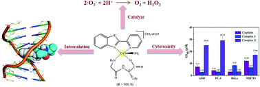
Two novel mononuclear mixed ligand copper(II) complexes, [Cu(Gly-L-val)(HPB)(H2O)]·ClO4·1.5H2O (1) and [Cu(Gly-L-val)(PBT)(H2O)]·ClO4 (2) (Gly-L-val = glycyl-L-valine, HPB = 2-(2′-pyridyl)benzimidazole, PBT = 2-(2′-pyridyl)benzothiazole), have been synthesized and characterized using various analytical and spectroscopic methods. The interactions of the complexes with DNA have been explored by viscometry, thermal denaturation, cyclic voltammetry (CV), agarose gel electrophoresis and spectroscopic means (UV absorption, circular dichroism (CD) and fluorimetry), as well as molecular docking techniques. These studies confirmed the mode of the complexes bound to calf thymus DNA (CT-DNA) through insertion with certain affinity (Kb = 3.211 × 10⁵ M−1 for 1 and 4.734 × 10⁴ M−1 for 2). In the fluorimetric experiments of thermodynamics (KSV = 1.145 × 10⁴ M−1 for 1 and 2.634 × 10³ M−1 for 2), the changes in enthalpy (ΔH > 0), entropy (ΔS > 0) and Gibbs free energy (ΔG < 0) in the interactions between the complexes with DNA suggested that the process occurred spontaneously through hydrophobic interactions. The complexes displayed oxidative cleavage of pBR322 plasmid DNA in the presence of ascorbic acid, probably induced by ˙OH as a reactive oxygen species. Furthermore, the molecular docking technique was applied to ascertain the mode of action for the complexes towards DNA. Moreover, superoxide dismutase (SOD) activity studies were performed using the photoreduction of nitroblue tetrazolium (NBT) under a non-enzymatic system and the antioxidant activities of 1 and 2 determined with IC50 values of 0.337 and 0.146 μM, respectively. The cytotoxicity of the Cu(II) complexes against A549, HeLa, PC-3 tumor cell lines and NIH3T3 (non-tumor cell line) was studied by an MTT assay and it was found that 1 exhibited better cytotoxicity against A549 and PC-3 than 2 and the widely used drug cisplatin.


Introduction

Nowadays, cancer has become one of the most serious and dreadful diseases, and the leading cause of death in the world.1 The clinical success of cisplatin for treating malignancies has attracted a lot of scientists to work towards the development of new metal complexes in order to overcome the drawbacks of cisplatin as an anticancer drug.2,3 Thus, a large number of transition metal complexes have been synthesized and screened for their antitumor activity.4–7 Copper(II) complexes are considered the most promising alternatives to cisplatin because copper is a bio-essential trace element8,9 and plays an important role in vital life processes including iron transport, energy metabolism and respiration.10 Encouragingly, Lena Ruiz and co-workers developed a series of medicines named Casiopeinas® based on copper(II) complexes, which have been approved for clinical trials as antitumor drugs.11,12 In addition, the unique spectroscopic and redox properties of copper(II) complexes compared to small organic compounds are beneficial to the cleavage DNA under physiological conditions because these features are crucial for generating the reactive oxygen species (ROS) necessary for DNA cleavage.13 The ligands HPB and PBT belong to the family of heterocyclic aromatic compounds, which are important fragments in medicinal molecules because of their wide range of biological and pharmaceutical activities such as antifungal,14,15 antibacterial,14,15 antitumor,7,14–16 anti-inflammatory17 and antiviral.18 Additionally, peptides can work as an ancillary ligand contributing to improve the biological compatibility and recognition of the complexes in the physiological system due to their structure being similar to protein. Importantly, some dipeptides such as glycyl-L-valine have been known for their clinical and nutritional importance,19,20 valine is the essential amino acid for the human body and has wide applications in the field of pharmaceutical and food industries,21 and hence, glycyl-L-valine can be a suitable research model as ancillary ligand. Therefore, it is of great interest and significance to explore the biochemical properties of the benzoheterocycle derivative based copper(II) complexes with peptides, in consideration of the possible synergistic effects.

As we all know, DNA plays a vital role in the life process. Extensive studies have proven that DNA is the primary intracellular target of most anticancer drugs. Since it contains all the genetic information required for cellular function and various mutagens can damage its replication machinery, which may block the division of cell and lead to programmed cell death.22,23 Thus, the study of interactions between small molecules and DNA is essential in new antitumor drug discovery research. Metal complexes are known to bind to DNA via covalent or non-covalent interactions, which include intercalative, electrostatic and groove binding modes.24 The trend of current research is preferably non-covalent binding.25 It is worth noting that there has been considerable research on the binding of metal complexes with DNA, but only limited thermodynamic parameters in regard the interaction of complexes to DNA have been measured. It is also important to study the thermodynamic relationship of DNA-complex interactions so as to thoroughly understand the driving forces behind the binding of complexes with DNA and to develop novel and effective DNA-targeted drugs.26

The present work stems from our continued interest in researching the DNA-binding interactions of aromatic heterocyclic derivatives based Cu(II)–dipeptide complexes.27,28 Herein, we have synthesized and characterized two new copper(II) complexes, [Cu(Gly-L-val)(HPB)(H2O)]·ClO4·1.5H2O (1) and [Cu(Gly-L-val)(PBT)(H2O)]·ClO4 (2). The interactions of the complexes with DNA were studied by multispectroscopic methods (UV, CD and fluorimetry), cyclic voltammetry, viscosity, thermal denaturation and agarose gel electrophoresis as well as molecular docking techniques. The thermodynamic relationship of the DNA-complex was revealed. Besides, the antioxidant activities of the complexes were evaluated using the photoreduction of nitroblue tetrazolium (NBT) assays and the in vitro cytotoxicity were assessed by an MTT assay against three human carcinoma cell lines (A549, HeLa, and PC-3) and mouse embryonic fibroblast (NIH3T3). We hope that the research results will be valuable in understanding the DNA-binding properties of the complexes as well as laying a foundation for the rational design of newer and more effective anticarcinogens that target DNA.

Experimental

Materials and instruments

The reagents and chemicals were obtained from commercial sources and used without further purification. The ligands HPB and PBT were synthesized using previously reported procedures.29,30 Glycyl-L-valine was purchased from Aladdin. Calf thymus DNA (CT-DNA) (stored at 4 °C) and ethidium bromide (EthBr) were purchased from Sigma. pBR322 DNA was obtained from MBI Fermentas (Lithuania). 3-(4,5-Dimethylthiazol-2-yl)-2,5-diphenyltetrazolium bromide (MTT), nitroblue tetrazolium (NBT), sodium azide (NaN3), riboflavin (VB2) and tetramethylethylenediamine (TEMED) were purchased from Sinopharm Chemical Reagent Co., Ltd. (Shanghai, China). Human cancer cell lines of HeLa (cervical), A549 (pulmonary), PC-3 (prostatic) and normal cell line of NIH3T3 (mouse embryonic fibroblast) were obtained from the Laboratory Animal Center of Sun Yat-Sen University (Guangzhou, China). Deionized water was used in all experiments.

All experiments involving CT-DNA were performed in Tris–HCl buffer solution (pH 7.2) containing 5 mM Tris–HCl and 50 mM NaCl. Solutions of CT-DNA in Tris–HCl buffer gave a ratio of UV absorbance at 260 and 280 nm (A260/A280) of ca. 1.8–1.9, indicating that the DNA was sufficiently free of protein.31 The concentration of DNA was determined by UV absorbance at 260 nm using the molar absorption coefficient of 6600 M−1 cm−1. DNA solutions were used after no more than 4 days.

Elemental analyses (carbon, hydrogen and nitrogen) were performed using a Vario EL elemental analyzer (Elementar, Germany). ESI-MS were carried out using a API4000 triple quadrupole mass spectrometer (AB Sciex, USA). Molar conductance was measured with a DDS-11A digital conductometer (LeiCi, Shanghai). Electron-spin-resonance (ESR) spectra for the complexes were obtained using a Bruker EMX A300 spectrometer (Bruker, Germany). IR (KBr discs, 4000–400 cm−1), UV-vis, CD and emission spectra were recorded using a VERTEX 70 FT-IR spectrometer (Bruker, Germany), Pharmacia 2550 spectrophotometer (Shimadzu, Japan), Chirascan CD spectropolarimeter (Applied Photophysics, UK) and Hitachi F-4500 fluorescence spectrophotometer (Hitachi, Japan), respectively. Cyclic voltammetry measurements were performed using a CHI 660A Electrochemical Workstation with a standard three-electrode system comprised of Pt-wire as the auxiliary electrode, a glass carbon electrode as the working electrode, and a saturated calomel electrode (SCE) as the reference electrode.

Synthesis of [Cu(Gly-L-val)(HPB)(H2O)]·ClO4·1.5H2O (1)

Gly-L-val (0.087 g, 0.5 mmol) dissolved in water (5 mL), which was deprotonated using a solution of NaOH (0.020 g, 0.5 mmol), was added to a solution of Cu(ClO4)2·6H2O (0.185 g, 0.5 mmol) with stirring. A solution of HPB (0.098 g, 0.5 mmol) in MeOH (20 mL) was added dropwise to the above solution with stirring continued at 50 °C for 2 h. The resulting solution was filtered and kept aside for slow evaporation at room temperature until a blue microcrystalline product was formed. It was then recrystallized from 80% MeOH.

Yield: 0.204 g (71%). Anal. calc. for C19H27N5O9.5ClCu (1): C, 39.58; H, 4.72; N, 12.15; found: C, 39.29; H, 4.70; N, 11.81. IR bands (KBr disc, cm−1): 3420 (b), 3322 (m), 3092 (m), 2960 (m), 1611 (s), 1459 (s), 1382 (m), 1101 (s), 624 (s), 440 (s). UV-vis (MeOH) [λmax/nm (ε/M−1 cm−1)]: 336 (25[thin space (1/6-em)]142), 664 (71.03). Molar conductance, ΛM−1 cm2 mol−1 (1.0 × 10⁻³ M, MeOH, 28 °C) = 113.8. ESI-MS (MeOH): m/z = 431.0 [Cu(Gly-L-val)(HPB)]+. ESR (MeOH): g = 2.2801, g = 2.0589.

Synthesis of [Cu(Gly-L-val)(PBT)(H2O)]·ClO4 (2)

Complex 2 was prepared by the same procedure used for 1, replacing HPB with PBT (0.106 g, 0.5 mmol).

Yield: 0.221 g (78%). Anal. calc. for C19H23N4O8ClSCu (2): C, 40.28; H, 4.09; N, 9.89; found: C, 40.51; H, 3.97; N, 10.06. IR bands (KBr disc, cm−1): 3424 (b), 3336 (m), 3093 (m), 2964 (m), 1625 (s), 1496 (s), 1389 (s), 1096 (s), 624 (s), 419 (s). UV-vis (MeOH) [λmax/nm (ε/M−1 cm−1)]: 308 (18[thin space (1/6-em)]694), 642 (66.75). Molar conductance, ΛM−1 cm2 mol−1 (1.0 × 10⁻³ M, MeOH, 28 °C) = 98.8. ESI-MS (MeOH): m/z = 450.2 [Cu(Gly-L-val)(PBT)]+. ESR (MeOH): g = 2.2798, g = 2.0651.

DNA binding experiments

Electronic absorption titration. Absorption spectra titration was carried out with fixed concentrations of the Cu(II) complexes (50 μM) with a gradually increase in the concentration of DNA. To eliminate the impact of absorbance changes due to DNA, an equal volume of DNA was added to both the sample and reference cells. After addition of DNA to the metal complexes, the resulting solutions were kept for 5 min to equilibrate at room temperature and then, the absorption spectra were recorded in the range of 200–500 nm. In order to quantitatively compare the DNA binding affinities, the intrinsic binding constant Kb was determined using the Wolfe–Shimmer equation:32
[DNA]/(εaεf) = [DNA]/(εbεf) + 1/Kb(εbεf)
where [DNA] is the concentration of DNA in the base pairs, εa, εf and εb correspond to the apparent extinction coefficient (Aobsd/[Cu]), the extinction coefficient of the free (unbound) and fully bound compound, respectively. From the plots of [DNA]/(εaεf) vs. [DNA], the binding constants Kb were given by the ratio of the slope to the intercept.
Fluorimetric studies. EthBr emits an intense fluorescence in the presence of DNA due to its strong intercalation between adjacent DNA base pairs. The fluorescence can be quenched when a second molecule is added to the solution and replaces EthBr. In the fluorescence quenching studies, the concentrations of DNA and EthBr were kept constant (10 μM and 8 μM, respectively), while varying the complex concentration from 0 to 11.7 × 10⁻⁵ M at three different temperatures (300, 305 and 310 K). The fluorescence spectra were measured using an excitation wavelength at 525 nm and emission wavelength in the range of 540 to 680 nm. For each addition, the solutions were kept for 10 min to equilibrate.
Viscosity experiments. Viscosity experiments were performed using an Ubbelohde viscometer maintained at a constant temperature (29.0 ± 0.1 °C) in a thermostatic water-bath. The flow time was measured using a digital stopwatch, each sample was measured three times and an average flow time was calculated. The data for (η/η0)1/3, where η and η0 represent the viscosity of CT-DNA (200 μM) in the presence and absence of the complexes, respectively, were plotted against r ([complex]/[DNA] = 0.0–0.35). The viscosity values were calculated according to the relationship η = (tt0)/t0, where t and t0 are the observed flow time in the presence and absence of the samples, respectively.

DNA melting experiments

DNA melting experiments were carried out using a Perkin-Elmer Lambda 35 spectrophotometer equipped with a Peltier temperature-controlling programmer (±0.1 °C). The absorbance at 260 nm was continuously monitored for solutions of CT-DNA (50 μM) in the absence and presence of the Cu(II) complexes (2.5 μM). The temperature was increased at a rate of 1 °C min−1 from 30 to 90 °C. The melting temperature (Tm) was taken as the mid-point of the hyperchromic transition. The data were presented as (AA1)/(AfA1) versus temperature, where A, A1 and Af are the observed, initial and final absorbance at 260 nm, respectively.

Circular dichroism (CD) studies

The CD spectra of CT-DNA (100 μM) were recorded in 5 mM Tris–HCl/50 mM NaCl buffer solution (pH 7.2) at room temperature by increasing the [complex]/[CT-DNA] ratios (r = 0.0, 0.2, 0.4, 0.6, 0.8 and 1.0). The test system was protected by a nitrogen atmosphere during the experiment. The spectra were measured as the average of three scans from 220 to 320 nm and subtracting the buffer background.

Cyclic voltammetry

All cyclic voltammetric experiments were carried out in a single compartment cell at 25 °C. The supporting electrolyte was comprised of 10 mM Tris–HCl/50 mM NaCl buffer solution (pH 7.2). The experiments were performed in 0.3 mM complex solutions in the absence and presence of CT-DNA. Before the experiments, all solutions were deoxygenated with N2 for 10 min and were kept under a N2 atmosphere throughout the experiments.

DNA cleavage experiments

The cleavage of supercoiled pBR322 plasmid DNA (250 ng) was monitored using agarose gel electrophoresis. The samples were incubated for 1 h at 37 °C in the dark. A loading buffer was added and electrophoresis performed at 100 V for 40 min in Tris–boric acid–EDTA (TBE) buffer (pH 8.3) using 0.8% agarose gel containing 5 μL GoldView. Control experiments were carried out using 50 μM ascorbic acid, 20 μM Cu(ClO4)2·6H2O and 20 μM ligands. After electrophoresis, the bands were visualized by UV light and photographed using a Bio-Rad Laboratories-Segrate Gel Imaging System.

The DNA cleavage mechanisms of the complexes were investigated in the presence of typical reactive oxygen species scavengers such as DMSO, tert-butyl alcohol and ethanol (hydroxyl radical), sodium azide (NaN3) (singlet oxygen) and superoxide dismutase (SOD) (superoxide anion radical). Each sample was treated and analyzed according to the procedure described above.

Molecular docking studies

The molecular docking studies were performed using the AutoDock Vina1.1.2 (ref. 33) program to investigate the interactions between the complexes and DNA containing iodinated 8-mer oligonucleotides d(5′-G-dIU-TGCAAC-3′) (PDB ID: 454D).34,35 This DNA fragment included a cavity between GC/GC consecutive base pairs. Meanwhile, the water molecules and substrate were deleted. Polar hydrogens were added and Gasteiger charges were calculated. The DNA was enclosed in a grid box with 60 × 60 × 60 points in which almost involved the entire DNA molecule and the binding site was located in the center of DNA molecule. Other parameters were the default settings. The structures of the complexes were sketched using Gaussian viewer. In addition, the full geometry optimizations were performed with the Gaussian09 program package using the hybrid density functional theory (B3LYP) method and 6-31G(D) basis set. All calculations were carried out on Dell T7500 server with dual XEON 5660 cores, RedHat linux operating system. Visualization of the docked pose was carried out using PyMol molecular graphics program.

Superoxide radical scavenging activity

The superoxide radical (O2˙) scavenging activity of the title complexes were determined using the modified nitroblue tetrazolium (NBT) photoreduction method.28 A non-enzymatic system of TEMED–VB2–NBT containing the tested complexes (0.05–0.80 μM), TEMED (100 μM), VB2 (6.80 μM) and NBT (93.20 μM) in 0.05 M phosphate buffer (pH 7.8) was applied. The reactions were monitored by detecting the concentration of the reduced NBT (blue formazan) at 560 nm. The inhibition ratio (I) of NBT reduction was calculated according to the following equation:
I (%) = (A0Ai)/A0 × 100
where A0 and Ai represent the slopes of the straight line of absorbance values as a function of time in the absence and presence of the title complexes, respectively. The IC50 value is the concentration of the complex, which causes 50% inhibition of NBT reduction. Each sample was measured in three parallel experiments.

In vitro cytotoxicity assays (MTT)

In vitro cytotoxicity tests were carried out using the MTT colorimetric assay. Three different human tumor cell lines of HeLa (cervical), A549 (pulmonary), PC-3 (prostatic) and mouse embryonic fibroblast NIH3T3 (normal cell line) were seeded into 96-well microtiter plates (1 × 10⁴ cells per well) and incubated overnight in a humidified atmosphere containing 5% CO2 at 37 °C. The tested compounds were dissolved in DMSO and diluted with RPMI 1640 medium supplemented with 10% fetal bovine serum (FBS), 100 units per mL penicillin and 50 units per mL streptomycin to the required concentrations (ranging from 1.56 to 100 μM) prior to use. Control wells were prepared by the addition of culture medium (200 μL). The plates were incubated in a 5% CO2 incubator at 37 °C for 48 h. Then, a stock MTT dye solution (20 μL, 5 mg mL−1) was added to each well after 4 h of incubation. The MTT formazan formed was then dissolved in 150 μL DMSO and the optical density (OD) of each well was then measured at 595 nm on a microplate spectrophotometer. The anti-growth effects of the tested compounds are expressed as GI50 values, which indicate the concentration of the compound that inhibits 50% of the cell growth. The GI50 value was calculated using the equation: 100 × (TiT0)/(CT0) = 50,36,37 where T0 and Ti indicate the OD values at time zero and after the treatment period, respectively, and C indicates the OD value measured in untreated cells (control) after an incubation period equal to the treatment period.

Results and discussion

Synthesis and characterization

The titled complexes were isolated by the reaction of copper(II) perchlorate hexahydrate with glycyl-L-valine and HPB/PBT in equimolar quantities using 80% MeOH as the solvent (Scheme 1). The complexes were characterized by elemental analyses, IR, UV-vis, ESI-MS, ESR and molar conductance measurements. The obtained results for the elemental analyses were found to be in good agreement with the calculated values, which confirmed the formation of the complexes. The molar conductance values of the two complexes in methanol indicate that the complexes are 1[thin space (1/6-em)]:[thin space (1/6-em)]1 type electrolytes.38 The ESI-MS of the complex 1 displayed a peak at m/z = 431.0 in methanol matching exactly with [Cu(Gly-L-val)(HPB)]+ (m/z = 450.2 for 2, [Cu(Gly-L-val)(PBT)]+).
image file: c6ra01868h-s1.tif
Scheme 1 Synthetic route for the ligands and complexes.

The electronic absorption spectra of the complexes were recorded in methanol at room temperature. The intense bands at 316 nm for 1 and 309 nm for 2 were assigned to the intra-ligand (π → π*) transitions of the HPB and PBT ligands, respectively. The broad and weak bands were observed at the lower frequency of 664 and 642 nm in the spectrum of 1 and 2, respectively, corresponding to the d → d transitions of copper(II) indicating the approximate square pyramidal geometry around copper(II).39

In the IR region, the peak near at 3420 cm−1 was attributed to the stretching vibration ν(–OH) of the water molecule. The band near 3322 cm−1 for 1 (3336 cm−1 for 2) was the stretching vibration ν(–NH) of the secondary amide in the dipeptide. In addition, the two bands at approximately 3092 and 2920 cm−1 for 1 (3093 and 2964 cm−1 for 2) were attributed to asymmetric νas(–NH2) and symmetric νs(–NH2) stretching frequencies, respectively. The absence of any band in the region of 1700–1750 cm−1 for the complexes suggests coordination of the carboxylate group of the dipeptide to the central copper ion. Besides, the difference values Δν = [νas(–COO) (1, 1611 cm−1; 2, 1625 cm−1) − νs(–COO) (1, 1382 cm−1; 2, 1389 cm−1)] > 200 cm−1 can be explained by the fact that the COO group was coordinated to the central Cu2+ in a unidentate manner.40 Moreover, the band at 1459 cm−1 for 1 (1496 cm−1 for 2) can be assigned to the ring stretching frequencies ν(C[double bond, length as m-dash]N) of the ligand (HPB/PBT), indicating that HPB/PBT was coordinated to the central Cu2+. The complexes showed very strong bands at 1101 cm−1 for 1 and 1096 cm−1 for 2, which can be attributed to the ν(Cl–O) of ClO4. The bands at 440 and 624 cm−1 for 1 (419 and 624 cm−1 for 2) most likely belong to the ν(Cu–N) and ν(Cu–O) stretching vibrations, respectively.

The X-band ESR spectra of the complexes were recorded in MeOH (100 K) at a frequency of 9.46 GHz under the magnetic field strength 3000 ± 1000 gauss using tetracyanoethylene (TCNE) as the field marker. The ESR spectra revealed an anisotropic signal with g = 2.2801, g = 2.0589, and A = 167 G for 1 and g = 2.2798, g = 2.0651, and A = 163 G for 2. The parameters g > g > ge (2.0023) revealed that the unpaired electron was located in the dx2y2 orbital of the Cu(II) ions, characteristic of local symmetry, indicating the nearly square pyramidal geometry of the complexes,41 which is in agreement with the results obtained from the electronic absorption spectra.

Therefore, from the above results, a tentative structure was proposed for the two Cu(II) complexes with an approximate square-pyramidal geometry where the four equatorial positions were occupied by HPB/PBT (N,N) and dipeptide (N,O), and the axial position was occupied by a H2O molecule.

Electronic absorption spectra studies

Electronic absorption spectroscopy is an efficient and widely employed method used to study the binding mode of metal complexes to DNA. In general, the non-covalent binding of small molecules to DNA involves three interaction modes: groove binding, intercalation and electrostatic interactions. Intercalative binding is the most effective mode for drugs targeted to DNA, which is associated with the DNA damage or antineoplastic activity of the complexes. The effect of hypochromism and red shifts can be observed with the intercalative mode of the complexes to DNA involving a stacking interaction between aromatic chromophore and the base pairs of DNA. The absorption spectra of the two complexes incubated with increasing concentrations of CT-DNA are given in Fig. 1. The absorption bands of complexes 1 and 2 decreased in molar absorptivity (hypochromism, 1: 28.9% and 2: 17.8%) as well as 1–2 nm bathochromism, which may be attributed to the intercalation between the heteroaromatic rings of the complexes and the DNA base pairs. The extent of the hypochromism commonly parallels the affinity of a compound binding to DNA and the strength of the intercalative interaction.42 The binding constants (Kb) were calculated as 3.211 × 10⁵ M−1 (1) and 4.734 × 10⁴ M−1 (2), respectively, which are smaller than that of the classical intercalator EthBr.43 The higher Kb value for 1 compared to 2 shows that the binding ability of 1 to DNA was stronger than 2. This may be related to the difference of the benzoheterocycle moiety in the complexes. In addition, when compared to 2, 1 exhibits a higher number of hydrogen bonds to DNA, as seen from the molecular docking studies (cf. below).
image file: c6ra01868h-f1.tif
Fig. 1 The electronic absorption spectra of 1 (a) and 2 (b) (50 μM) in the absence and presence of CT-DNA. Inset: linear plots for the intrinsic DNA binding constant (Kb).

Fluorescence studies

Fluorescence spectroscopy is employed to investigate the interaction characteristics between a chromophore and other compounds. The fluorescence quenching efficiency was evaluated by the Stern–Volmer constant (KSV) according to the classical Stern–Volmer equation:
F0/F = 1 + KSV[Q]
where F0 and F correspond to the fluorescence intensities of EthBr–DNA in the absence and presence of the complexes, respectively, and [Q] is the concentration of the complexes. KSV is a linear Stern–Volmer quenching constant obtained from the linear regression of F0/F with [Q]. As shown in Fig. 2, the emission band of the EthBr–DNA system decreased in intensity with an increase in the concentration of the two complexes, which suggested that the complexes could displace EthBr at their DNA binding sites, which is characteristic for the intercalative binding of the complexes to DNA. The obtained KSV values at 300 K for complexes 1 and 2 were found to be 1.145 × 10⁴ M−1 and 2.634 × 10³ M−1, respectively. Complex 1 exhibits a stronger DNA binding affinity when compared with 2. The result was consistent with that of absorption spectroscopic studies.

image file: c6ra01868h-f2.tif
Fig. 2 The emission spectra of EthBr (8 μM) bound to CT-DNA (10 μM) in the absence and presence of 1 (a) and 2 (b) in 5 mM Tris–HCl/50 mM NaCl buffer (pH 7.2) at 300 K with λex = 525 nm. Inset: the Stern–Volmer quenching curve.

Fluorescence quenching studies

Fluorescence quenching refers to the process in which the fluorescence intensity of the fluorophore decreases after interaction with a quencher, including collisional quenching, excited-state reactions, energy transfer ground-state complex formation and molecular rearrangements. The mechanisms of quenching are divided into two types: dynamic and static quenching. The two mechanisms can be distinguished according to the relationship of temperature dependence of the KSV. In general, dynamic quenching depends on diffusion, higher temperatures lead to larger diffusion coefficients, which cause an increased collision probability and the KSV can be increased upon increasing the temperature. In contrast, increased temperatures decreases the compounds stability and thus, lower values of KSV are obtained in the static quenching.44 As seen in Fig. 3, the Stern–Volmer plots were linear, which showed that only one type of quenching process occurs, either dynamic or static quenching. Obviously, the KSV increased with temperature (1.145 × 10⁴ M−1 (300 K), 1.173 × 10⁴ M−1 (305 K), 1.203 × 10⁴ M−1 (310 K) for 1 and 2.634 × 10³ M−1 (300 K), 3.102 × 10³ M−1 (305 K), 3.474 × 10³ M−1 (310 K) for 2, Table 1), indicating that the fluorescence quenching process was a dynamic quenching mechanism.
image file: c6ra01868h-f3.tif
Fig. 3 Plots of F0/F versus the concentration of the Cu(II) complex for the binding of 1 (a) and 2 (b) with DNA at different temperatures.
Table 1 The quenching constants and thermodynamic parameters obtained for the binding of the complexes to DNA at different temperatures
Complex T (K) KSV (M−1) R2 ΔG0 (kJ mol−1) ΔH0 (kJ mol−1) ΔS0 (J mol−1 K−1)
1 300 1.145 × 10⁴ 0.982 −23.310 3.821 90.438
305 1.173 × 10⁴ 0.991 −23.760 3.821 90.438
310 1.203 × 10⁴ 0.988 −24.215 3.821 90.438
2 300 2.634 × 10³ 0.994 −19.645 21.402 136.823
305 3.102 × 10³ 0.998 −20.387 21.402 136.823
310 3.474 × 10³ 0.996 −21.013 21.402 136.823


Thermodynamic studies

In order to have a better understanding of the driving forces behind the binding of metal complexes to DNA, the thermodynamic parameters (such as enthalpy (ΔH), entropy (ΔS) and Gibbs free energy (ΔG)) with three different temperatures were obtained and analyzed. The main interaction forces in regard to non-covalent binding contain hydrogen bonding interactions, van der Waals forces, electrostatic interactions and hydrophobic interactions.45 If ΔH > 0 and ΔS > 0, the main force is hydrophobic interactions; if ΔH < 0 and ΔS < 0, the major force is the van der Waals forces or hydrogen bonding; if ΔH < 0 or ΔH ≈ 0 and ΔS > 0, electrostatic forces play the major role in the reaction.46 The values of ΔH, ΔS and ΔG for a binding reaction were calculated from the Vant't Hoff equation and Gibbs–Helmholtz equation:47
ln[thin space (1/6-em)]K = −ΔH/RT + ΔS/R

ΔG = ΔHTΔS = −RT[thin space (1/6-em)]ln[thin space (1/6-em)]K
where K is the Stern–Volmer quenching constant at the corresponding temperature and R is the gas constant. The ΔH can be regarded as a constant if the temperature does not vary significantly. The thermodynamic parameters are listed in Table 1. The positive ΔH and ΔS values suggested that the binding of the complexes to DNA is an endothermic and entropy increasing process. In addition, the hydrophobic interactions play a major role in this process. Moreover, the negative value of ΔG reveals that the interaction process was spontaneous. The binding process of the complexes with CT-DNA was entropy-driven due to |ΔH| < |TΔS|.

Viscosity measurements

The viscosity of CT-DNA upon treatment with varying concentrations of the complexes was measured to clarify the interaction nature between the complexes and DNA. As it is sensitive to the change in the length of the DNA molecule, hydrodynamic measurement (i.e. viscosity) is regarded as the least ambiguous and the most critical tests of a DNA binding mode in solution in the absence of crystallographic or NMR structural data.48 The intercalation mode will cause an increase in the relative viscosity of DNA, but the non-intercalation binding mode has little effect on the relative viscosity of DNA during the binding process.49,50 As shown in Fig. 4, the relative viscosity of DNA steadily increases upon increasing the concentration of the complexes. This may be explained by the fact that the complexes can insert between the DNA adjacent base pairs, leading to an increase in the separation of the base pairs at the insertion sites and as a consequence, an increase in the overall DNA contour length. The increased degree of the viscosity of DNA related to the interaction is in the order 1 > 2, indicating that the DNA binding affinity of 1 was greater than 2. This was consistent with the results of the absorption and emission spectral studies.
image file: c6ra01868h-f4.tif
Fig. 4 The effect of increasing amounts of EthBr and the complexes on the relative viscosity of CT-DNA in Tris–HCl buffer at pH 7.2 at 29 ± 0.1 °C. Conditions: [DNA] = 2.0 × 10⁻⁴ M and [complex]/[DNA] = 0, 0.05, 0.10, 0.15, 0.20, 0.25, 0.30 and 0.35.

DNA thermal denaturation studies

The hydrogen bonding and base stacking interactions between the strands of the double helix in DNA can be disrupted by heating, resulting in the separation of the double helix into two single strands. DNA melting experiments can give further insight into conformational changes of DNA when the temperature is raised and offers information about the interaction strength of the complexes with DNA.51 The melting temperature (Tm) of DNA is defined as the temperature at which half of the double helical DNA strands are unfolded to single strands.52 Generally, intercalation of natural or synthesized organic- and metallo-intercalators into the DNA double helix increases the thermal stability of the helix and thereby causes an appreciable enhancement in the Tm of DNA. However, groove binding leads to an imperceptible variation in the value of Tm. The melting profiles of CT-DNA in the absence and presence of the Cu(II) complexes are provided in Fig. 5. The Tm of free CT-DNA was determined to be 60.1 °C. The melting point increased by 10.8 °C for complex 1 and 5.1 °C for complex 2 at a concentration ratio of [Cu]/[DNA] = 0.05, respectively. The obvious increases (∼5 or 11 °C) in the Tm indicate the intercalative binding of the two complexes to DNA. In addition, the interaction strength of 1 was greater than 2, in conformity with the above results obtained with the absorption spectra and viscosity measurements.
image file: c6ra01868h-f5.tif
Fig. 5 The melting curves of CT-DNA in the absence (■) and presence of complexes 1 (●) and 2 (▲). [Cu] = 2.5 μM and [DNA] = 50 μM.

Circular dichroism spectra

Circular dichroism (CD) is a powerful technique used to monitor the possible conformational changes of biomolecules (DNA or proteins) upon interaction with metal complexes, as the CD signals are quite sensitive to the interaction mode. The observed CD spectrum of CT-DNA exhibited a positive band at 274 nm and a negative band at 246 nm, owing to the π–π stacking of base pairs and right-hand helicity, respectively, which is characteristic of the B-form DNA.53 As shown in Fig. 6, the intensities of both the positive and negative bands decreased significantly (shifting to zero levels) with an increasing [complex]/[DNA] ratio, with the former being affected slightly more than the latter. Meanwhile, the positive and negative bands both showed red shifts (6–7 nm and 2–3 nm for the positive and negative bands, respectively). This suggested that the interactions of the complexes with DNA disturbed the right handed helicity and base stacking of DNA and thus, induced certain conformational changes in the B-DNA, such as the conversion from a B → C like structure.54 The larger decrease in the positive bands indicated that the complexes intercalated into the DNA base pairs and thereby unwind the double helix.55,56 Moreover, the changing intensity of 1 exceeded 2, which was in agreement with the results obtained by the methods described above.
image file: c6ra01868h-f6.tif
Fig. 6 The CD spectra of CT-DNA in the absence and presence of 1 (a) and 2 (b) in 5 mM Tris–HCl/50 mM NaCl buffer at room temperature. Conditions: [DNA] = 1.0 × 10⁻⁴ M and [1 or 2]/[DNA] = 0.0, 0.2, 0.4, 0.6, 0.8 and 1.0.

Redox behavior

In order to further understand the binding nature of the Cu(II) complexes to DNA, electrochemical methods were employed to study the redox behavior between the metal complexes and DNA. The typical cyclic voltammograms of the copper(II) complexes in the absence and presence of CT-DNA are shown in Fig. 7, and the related data are summarized in Table 2.
image file: c6ra01868h-f7.tif
Fig. 7 The cyclic voltammograms for complex 1 (a) and 2 (b) in the absence (solid line) and presence (dash line) of CT DNA. [1 or 2] = 0.3 mM, [DNA]/[complex] = 3 and scan rate = 200 mV s−1.
Table 2 The electrochemical parameters obtained for the interactions of DNA with the complexes
Complex Epa/V Epc/V ΔEp/V E1/2/V Ipa/μA Ipc/μA Ipa/Ipc
1 0.236 −0.140 0.376 0.048 3.862 −3.691 1.046
1 + CT-DNA 0.363 −0.133 0.496 0.115 3.227 −3.235 0.998
2 0.194 0.087 0.107 0.141 2.469 −2.654 0.930
2 + CT-DNA 0.212 0.087 0.125 0.150 2.142 −2.084 1.028


The ratio of the oxidation peak current to reduction peak current (Ipa/Ipc) ≈ 1 and indicated that the redox process was quasi-reversible. No new peaks appeared after the addition of CT-DNA to the complexes with R ([DNA]/[complex]) = 3. Complexes 1 and 2 all experienced a significant reduction in Ipa and Ipc, indicating that the complexes are bound strongly to DNA. According to Kelly and coworkers, the complex was bound to DNA and the product was non-electroactive, decreasing the concentration of electroactive species in solution resulting in a decrease in the peak currents.57 The observed decrease in the voltammetric current in the presence of DNA was attributed to slow diffusion of the complexes bound to DNA. Meanwhile, the anodic peak potential (Epa) and formal potential (E1/2) showed positive shifts. Generally, when the metal complex binds DNA via electrostatic interactions, the electrochemical potential of the complex will shift in the negative direction. If it intercalates into DNA, the potential will shift positively.58 Therefore, the positive shifts in the CV peak potentials of the complexes indicated an intercalative binding mode of the complexes to DNA. This binding was considered principally due to hydrophobic interactions.59

The separation between E0′b and E0′f can be used to estimate the ratio of binding constants of the reduced and oxidized forms to DNA using the following equation:54

E0′bE0′f = 0.059[thin space (1/6-em)]log[K+/K2+]
where E0′fand E0′b are the formal potentials of the Cu(I)/Cu(II) couple in the free and bound species, respectively, and K+ and K2+ are the corresponding binding constants for the binding of the Cu(I) and Cu(II) species to DNA, respectively (Scheme 2). The values of K+/K2+ were 13.6 for 1 and 1.4 for 2, suggesting that the interaction of Cu(I) form with DNA was stronger than Cu(II) form. The more prominent decrease in the peak currents and greater shift in the peak potentials observed for 1 over 2 upon the addition of DNA indicate that the binding affinity of the complexes to DNA follows the order of 1 > 2. The result was consistent with the experiment data described above.


image file: c6ra01868h-s2.tif
Scheme 2

Chemical nuclease activities

In order to assess the chemical nuclease activities of the complexes for DNA strand scission, pBR322 DNA was incubated with the complexes and the cleavage reactions were monitored by agarose gel electrophoresis. When plasmid DNA was subjected to electrophoresis, the fastest migration will be observed for the intact supercoiled DNA (Form I). If scission occurs on one strand, the supercoiled DNA will relax to generate a slower-moving open circular form (Form II). Once both strands are cleaved, a linear form (Form III) will be generated that migrates between Form I and Form II.60 As shown in Fig. 8, control experiments performed using only ascorbic acid, Cu(ClO4)2·6H2O or ligands alone in separate lanes showed no prominent cleavage of DNA. Meanwhile, no apparent DNA cleavage was observed for the complexes (20 μM) without ascorbic acid (lane 6). The complexes converted Form I to Form II and III with an increase in the concentration of the complexes in the presence of 50 μM ascorbic acid (lanes 7–10), providing clear evidence of direct double-strand DNA cleavage. The double-strand breaks are thought to be more significant than single-strand breaks as sources of cell lethality, because they are apparently less readily repaired by DNA repair mechanisms.61 Under identical experimental conditions, complex 1 shows a higher cleavage ability (Fig. 9), which was traced to its higher DNA binding affinity and Cu(II)/Cu(I) redox potential (cf. above).
image file: c6ra01868h-f8.tif
Fig. 8 Cleavage of plasmid pBR322 DNA (250 ng) by 1 (a) and 2 (b) after 1 h of incubation at 37 °C. Lane 1, DNA control; lane 2, DNA + ascorbic acid (50 μM); lane 3, DNA + Cu(ClO4)2·6H2O (20 μM); lane 4, DNA + HPB/PBT (20 μM); lane 5, DNA + Gly-L-val (20 μM); lane 6, DNA + 1 or 2 (20 μM); lanes 7–10, DNA + ascorbic acid (50 μM) + 1 or 2 (5, 10, 15 and 20 μM, respectively).

image file: c6ra01868h-f9.tif
Fig. 9 A histogram of the percentage of degraded DNA (lanes 7–10).

Copper complexes can cleave DNA through an oxidative mechanism or hydrolytic mechanism. In the process of oxidative DNA cleavage, it may lead to the formation of reactive hydroxyl radical (˙OH), superoxide anion radical (O2˙) and/or singlet oxygen (1O2) species, which cause damage to the base and/or sugar of DNA.62 To reveal among the reactive oxygen species (ROS), which are responsible for the DNA cleavage, ROS scavengers were used and the results are illustrated in Fig. 10. When the hydroxyl radical (˙OH) scavengers (DMSO, tert-butyl alcohol, and ethanol) were added to the reaction mixture respectively, inhibition of DNA cleavage was observed revealing that the cleavage reaction involved ˙OH. The presence of NaN3 (1O2 quencher) didn't significantly reduce the efficiency of DNA cleavage, ruling out the possibility of the involvement of diffusible 1O2 or singlet oxygen-like entities in the cleavage reaction. Furthermore, DNA cleavage was promoted by adding superoxide dismutase (O2˙ scavenger) to the reaction mixture (lane 8) and free SOD did not exhibit any DNA cleavage (lane 2), which validates that O2˙ could be involved in the DNA strand scission in a round-about way, in which the complexes can catalyze the dismutation O2˙ to generate H2O2 and O2. Moreover, it has been known that the Cu(I) species bind to DNA with an affinity higher than Cu(II) species from the above electrochemical test. Thus, the DNA was made more accessible for ROS (˙OH) produced by a Fenton type reaction,63 resulting in higher DNA cleavage.


image file: c6ra01868h-f10.tif
Fig. 10 Cleavage of plasmid pBR322 DNA (250 ng) by 1 (a) and 2 (b) in the presence of different conventional reactive oxygen species scavengers after 1 h of incubation at 37 °C. Lane 1, DNA control; lane 2, DNA + ascorbic acid (50 μM) + SOD (15 units); lane 3, DNA + ascorbic acid (50 μM) + 1 or 2 (10 μM); lanes 4–8, DNA + ascorbic acid (50 μM) + 1 or 2 (10 μM) + [DMSO (0.2 M), tert-butyl alcohol (0.2 M), EtOH (0.2 M), NaN3 (0.2 M) and SOD (15 units), respectively].

Molecular docking analysis

The molecular docking technique is an attractive scaffold to better understand the interaction of a macromolecule (receptor) and a small molecule (ligand). It can contribute to rational drug design and mechanistic studies by placing a small molecule into the binding site of the DNA target specific region mainly in a non-covalent mode.64 As shown in Fig. 11, the resulting model showed that 1 and 2 could intercalate into the cavity between GC/GC consecutive base pairs through heteroaromatic rings and stabilized by hydrophobic interactions and hydrogen bonding. There are certain hydrogen-bonding interactions between complex 1 and DNA, such as complex 1: H16⋯454D:DG-4:N7 (1.8 Å), complex 1: H18⋯454D:DC-13:O4′ (2.6 Å), complex 1: H19⋯454D:DC-5:N4 (2.6 Å) and complex 1:H19⋯454D:DG-12:O6 (1.8 Å). In addition, the hydrophobic interactions are the predominant forces between complex 2 and DNA in the absence of hydrogen bonding. The resulting relative binding energy of docked 1 and 2 with DNA were found to be −34.31 and −30.12 kJ mol−1, respectively. The more negative the relative binding energy, the more potent the binding as between complex and DNA. Thus, the results indicated that 1 binds to the DNA stronger than 2, which correlated well with the experimental DNA binding results.
image file: c6ra01868h-f11.tif
Fig. 11 The molecular docked models of the complexes with DNA (a: complex 1; b: complex 2). The hydrogen bonds between 1 and DNA are represented using yellow dashed lines.

Antioxidant activity

The superoxide anion radical (O2˙) is one of the most reactive species of ROS, which can result in cell membrane and DNA damage, aging, chromosomal aberrations, carcinogenesis and further initiate or propagate the development of many diseases.65,66 Therefore, it's important for further investigation of the O2˙ scavenging activities of the metal complexes. As shown in Fig. 12, the inhibition ratio was concentration-dependent and increased with the sample concentration. 1 and 2 exhibited excellent antioxidant activity with IC50 values of 0.337 and 0.146 μM, respectively. This can be related to the flexible geometry around the center Cu2+ contributing to the O2˙ approach, with fast exchange of the weakly coordinated axial H2O molecule.67 Moreover, the electrons of the nitrogen heteroaromatic ligands, such as pyridine and thiazole, can facilitate the stability of the complex – O2˙ interactions in favour of high SOD activity. The complexes are potent SOD mimics due to their significantly lower molecular weight when compared with that of the native Cu, Zn-SOD (MW 32[thin space (1/6-em)]000 Da), although the activities of the complexes are slightly lower than the SOD (IC50 ∼ 0.04 μM).68 As shown in Scheme 3, the mechanism for the complexes scavenging O2˙ was considered to be the initial formation of a six-coordinate octahedral adduct, which is quite unstable due to the influence of the Jahn–Teller effect69 then, the rapid internal transformation of Cu(II) and Cu(I) made possible via electron transfer between copper and the reactive oxygen radical anion, and eventually the reaction of catalyzing the dismutation of O2˙ was realized. The redox property of copper plays a crucial role in this process. In addition, an adequate Cu(II)/Cu(I) redox potential between E0 = −0.405 V vs. SCE for O2/O2˙ and E0 = 0.645 V vs. SCE for O2˙/H2O2 was considered necessary for the effective catalysis of the superoxide radical.70 From the above results obtained from the electrochemical experiments, the redox potentials of the complexes were all in the allowed ranges of an SOD mimic, hence both of them have relatively high SOD-like activity. Meanwhile, the difference in Cu(II)/Cu(I) redox potential provides a possible explanation of the SOD-like activity order of the complexes: 1 < 2.70 Hence, the present work would be helpful in developing new potential antioxidants and new therapeutic reagents for some diseases.66
image file: c6ra01868h-f12.tif
Fig. 12 The effect of 1 (a) and 2 (b) on the photoreduction of NBT and their corresponding plots (c) for percentage inhibition versus the Cu(II) complex concentration.

image file: c6ra01868h-s3.tif
Scheme 3 The possible mechanism of the SOD-like activity of the Cu(II) complexes.

Cytotoxic assays

The in vitro cytotoxic activities of the complexes were evaluated against cell lines of HeLa (cervical), A549 (pulmonary), PC-3 (prostatic) and the control cell line of NIH3T3 (mouse embryonic fibroblast) by an MTT assay. The GI50 values for the cell lines incubated with the compounds are summarized in Table 3. Complex 1 exhibited higher in vitro anti-proliferative effects against A549 and PC-3 than cis-platin, one of the most commonly used anticancer drugs against malignancies. In addition, 2 displayed lower anti-proliferative activity than cis-platin, but its anti-proliferative activity against HeLa cells is the highest among the selected tumor cell lines. Additionally, 1 displayed higher anti-proliferative effects against tumor cell lines (A549 and PC-3) than the normal cell line (NIH3T3). Hence, the above results make a clear indication that 2 was active on specific cancer cells and 1 has the better potential to act as an effective antitumor agent against A549 and PC-3. The relative better anti-proliferative effects of 1 may be attributed to its stronger ability to bind and cleave DNA when compared with 2.
Table 3 The antiproliferative activity (GI50 values) of the complexes with different cell lines
Complex GI50 values (μM)
A549 PC-3 HeLa NIH3T3
1 2.84 ± 0.19 3.07 ± 0.21 8.42 ± 0.93 6.63 ± 0.15
2 25.03 ± 2.61 29.14 ± 3.29 3.18 ± 0.25 17.06 ± 2.33
Cisplatin 7.17 ± 1.12 4.18 ± 0.34 3.04 ± 0.27 12.09 ± 1.06


Conclusions

Two new mononuclear mixed ligand copper(II) complexes with the compositions [Cu(Gly-L-val)(HPB)(H2O)]·ClO4·1.5H2O (1) and [Cu(Gly-L-val) (PBT)(H2O)]·ClO4 (2) have been prepared and characterized using various analytical and spectroscopic methods. Their DNA-binding behaviors were examined by UV, fluorimetry, CD, viscosity measurements, thermal denaturation and cyclic voltammetric experiments as well as molecular docking techniques. The corroborative results of these experiments validated that the copper(II) complexes bind to DNA via non-covalent interactions with an intercalative mode. On the basis of the fluorimetric experiments, we found that the interaction processes of the complexes with DNA were dynamic quenching and occurred spontaneously through hydrophobic interactions. Besides, the complexes exhibited efficient activity for the oxidative cleavage of DNA at ∼5 μM and were capable of double strand cleavage within a certain concentration range in the presence of ascorbic acid, probably induced by ˙OH formation. Furthermore, the complexes also displayed anticancer properties against three selected human tumor cell lines. Especially, 1 displayed higher anti-proliferative effects against A549 and PC-3 than that of the widely used anti-cancer drug cisplatin. Meanwhile, the cytotoxicity correlated with the DNA binding and cleavage activities of the complexes and showed the sequence 1 > 2. As a whole, the biochemical properties of the Cu(II) complexes were linked, not isolated. This work will be valuable in the development probes of DNA structure and potential anticancer drugs.

Acknowledgements

We acknowledge the Program of Natural Science Foundation of Guangdong Province (2015A030313423) and Guangdong Province College Students' Innovation and Entrepreneurship Training Program (No. 1056413042) for financial support.

References

  1. R. Siegel, J. Ma, Z. Zou and A. Jemal, Ca-Cancer J. Clin., 2014, 64, 9–29 CrossRef PubMed.
  2. K. Zheng, L. Jiang, Y. T. Li, Z. Y. Wu and C. W. Yan, RSC Adv., 2015, 5, 51730–51744 RSC.
  3. G. Bologna, P. Lanuti, P. D Ambrosio, L. Tonucci, L. Pierdomenico, C. D. Emilio, N. Celli, M. Marchisio, N. D. Alessandro, E. Santavenere, M. Bressan and S. Miscia, Biometals, 2014, 27, 575–589 CrossRef PubMed.
  4. A. Basu, D. Thiyagarajan, C. Kar, A. Ramesh and G. Das, RSC Adv., 2013, 3, 14088–14098 RSC.
  5. I. Correia, S. Roy, C. P. Matos, S. Borovic, N. Butenko, I. Cavaco, F. Marques, J. Lorenzo, A. Rodríguez, V. Moreno and J. C. Pessoa, J. Inorg. Biochem., 2015, 147, 134–146 CrossRef CAS PubMed.
  6. M. J. Li, T. Y. Lan, X. H. Cao, H. H. Yang, Y. P. Shi, C. Q. Yi and G. N. Chen, Dalton Trans., 2014, 43, 2789–2798 RSC.
  7. S. K. Mal, M. Mitra, G. Kaur, V. M. Manikandamathavan, M. S. Kiran, A. R. Choudhury, B. U. Nair and R. Ghosh, RSC Adv., 2014, 4, 61337–61342 RSC.
  8. W. Kaim and J. Rall, Angew. Chem., Int. Ed., 1996, 35, 43–60 CrossRef CAS.
  9. R. Borthakur, A. Kumar, A. Lemtur and R. A. Lal, RSC Adv., 2013, 3, 15139–15147 RSC.
  10. C. Marzano, M. Pellei, F. Tisato and C. Santini, Anti-Cancer Agents Med. Chem., 2009, 9, 185–211 CrossRef CAS PubMed.
  11. M. E. Bravo-Gómez, J. C. García-Ramos, I. Gracia-Mora and L. Ruiz-Azuara, J. Inorg. Biochem., 2009, 103, 299–309 CrossRef PubMed.
  12. A. I. Valencia-Cruz, L. I. Uribe-Figueroa, R. Galindo-Murillo, K. Baca-López, A. G. Gutiérrez, A. Vázquez-Aguirre, L. Ruiz-Azuara, E. Hernández-Lemus and C. Mejía, PLoS One, 2013, 8, e54664 CAS.
  13. S. Anbu and M. Kandaswamy, Inorg. Chim. Acta, 2012, 385, 45–52 CrossRef CAS.
  14. R. M. Kumbhare, T. L. Dadmal, R. Pamanji, U. B. Kosurkar, L. R. Velatooru, K. Appalanaidu, Y. K. Rao and J. V. Rao, Med. Chem. Res., 2014, 23, 4404–4413 CrossRef CAS.
  15. D. Seenaiah, P. R. Reddy, G. M. Reddy, A. Padmaja, V. Padmavathi and N. S. Krishna, Eur. J. Med. Chem., 2014, 77, 1–7 CrossRef CAS PubMed.
  16. P. Xiang, T. Zhou, L. Wang, C. Y. Sun, J. Hu, Y. L. Zhao and L. Yang, Molecules, 2012, 17, 873–883 CrossRef CAS PubMed.
  17. K. C. S. Achar, K. M. Hosamani and H. R. Seetharamareddy, Eur. J. Med. Chem., 2010, 45, 2048–2054 CrossRef CAS PubMed.
  18. Y. F. Li, G. F. Wang, P. L. He, W. G. Huang, F. H. Zhu, H. Y. Gao, W. Tang, Y. Luo, C. L. Feng, L. P. Shi, Y. D. Ren, W. Lu and J. P. Zuo, J. Med. Chem., 2006, 49, 4790–4794 CrossRef CAS PubMed.
  19. S. Mandal, G. Das and H. Askari, New J. Chem., 2015, 39, 5208–5217 RSC.
  20. R. Steininger, J. Karner, E. Roth and K. Langer, Metabolism, 1989, 38, 78–81 CrossRef CAS PubMed.
  21. T. Bartek, B. Blombach, E. Zoennchen, P. Makus, S. Lang, B. J. Eikmanns and M. Oldiges, Biotechnol. Prog., 2010, 26, 361–371 CAS.
  22. R. Singh, M. Afzal, M. Zaki, M. Ahmad, S. Tabassum and P. K. Bharadwaj, RSC Adv., 2014, 4, 43504–43515 RSC.
  23. R. S. Kumar and S. Arunachalam, Eur. J. Med. Chem., 2009, 44, 1878–1883 CrossRef CAS PubMed.
  24. S. K. Jana, S. K. Seth, H. Puschmann, M. Hossain and S. Dalai, RSC Adv., 2014, 4, 57855–57868 RSC.
  25. G. J. Chen, X. Qiao, P. Q. Qiao, G. J. Xu, J. Y. Xu, J. L. Tian, W. Gu, X. Liu and S. P. Yan, J. Inorg. Biochem., 2011, 105, 119–126 CrossRef CAS PubMed.
  26. J. B. Chaires, Curr. Opin. Struct. Biol., 1998, 8, 314–320 CrossRef CAS PubMed.
  27. X. B. Fu, G. T. Weng, D. D. Liu and X. Y. Le, J. Photochem. Photobiol., A, 2014, 276, 83–95 CrossRef.
  28. X. B. Fu, D. D. Liu, Y. Lin, W. Hu, Z. W. Mao and X. Y. Le, Dalton Trans., 2014, 43, 8721–8737 RSC.
  29. S. M. Yue, H. B. Xu, J. F. Ma, Z. M. Su, Y. H. Kan and H. J. Zhang, Polyhedron, 2006, 25, 635–644 CrossRef CAS.
  30. S. Tzanopoulou, I. C. Pirmettis, G. Patsis, C. Raptopoulou, A. Terzis, M. Papadopoulos and M. Pelecanou, Inorg. Chem., 2006, 45, 902–909 CrossRef CAS PubMed.
  31. M. E. Reichmann, S. A. Rice, C. A. Thomas, P. Doty and M. Afzal, J. Am. Chem. Soc., 1954, 76, 3047–3053 CrossRef CAS.
  32. A. Wolfe, G. H. J. Shimer and T. Meehan, Biochemistry, 1987, 26, 6392–6396 CrossRef CAS PubMed.
  33. O. Trott and A. J. Olson, J. Comput. Chem., 2010, 31, 455–461 CAS.
  34. C. L. Kielkopf, K. E. Erkkila, B. P. Hudson, J. K. Barton and D. C. Rees, Nat. Struct. Mol. Biol., 2000, 7, 117–121 CAS.
  35. W. Hu, S. W. Deng, J. Y. Huang, Y. M. Lu, X. Y. Le and W. X. Zheng, J. Inorg. Biochem., 2013, 127, 90–98 CrossRef CAS PubMed.
  36. M. D. Coskun, F. Ari, A. Y. Oral, M. Sarimahmut, H. M. Kutlu, V. T. Yilmaz and E. Ulukaya, Bioorg. Med. Chem., 2013, 21, 4698–4705 CrossRef CAS PubMed.
  37. A. Monks, D. Scudiero, P. Skehan, R. Shoemaker, K. Paull, D. Vistica, C. Hose, J. Langley, P. Cronise and A. Vaigro-Wolff, J. Natl. Cancer Inst., 1991, 83, 757–766 CrossRef CAS PubMed.
  38. W. J. Geary, Coord. Chem. Rev., 1971, 7, 81–122 CrossRef CAS.
  39. P. R. Reddy, N. Raju and B. Satyanarayana, Chem. Biodiversity, 2011, 8, 131–144 CAS.
  40. K. Nakamoto, Infrared and Raman Spectra of Inorganic and Coordination Compounds (Part B), John Wiley and Sons, New York, 6th edn, 2009, vol. 58, p. 65 Search PubMed.
  41. B. J. Hathaway, J. Chem. Soc., Dalton Trans., 1972, 1196–1199 RSC.
  42. V. K. Verma, A. K. Asatkar, T. A. Jain, S. K. Tripathi, R. Singh, P. B. Hitchcock, S. Nigam and S. K. Gupta, Polyhedron, 2009, 28, 2591–2598 CrossRef CAS.
  43. J. B. LePecq and C. Paoletti, J. Mol. Biol., 1967, 27, 87–106 CrossRef CAS PubMed.
  44. B. L. Fei, W. S. Xu, H. W. Tao, W. Li, Y. Zhang, J. Y. Long, Q. B. Liu, B. Xia and W. Y. Sun, J. Photochem. Photobiol., B, 2014, 132, 36–44 CrossRef CAS PubMed.
  45. I. M. Klotz, Ann. N. Y. Acad. Sci., 1973, 226, 18–35 CrossRef CAS PubMed.
  46. H. A. Azab, E. M. Mogahed, F. K. Awad, R. M. A. El Aal and R. M. Kamel, J. Fluoresc., 2012, 22, 971–992 CrossRef CAS PubMed.
  47. N. Shahabadi, Z. M. Kalar and N. H. Moghadam, Spectrochim. Acta, Part A, 2012, 96, 723–728 CrossRef CAS PubMed.
  48. S. Satyanarayana, J. C. Dabrowiak and J. B. Chaires, Biochemistry, 1993, 32, 2573–2584 CrossRef CAS PubMed.
  49. S. Satyanarayana, J. C. Dabrowiak and J. B. Chaires, Biochemistry, 1992, 31, 9319–9324 CrossRef CAS PubMed.
  50. I. Haq, P. Lincoln, D. Suh, B. Norden, B. Z. Chowdhry and J. B. Chaires, J. Am. Chem. Soc., 1995, 117, 4788–4796 CrossRef CAS.
  51. L. Xu, N. J. Zhong, Y. Y. Xie, H. L. Huang, G. B. Jiang and Y. J. Liu, PLoS One, 2014, 9, e96082 Search PubMed.
  52. S. Das and G. S. Kumar, J. Mol. Struct., 2008, 872, 56–63 CrossRef CAS.
  53. V. I. Ivanov, L. E. Minchenkova, A. K. Schyolkina and A. I. Poletayev, Biopolymers, 1973, 12, 89–110 CrossRef CAS PubMed.
  54. S. Mahadevan and M. Palaniandavar, Inorg. Chem., 1998, 37, 693–700 CrossRef CAS.
  55. X. Y. Xu, D. D. Wang, X. J. Sun, S. Y. Zeng, L. W. Li and D. Z. Sun, Thermochim. Acta, 2009, 493, 30–36 CrossRef CAS.
  56. G. Y. Li, K. J. Du, J. Q. Wang, J. W. Liang, J. F. Kou, X. J. Hou, L. N. Ji and H. Chao, J. Inorg. Biochem., 2013, 119, 43–53 CrossRef CAS PubMed.
  57. J. M. Kelly, E. G. Lyons, J. M. V. Putten and R. E. Smyth, Analytical Chemistry, Symposium Series, Electrochemistry, Sensors and Analysis, Elsevier, Amsterdam, 1986, vol. 25, p. 205 Search PubMed.
  58. M. T. Carter, M. Rodriguez and A. J. Bard, J. Am. Chem. Soc., 1989, 111, 8901–8911 CrossRef CAS.
  59. K. Nagaraj, K. S. Murugan, P. Thangamuniyandi and S. Sakthinathan, RSC Adv., 2014, 4, 56084–56094 RSC.
  60. J. K. Barton and A. L. Raphael, J. Am. Chem. Soc., 1984, 106, 2466–2468 CrossRef CAS.
  61. L. F. Povirk and M. J. Austin, Mutat. Res., 1991, 257, 127–143 CAS.
  62. M. Shamsi, S. Yadav and F. Arjmand, J. Photochem. Photobiol., B, 2014, 136, 1–11 CrossRef CAS PubMed.
  63. M. Gulumian and J. A. Van Wyk, Chem.–Biol. Interact., 1987, 62, 89–97 CrossRef CAS PubMed.
  64. R. Rohs, Nucleic Acids Res., 2005, 33, 7048–7057 CrossRef CAS PubMed.
  65. Q. Wang, Z. Y. Yang, G. F. Qi and D. D. Qin, Eur. J. Med. Chem., 2009, 44, 2425–2433 CrossRef CAS PubMed.
  66. Y. J. Liu, Z. H. Liang, Z. Z. Li, C. H. Zeng, J. H. Yao, H. L. Huang and F. H. Wu, Biometals, 2010, 23, 739–752 CrossRef CAS PubMed.
  67. X. Y. Le, S. R. Liao, X. P. Liu and X. L. Feng, J. Coord. Chem., 2006, 59, 985–995 CrossRef CAS.
  68. H. Fu, Y. H. Zhou, W. L. Chen, Z. G. Deqing, M. L. Tong, L. N. Ji and Z. W. Mao, J. Am. Chem. Soc., 2006, 128, 4924–4925 CrossRef CAS PubMed.
  69. A. M. Ramadan and M. M. El-Naggar, J. Inorg. Biochem., 1996, 63, 143–153 CrossRef CAS.
  70. D. F. Li, S. Li, D. X. Yang, J. H. Yu, J. Huang, Y. Z. Li and W. X. Tang, Inorg. Chem., 2003, 42, 6071–6080 CrossRef CAS PubMed.

This journal is © The Royal Society of Chemistry 2016
Click here to see how this site uses Cookies. View our privacy policy here.