DOI:
10.1039/C6RA01866A
(Paper)
RSC Adv., 2016,
6, 51310-51319
Surface functionalization of natural lignin isolated from Aloe barbadensis Miller biomass by atom transfer radical polymerization for enhanced anticancer efficacy†
Received
21st January 2016
, Accepted 9th May 2016
First published on 11th May 2016
Abstract
Lignin (LIG), one of the major natural polymers in the biomass is widely used for various industrial and biomedical applications, mainly in its modified form of grafted lignin. The present study focuses on the isolation of lignin from Aloe barbadensis Miller by a microwave extraction technique and its applicability as a medicinal biomass. The isolated lignin was used for the synthesis of a drug carrier by grafting with methacrylate (MA). Lignin grafted methacrylate was synthesised (LIG-g-MA) via atom transfer radical polymerization (ATRP). 5-Fluorouracil (5-FU) was used as a model cancer drug and it was encapsulated on the hollow nanocarrier by the emulsion/solvent evaporation technique. While NMR spectroscopy elucidates the structure of the isolated lignin, FT-IR and XRD techniques address the binding nature and crystalline character of 5-FU loaded carrier. The surface morphology of the isolated lignin, grafted lignin and drug-loaded carrier was confirmed with SEM and TEM techniques. The encapsulation efficiency and in vitro drug release were studied to determine the efficiency of the carrier. The cell viability and cytotoxicity effect of 5-FU loaded on LIG-g-MA and unloaded LIG-g-MA were performed against MCF-7 and VERO cell lines. In short, the modified natural lignin based on LIG-g-MA hollow-nanofibrous material has been shown to be a potentially useful biocompatible nanocarrier in chemotherapy for cancer treatment.
1. Introduction
Polymeric materials are widely used in manufacturing biomedical devices because of their viable permeability, good mechanical properties, tunable elasticity and self-assembly behavior. Over the past few decades, polymer-based composites and nanocomposites have out stripped metal based synthetic materials in a number of usages.1–3 And for now, there is a serious demand for new bio-based and renewable materials and composites for diverse applications. This necessitates the isolation of various natural polymers from different sources like, chitosan from crab shells,4 gelatin from seaweeds,5 gluten biopolymer from wheat,6 surface modified lignin nanofiber mats,7 biocomposites from waste egg shells8 etc., which between them possess an enormous range of applications. The versatile applications of natural biopolymers in technology and industry include energy storage in batteries, structural composites, biomedical devices, synthesis of unconventional electrode materials and super capacitors.9–11 In particular, biopolymers in therapeutics have several advantages in the biomedical field. These include easy conjugation for active targeting, particularly for hydrophobic anticancer drugs, high drug loading capacity in the hydrophobic core, and rapid cellular uptake facilitated by their nanosize characteristics.12 The most commonly used natural polymers in the biomedical area are chitosan, gelatin, sodium alginate, albumin, lignin etc. Recently, in applications such as drug vehicles,13–15 entrapment of enzymes,16 tissue regeneration17 and implantable devices,18,19 biomaterials that assemble a combination of specific medical requirements were desired.
Lignin is exploited as a waste product from plant sources, and it is the second most abundant, inexpensive, easily available and low-cost polymer. The use of lignin in many fields is a developing area of exploration.20 Lignin has three different types of phenyl propane units which make the polymer more rigid and with three-dimensional networks. The aromatic macromolecule of lignin is derived from plants and changes its properties like hydrophobicity, stiffness, and crystallinity depending on the humidity, temperature and UV exposure.21–23 Thus, chemical modification of lignin has been practised to alleviate the drawbacks and to expand its applications, especially in pharmaceutical research. Lignin is more impervious to many types of natural assault than cellulose and other polysaccharides. In vitro, lignin has been reported to have antimicrobial, antifungal action and cell reinforcement effects.24 Among the various modification techniques, graft copolymerization is the most suitable to modify appropriate materials.25 By using radical polymerization, condensation polymerization, and even a chemo-enzymatic approach, graft polymers have emerged as being more effective for biomedical applications.26–31 In order to improve the miscibility and compatibility of lignin within the macromolecular chains, grafting chains which are chemically identical or similar to the host polymer has been performed. Since there has been a great deal of interest in chemical modification, it opens the door for the use of “click-chemistry” to obtain multilayered polymers (cross-linked) that can be degraded by hydrolysis. There has been growing attention on making use of lignin's hydrophobic polyol structure to develop novel lignin-based functional materials.32 Recently, Sivasankarapillai et al. investigated the effect of toughening agents for designing lignin co-polymeric materials.33 They have developed a highly branched lignin-based poly(ester-amine) copolymer by reaction of triethanolamine (TEA) with adipic acid (AA). Lignin appears to be especially compatible with polyester materials, both as a reactant to form covalently bonded lignin composites and additionally in forming lignin–polyester blends. In this work, we extracted lignin from Aloe barbadensis Miller by a microwave extraction technique and the isolated lignin was grafted with methacrylate (MA) via atom transfer radical polymerization (ATRP). The investigation was carried out to explore the influence of the lignin grafted methacrylate (LIG-g-MA) polymers on the surface morphology, mechanical properties, and biocompatibility of the resulting hollow-nanofibre, establishing the potential for 5-fluorouracil delivery. For effective treatment of breast cancer, 5-FU is the first line chemotherapeutic agent. 5-FU applies its impact through obstruction with the nucleoside digestion system which can be joined into RNA and DNA, prompting cytotoxicity and cell death. Along these lines, to promote the viability and safety of 5-FU, different drug vehicles, including micro and nanomaterials formulating 5-FU have been assessed.34
2. Materials and methods
2.1. Materials
Lignin, methacrylate, ammonium persulphate (APS) and sodium lauryl sulphate (SLS) were purchased from Sigma-Aldrich, Mumbai, India. 5-Fluorouracil was received from Alfa-Aesar, Mumbai, India. Solvents such as dimethyl sulfoxide (DMSO), chloroform (CHCl3) etc. were procured from HiMedia laboratories, India. All the reagents were of analytical grade and used without further purification. Water used throughout the experiments was purified by double distillation.
2.2. Isolation of lignin
The lignin was isolated on the basis of the requirement and application feature. The lignin from Aloe barbadensis Miller was extracted by the following procedure. Aloe barbadensis Miller was first collected, washed with deionized water, air-dried, and finally dried under reduced pressure at room temperature. The dried material (30 g) was refluxed with 1 L of 1 N NaOH for 1 h under N2 atmosphere to remove tannins and neutralized with deionized water. Aloe barbadensis Miller (25 g) was introduced into a round bottom flask, with 50 mL water solution containing 95% ethanol (10 mL) and 2 N sulfuric acid (2 mL). The whole mixture in the round bottom flask was closed with a cork, and evenly placed in a rotor and thoroughly mixed by keeping the temperature in the range of 300–400 °C for 30 min. The isolated lignin obtained was then introduced into a microwave solvent extraction system; model MES-1000 purchased from CEM Corporation. The MES-1000 was furnished with an alternating turntable drive system rotating 360° with 12 sample vessel holders. It delivers selectable power up to 950 W at a frequency of 2.45 GHz at full power. At the end of the reaction, samples were allowed to cool down to room temperature (1 h). The solid residue (lignin pulp) was isolated by filtration, washed with an aqueous ethanol solution (same concentration as for the reaction), dried at 60 °C by using an oven, and the amount isolated was estimated gravimetrically. Ethanol was evaporated from the remaining filtrate fraction using a rotary evaporator at 40 °C and under vacuum, and deionized water was added to the concentrated filtrate to precipitate lignin. Lignin was then vacuum-filtered, washed with deionized water, and dried at 60 °C. For better accuracy, each experiment was conducted in duplicate.
2.3. Structural characterization of extracted lignin by NMR spectroscopy
The purified acetylated lignin was dissolved in DMSO and its structure was determined by the proton (1H) and 13C NMR spectra. The 1H and 13C spectra were observed at 25 pulse 40° on a Bruker (Avance) NMR spectrometer (400 MHz proton frequency) equipped with a TCI cryoprobe and also Z-gradient computers. For 1D–1H NMR spectra, a total of 1254 material moments were carried out at 100 MHz. An exponential spectra response beside a stripe-broadening element of 0.3 Hz was applied anterior to Fourier metamorphosis. The resulting spectra were manually phased and moreover the baseline was corrected.
2.4. Preparation of MA-grafted LIG polymer
LIG-g-MA polymer was synthesized by graft copolymerization of MA onto the LIG backbone in the presence of APS as a radical initiator35 (Scheme 1). In a typical experiment, 3 mL of MA was dissolved in a three-necked reactor which contained LIG water solution (4 mg mL−1). Before the reaction, 100 mg of APS was dissolved in 5.0 mL double distilled water. The APS solution was added into the MA containing lignin solution. The temperature of the reactor was controlled by a thermostated water bath at 80 °C and the reaction mixture was stirred for 3 h under N2 atmosphere. Then the polymer was poured into ethanol to remove unreacted LIG whereas the LIG-g-MA settled down and was separated and dried at 37 °C for 24 hours.
 |
| | Scheme 1 Schematic representation of the synthesis of grafted lignin with methacrylate APS by radical scavenging technique. | |
2.5. Preparation of polymer nanocarrier and drug loaded carrier
The oil-in-water (O/W) emulsion/solvent evaporation technique was used to prepare polymer nanocarrier by taking 100 mg LIG-g-MA polymer dissolved in 1![[thin space (1/6-em)]](https://www.rsc.org/images/entities/char_2009.gif) :4 DMSO:CHCl3 solvents. 50 mg of sodium lauryl sulphate used as a surfactant was dissolved in 50 mL H2O. Then the polymer solution was added to the surfactant solution with continuous stirring at 1000 rpm to achieve the O/W emulsion. The emulsified system was then magnetically stirred for the evaporation of the organic solvent for 3 h. After the evaporation of the organic solvent, the solution was centrifuged at 4000 rpm for 15 min. Finally, the particles were dried at 37 °C for 24 hours. The same procedure was followed for the encapsulation of 5-FU on the LIG-g-MA hollow-nanocarrier, except that 10 mg of 5-FU was added to the polymer solution in the organic phase before surfactant solution was added.
:4 DMSO:CHCl3 solvents. 50 mg of sodium lauryl sulphate used as a surfactant was dissolved in 50 mL H2O. Then the polymer solution was added to the surfactant solution with continuous stirring at 1000 rpm to achieve the O/W emulsion. The emulsified system was then magnetically stirred for the evaporation of the organic solvent for 3 h. After the evaporation of the organic solvent, the solution was centrifuged at 4000 rpm for 15 min. Finally, the particles were dried at 37 °C for 24 hours. The same procedure was followed for the encapsulation of 5-FU on the LIG-g-MA hollow-nanocarrier, except that 10 mg of 5-FU was added to the polymer solution in the organic phase before surfactant solution was added.
2.6. Characterization studies
2.6.1. FTIR analysis. A small quantity of drug carrier and drug-loaded carrier were separately mixed with 200 mg KBr and compressed to form pellets. These pellets were scanned on a Fourier transform infrared spectrometer (Spectrum GX-1, PerkinElmer, USA), in the spectral region of 4000–400 cm−1.
2.6.2. Fluorescence spectra. The fluorimetric analysis was performed to determined the changes caused by 5-FU addition on the carrier at different time intervals by measuring the absorption of the supernatant using spectrofluorimetry (G9800A, Agilent technologies, India).
2.6.3. XRD analysis. The X-ray diffraction (XRD) patterns were obtained for the pure drug (5-FU), pure lignin, polymer carrier and the 5-FU loaded polymer carrier. For this, the samples were exposed to a monochromatic nickel-filtered copper radiation (40 kV, 30 mA) in a wide-angle X-ray diffractometer (XPERT-PRO system) with 2θ ranging between 10–50°.
2.6.4. Surface morphology analysis. The morphology and surface appearance of the particles (isolated lignin, before and after the drug loading the LIG-g-MA carrier) were analyzed by scanning electron microscopy (SEM) (VEGA3SB, TESCAN, Czech Republic) and transmission electron microscope (TEM) (Technai G2-TF 20 (FEI) microscope). For SEM analysis, the samples were prepared as a suspension and one drop of it was placed on a glass surface and dried at 37 °C. Then the plate was mounted onto an aluminium stub using double-sided carbon adhesive tape and coated with gold using an ion sputterer. The coating was achieved at 25 mA for at least 60 seconds. Scanning was performed under high vacuum and ambient temperature with a beam voltage of 20–30 kV. For TEM determination, 5 μL of sample was placed on the TEM grid (prod no. 01810) and dried at 37 °C. The TEM microscope was operated at 200 KV.
2.7. Encapsulation efficiency
The hollow-nanocarrier suspensions were separated by centrifugation at 4000 rpm for 15 min and the drug encapsulation efficiency (EE) of the drug on the carrier was evaluated by measuring the absorption of the supernatant using a UV spectrophotometer (Jasco V630, India). The corresponding calibration curves were calculated by testing the supernatant of blank carrier. The absorption of 5-FU was measured at the λmax value of 266 nm. All the measurements were performed in triplicate, and the mean values were reported.
2.8. In vitro drug release studies
In vitro drug release profiles of 5-FU from drug-loaded polymer nanocarrier were investigated for 270 min in the PBS solution of pH 6.8 and pH 2.4. The polymer nanocarrier (30 mg) and 5 mL of release medium were put into a dialysis tube (MWCO: 12000 Da). The dialysis tube was placed in 50 mL of double distilled water at 37 °C and stirred continuously at 500 rpm. At specific time intervals, 2 mL solution was withdrawn from the outer compartment and replaced with fresh double distilled water (2 mL). The concentration of the released 5-FU was determined by UV spectrophotometer at λmax 266 nm. The analysis was performed in triplicate for each sample, for more accuracy.
2.9. Cell viability and cytotoxicity effect on MCF-7 and VERO cell lines
Cell viability and cytotoxicity effect against breast cancer cell line MCF-7 and normal VERO cell lines were determined by using a 3-[4,5-dimethylthiazol-2-yl]-2,5-diphenyl-tetrazolium bromide (MTT)-based cytotoxicity assay.36,37 Dilutions of 5-FU in the growth medium were prepared by placing 100 μL of 5-FU solution into the first well and serially diluting to obtain nine different concentrations. After 7 days of incubation, MCF-7 cell-lines or VERO cell line were treated with various concentrations of the 5-FU blank, LIG-g-MA and 5-FU loaded LIG-g-MA carrier. Surviving cell numbers were determined indirectly by MTT dye reduction. In brief, the plates were incubated at 37 °C for 7 days in 5% CO2 atmosphere and microscopic examination was carried out at every 24 h interval. After 7 days, drug solutions in the wells were discarded and 50 μL of MTT in PBS was added to each well. The plates were gently shaken and incubated for 3 h at 37 °C in a 5% CO2 atmosphere. The supernatant was removed and 100 μL of propanol was added and the plates were gently shaken to solubilize the formed formazan. The absorbance was measured using a micro-plate reader at a wavelength of 540 nm and it was performed in triplicate. The percentage of growth inhibition was calculated using the standard formula. The cytotoxicity effect of the MCF-7 cells was assessed by confocal microscopy (Olympus IX 81 under DU897 mode). MCF-7 cells were developed on glass-based dishes for 24 hours prior to 5-FU blank, LIG-g-MA and 5-FU loaded LIG-g-MA carrier treatment. After treatment, the cells were washed three times with PBS to remove unbound sample. The cells were re-dyed with tubulin and actin stains, and were observed under a confocal microscope.
3. Results and discussion
In the area of biomedical engineering, biomaterials are used to treat, enhance the activity (or) replace any part or function of the body. The LIG, isolated from the Aloe barbadensis Miller was analyzed by nuclear magnetic resonance (NMR) spectroscopy for its structural characterization. The utilization of lignin for a range of natural and industrial purposes is dependent on the analysis of lignin and the characteristics of this polymer. NMR is a versatile technique for the structural analysis since it provides information about the structural features and structural transformation of lignin.
3.1. Structural characterization
The 1H and 13C NMR characterization of isolated lignin and commercial lignin were examined by using its acetylated derivative and the spectra are shown in Fig. 1 and S1.† Both 1H and 13C NMR analysis for both lignin samples are usually difficult to predict due to the overlap of signals in short chemical shift ranges. The isolated lignin showed 1H NMR (d6-DMSO, δ, ppm): 1.26 (hydrocarbon contaminant), 2.01 (aliphatic acetate), 2.28 (aromatic acetate), 2.62 (benzylic protons in β–β structures), 3.81 (protons in methoxyl groups), 4.27 (Hγ in several structures), 4.39 (Hγ in primarily β-O-4 and β-5 structures), 4.65 (Hβ in β-O-4 structures), 4.80 (inflection due to Hα in pinoresinol units), 5.49 (Hα in β-5 structures), 6.06 (Hα in β-O-4 structures), 6.93 (aromatic protons), 7.41 (aromatic protons in benzaldehyde units and vinyl protons adjacent to aromatic rings in cinnamaldehyde units), 7.53 (aromatic protons in benzaldehyde units), 9.64 (formyl protons in cinnamaldehyde units), 9.84 (formyl protons in benzaldehyde units). 13C NMR (d6 DMSO, δ, ppm): 15–40 (CH3 and CH2 saturated aliphatic chain), 53.4 (C-β in β-5 unit), 53.9 (C-β in β-β unit), 55.6 (C in Ar–O–CH3), 60.2–86.6 (Cα–Cγ), 110.4–111.1 (C-2 in G units), 114.7–115.1 (C-5 in G units), 118.4–119.9 (C-6 in G units), 122.6 (C-1 and C-6 in Ar–CO–C–C units), 125.9 (C-5, C-5′ in non-etherified 5–5 units), 132.4 (C-5 and C-5′ in etherified 5–5 units), 134.6 (C-1 in etherified units), 143.3 (C-4 in ring B of β-5 units), 145.0 (C-4 and C-4′ of etherified 5–5 units), 146.8 (C-4 in etherified G units), 149.1 (C-3 in etherified G type β-O-4 units), 151.3 (C-4 in etherified G units with α-C![[double bond, length as m-dash]](https://www.rsc.org/images/entities/char_e001.gif) O), 156.4 (C-4 in H units), 166.2 (CO in Ar–COOH), 169.4 (ester CO in R′–O–CO–CH3), 191.6 (CO in Ar–CHO), 193.4 (CO in Ar–CHCH–CHO). The same chemical shift ranges were observed for 1H and 13C NMR of commercial lignin (Fig. S1†). The surface charges of the LIG-g-MA and drug loaded LIG-g-MA were determined using zeta potential analyzer and the results are given in the ESI (Fig. S2 and S3†). The surface charge of the LIG-g-MA and drug loaded LIG-g-MA were −14.0 mV and −30.01 mV respectively. The more stable nature of the drug loaded hollow-vehicle compared to unloaded vehicle was confirmed by the zeta potential.15
O), 156.4 (C-4 in H units), 166.2 (CO in Ar–COOH), 169.4 (ester CO in R′–O–CO–CH3), 191.6 (CO in Ar–CHO), 193.4 (CO in Ar–CHCH–CHO). The same chemical shift ranges were observed for 1H and 13C NMR of commercial lignin (Fig. S1†). The surface charges of the LIG-g-MA and drug loaded LIG-g-MA were determined using zeta potential analyzer and the results are given in the ESI (Fig. S2 and S3†). The surface charge of the LIG-g-MA and drug loaded LIG-g-MA were −14.0 mV and −30.01 mV respectively. The more stable nature of the drug loaded hollow-vehicle compared to unloaded vehicle was confirmed by the zeta potential.15
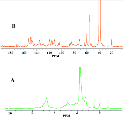 |
| | Fig. 1 NMR spectrum (A) 1H, (B) 13C of acetylated derivative of isolated lignin from Aloe barbadensis Miller. | |
3.2. FTIR analysis
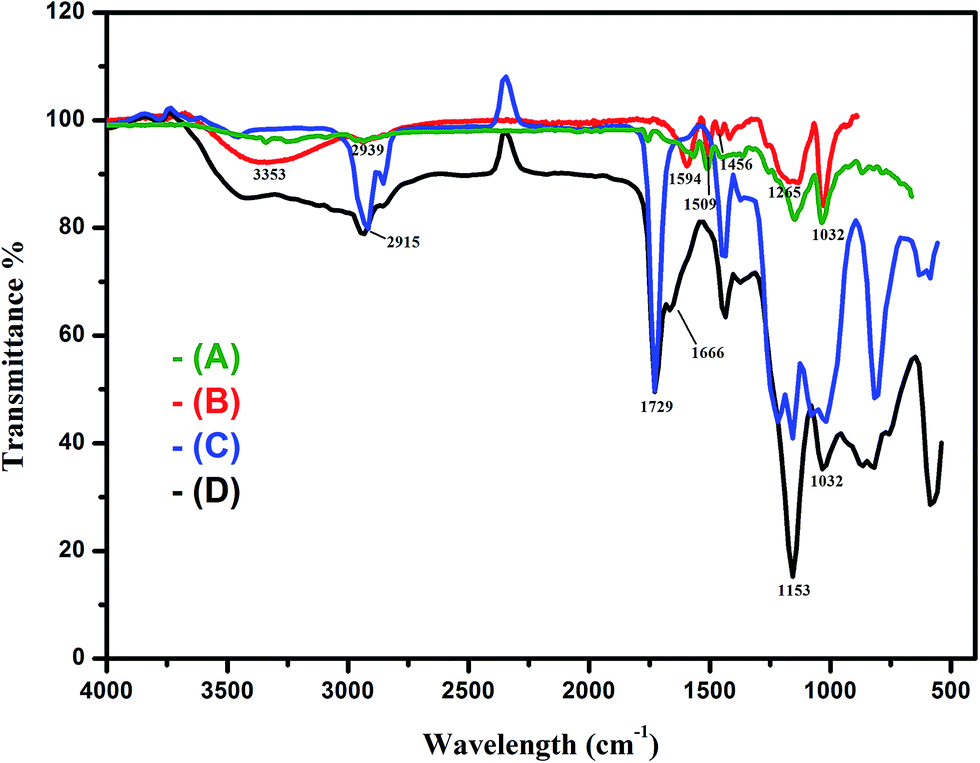
FT-IR spectroscopy analysis of commercial lignin, lignin, grafted lignin and 5-FU loaded lignin carrier showed interesting characteristics (Fig. 2). From the figure the characteristic peaks of commercial and isolated LIG were observed at 3353, 2939, 1594, 1509, 1456, 1265 and 1032 cm−1, which indicate the presence of O–H, C–H stretching vibrations, aromatic stretching band, aromatic ring, C–H deformation, C–O stretching, aromatic (phenyl) and C–O deformation (methoxyl group) respectively.38 The peak observed at 3353 cm−1 is strong evidence for the –OH stretching vibrations in pure LIG. This is caused by the presence of alcoholic and phenolic hydroxyl groups of LIG.39 The isolated lignin was well correlated with commercial lignin and it was confirmed by the FTIR spectrum of commercial lignin (Fig. 2A and B). However, in the FT-IR spectrum of LIG-g-MA polymer, the –OH peak assigned to lignin was absent. This is attributed to the grafting of methacrylate units on the lignin –OH groups. The other characteristic peaks at 1728, 1451 and 1154 cm−1 respectively correspond to the CO, C–H, and C–O groups of LIG-g-MA polymer. Normally the FT-IR absorption bands for the lignin include the aromatic skeletal vibrations as well. For instance, among the bands at 1606, 1507 and 1434 cm−1, the aromatic semicircle vibration (a vibration involving both C–C stretching and a change of the H–C–C bond angle) is assigned to that at 1507 cm−1.40 The FT-IR spectrum of 5-FU loaded LIG-g-MA confirms the binding of 5-FU onto the surface of the LIG-g-MA carrier. This is justified by the amide band at 1153 cm−1 (ref. 41) and also the distinct bands at 1729 and 1666 cm−1 due to the imide stretch and aromatic ring in 5-FU.42 In addition, for the co-polymer carrier, the –OCH3 peak at 2832 cm−1 was shifted to a lower region. This is due to the steric hindrance caused by the addition of drug molecule. The intense characteristic peaks of 5-FU loaded carrier observed at the same position, also indicate the impermanent interactions occurring between the drug and LIG-g-MA.
 |
| | Fig. 2 FT-IR spectra of (A) commercial lignin, (B) isolated pure lignin, (C) blank LIG-g-MA and (D) 5-FU loaded LIG-g-MA hollow-nanocarrier. | |
3.3. Fluorescence spectra
The fluorescent emission behavior was monitored for the 5-FU loaded LIG-g-MA hollow-nanocarrier at different time intervals. The spectra analysis was performed in the 470–510 nm range and typical emission was observed at 490 nm. The fluorescence quenching of 5-FU loaded LIG-g-MA carrier is shown in Fig. 3. Here the intensity decreases gradually with respect to time representing the photo-induced intermolecular electron transfer from 5-FU to the LIG-g-MA. This is considered as the quenching mechanism and the fluorescence quenching of LIG-g-MA by 5-FU can occur by any of the following four pathways; (i) the formation of (excited) charge transfer complex, (ii) energy transfer, (iii) proton transfer or (iv) electron transfer.43 Also, the intensity of fluorescence decreases rapidly up to 180 min. This is attributed to the successful 5-FU loaded LIG-g-MA carrier formation within 3 hours. The strong quenching interaction between 5-FU and LIG-g-MA confirms the encapsulation of 5-FU on the carrier as well. Thus, this technique provides strong evidence of the 5-FU encapsulation onto the LIG-g-MA hollow-nanocarrier.
 |
| | Fig. 3 Fluorescence quenching of LIG-g-MA by 5-FU in nano formulation. | |
3.4. XRD analysis
The X-ray diffraction pattern is a primary characterization tool for obtaining crystal structure, crystallite size, and strain. To study the characteristics of the drug in the polymer nanocarrier, the X-ray diffraction (XRD) pattern of pure 5-FU, LIG, blank LIG-g-MA hollow-nanocarrier and 5-FU loaded LIG-g-MA hollow-nanocarrier were studied (Fig. 4). The XRD patterns of pure 5-FU, LIG-g-MA, blank LIG-g-MA carrier and 5-FU loaded LIG-g-MA carrier provide evidence for the formation of the nanocarrier and drug loading on the carrier. 5-FU showed some characteristic intense peaks due to its crystalline nature (Fig. 4A). The broad peaks of LIG-g-MA polymer indicate its amorphous nature (Fig. 4B), whereas when the co-polymer was converted into the nano range, the peaks at 2θ values 17°, 33° and 37° shows its partially crystalline nature (Fig. 4C).44 After the encapsulation of the 5-FU drug on LIG-g-MA polymer hollow-nanocarrier, the peaks obtained were sharper and again confirm that the encapsulation of the drug encourages the order of the system.
 |
| | Fig. 4 X-Ray diffraction pattern images of (A) pure 5-FU, (B) LIG, (C) blank LIG-g-MA hollow-nanocarrier (D) 5-FU loaded LIG-g-MA nanocarrier. | |
3.5. Surface morphology analysis
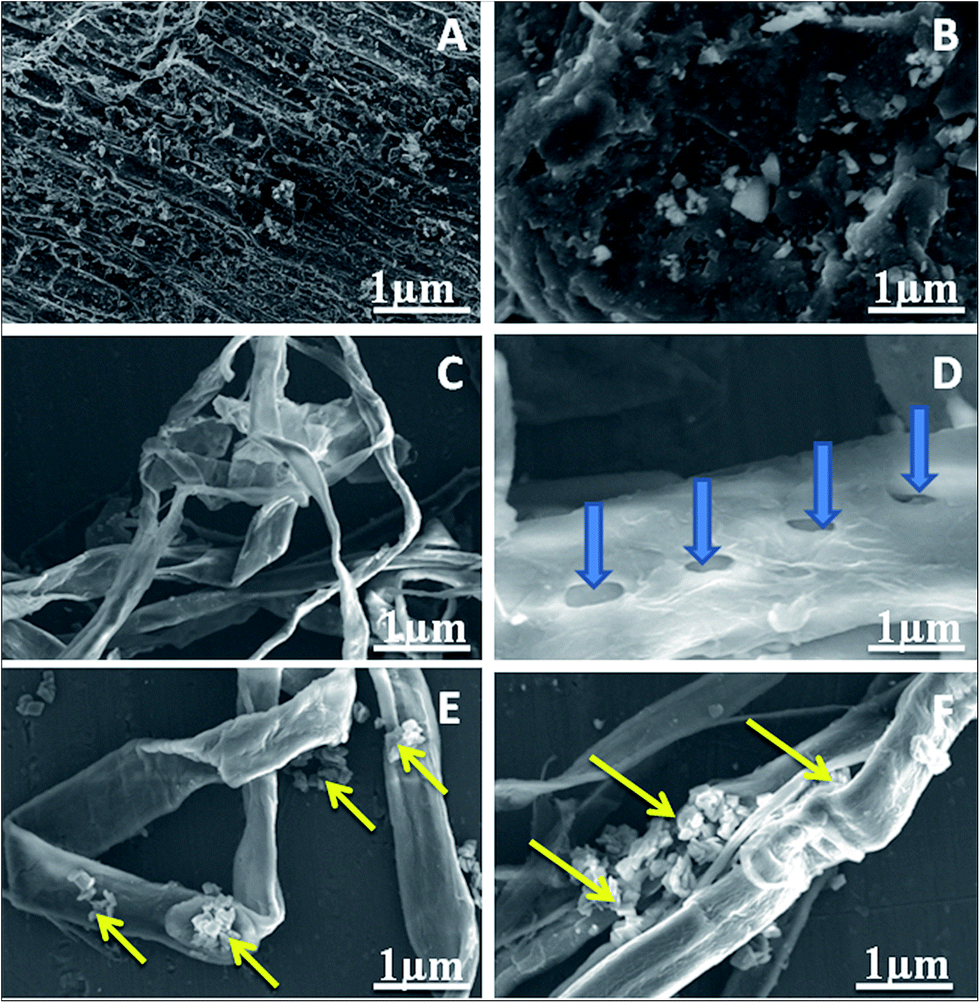
The morphology and microstructure of the isolated lignin, MA-grafted lignin, and 5-FU loaded LIG-g-MA hollow-nanocarrier were investigated using SEM and TEM techniques. The SEM analysis provides information on the morphology and structural information of the isolated lignin, MA-grafted lignin and 5-FU loaded LIG-g-MA carrier as illustrated in Fig. 5A–F. The SEM image of as isolated lignin from Aloe barbadensis Miller revealed a rough morphology (Fig. 5A). Later the lignin was washed with ethanol and water; it is rehabilitated to a smooth surface (Fig. 5A and B). Chemical or mechanical treatments on lignin change its particle sizes and make the surface smoother or rougher depending on each step of the treatment.45 The surface morphology of the LIG-g-MA polymer carrier is shown in Fig. 5C and D. The grafted lignin showed a highly branched and folded nanofibrous structure and this system is involved in intermolecular folding to form the nanofibrous network. The folding nature of the grafted lignin comes from the hydrogen and non-covalent bonding present in it. Furthermore, previous work by Makoto et al.25 on lignin blended with renewable resource-based cellulose acetate through iodine treatment established the influence of iodine on the formation of thin carbon fibers with diameters around 250 nm. The folded and branched structure46 as well as the nanotube nature47 of the lignin carrier make it responsive to external stimuli for the encapsulation of drug on the carrier. This makes the grafted lignin an ideal drug vehicle for targeted delivery into cells. Every single fiber has a number of porous sites; which help in the loading and release of drug in a controlled approach (Fig. 5D). The SEM image of the grafted LIG-g-MA polymer confirms it as a perfect drug vehicle for encapsulation of the drug as more 5-FU was encapsulated and entrapped on the carrier (Fig. 5E and F). The increase in concentration of 5-FU increases its loading. Good correlation was observed in TEM images for the nature of LIG-g-MA carrier and drug loaded LIG-g-MA carrier. The TEM image clearly shows the formation of hollow-fibers and entrapment of the drug on the fibrous hollow nanocarrier (Fig. 6).
 |
| | Fig. 5 SEM images of (A) as isolated lignin, (B) EtOH/H2O treated lignin, (C) grafted lignin with MA, (D) single hollow-nanocarrier LIG-g-MA, and (E) & (F) 5-FU loaded LIG-g-MA hollow-nanocarrier with two different concentrations (4 mg and 10 mg) respectively. | |
 |
| | Fig. 6 TEM images of (A) LIG-g-MA hollow-nanocarrier and (B) 5-FU loaded LIG-g-MA hollow-nanocarrier. The blue arrows represent the nature hollow fiber in the nanocarrier and green arrows represent the entrapment of 5-FU on the carrier. | |
3.6. Encapsulation efficiency
The hollow-nanocarrier encapsulated with 5-FU was successfully prepared by O/W emulsification technique. The decision to use this particular method was made based on the drug and polymer solubility i.e. 5-FU is hydrophobic and the co-polymer is soluble in CHCl3/DMSO. The encapsulation efficiency (EE) of the LIG-g-MA polymer is represented in Fig. S5.† The absorption intensity decreases gradually indicating a decrease in concentration of 5-FU in solution with time. This study also confirms the successful loading of 5-FU in the LIG-g-MA carrier. It is also observed that the EE of the formulations was significantly influenced by the formulation parameters. For instance, the EE increased as the cross-linking time increased. The initial concentration of the drug and the nature of the polymer composites play an important role in the drug encapsulation within the polymer nanocarrier as well. When the cavity groups in the carrier increased, the encapsulation also increased.
3.7. In vitro drug release
Cancer therapy is a widely investigated subject and many researchers are developing 5-FU model cancer drugs. The solubility of 5-FU is pH-dependent, which thus impacts on the medication discharge rate and last discharge sum. The in vitro drug releasing properties of 5-FU loaded LIG-g-MA nanocarrier were evaluated using the dialysis bag diffusion technique using pH 2.4 and 6.8 solutions at different time intervals. Fig. S8† shows that the releasing pattern shows a regular increase in releasing rate over a particular period. The sustainable release of 5-FU from the nanocarrier was evidence for the regular packing of 5-FU in the carrier. Various parameters like polymer degradation, drug binding nature, pH and drug solubility also influence the releasing rate of 5-FU. More specifically, the λmax value indicates the unmodified structure of 5-FU during encapsulation and its release. The releasing properties of 5-FU were observed to be fast at pH 2.4 compared to pH 6.8.
3.8. Cell viability against MCF-7 and VERO cell lines
Cell viability of the nanocarrier was checked with the conventional MTT reduction assay. For this, the MCF-7 cells were incubated with 5-FU, blank LIG-g-MA and 5-FU loaded LIG-g-MA hollow-nanocarrier at various concentrations for 7 days. The percentage of inhibition of various composites at different times in various concentrations is illustrated in Fig. S9.† The results indicate that the 5-FU loaded hollow-nanocarrier and 5-FU, imparted cytotoxicity to MCF-7 cells and inhibited the cell's growth rate depending on the concentration. The growth inhibition rates for both the 5-FU loaded carrier and released free 5-FU drug were nearly equal. This is an indication of the maximum level of release of 5-FU from the loaded nanocarrier. The IC50 values for free 5-FU and 5-FU loaded LIG-g-MA carrier were respectively 49 and 55 μg. These results indicate that the LIG-g-MA carrier is a potential delivery system and 5-FU can constantly be released in an active form from the system. Thus, from the obtained results it is evident that the fibrous composite nanocarrier is fruitful in increasing the cytotoxicity as well as the release behavior of 5-FU. These observations are in good agreement with the previous studies reporting the nanoformulation of chitosan/siRNA, nanoparticles encapsulated in PLGA nanofibres for siRNA delivery,48 gene delivery to human cells by lignin nanotubes47 and indomethacin (IDN) and aspirin delivery using magnetic polymeric composite nanofibres.49
3.9. Cytotoxicity effects against MCF-7 and VERO cell lines
Fig. 7 and 8 demonstrate the cell viability of the 5-FU encapsulated nanocarrier system against the MCF-7 and VERO cell lines at 1, 3 and 7 day intervals. The effects of cytotoxicity for the MCF-7 and VERO cell lines were estimated for 5-FU encapsulated LIG-g-MA, LIG-g-MA and free 5-FU during three experimental day intervals. As observed from the images, the cytotoxicity effect increased with increase in the number of days and thus shows a time-dependent activity. However, there was no impact on LIG-g-MA carrier, due to its good biocompatibility. The 5-FU stacked polymeric carrier LIG-g-MA showed raised impact relative to free 5-FU drug against the tumor cell lines. This may due to the potential of LIG-g-MA carrier to deliver drugs directly to the cells and tissues as they have the ability to perforate the membrane of the cells and pass into the cellular components without causing apparent cell damage.50 The 5-FU loaded LIG-g-MA carrier and 5-FU exhibited increased cytotoxicity with number of days. Nevertheless, all the samples had no effect against VERO normal cells (Fig. 8), with a slight negative effect for 5-FU compared to others.
 |
| | Fig. 7 Morphological study of 5-FU, blank LIG-g-MA and 5-FU loaded LIG-g-MA hollow-nanocarrier seeded on MCF-7 cells at three experimental day intervals. | |
 |
| | Fig. 8 Morphological study of blank LIG-g-MA, 5-FU loaded LIG-g-MA hollow-nanocarrier and 5-FU seeded on normal VERO cells at three experimental day intervals. | |
4. Conclusions
We demonstrate the potential application of lignin in the biomedical area, especially for cancer therapy. The lignin was isolated from Aloe barbadensis Miller biomass by an environmentally friendly and commercially feasible process. Chemically modified hollow-nanocarrier lignin was synthesized and the modified lignin was characterized by various techniques like FTIR, XRD and emission spectra. The surface morphology confirmed the capability of the modified lignin to carry the 5-fluorouracil anticancer drug. This was again proved by the encapsulation efficiency and in vitro drug delivery data. Furthermore, the cell feasibility and cytotoxicity effects of the drug loaded LIG-g-MA hollow-nanocarrier against MCF-7 and VERO cell lines exhibited its applicability in cancer chemotherapy. Consequently, this system shows great promise with regard to the circumvention of the present limitations of lignin in the management of cancer diseases.
Acknowledgements
M. Rajan, is thankful to the University Grants Commission (UGC), Government of India, for providing financial assistance under the scheme of “UGC-BSR Research Start-Up Grants” (Ref. No. F. 30-21/20014 (BSR)). The authors would like to extend their sincere appreciation to the Deanship of Scientific Research at King Saud University for its funding of this research through the Research Group project No. RGP-1435-057.
References
- C. Zhang, H. Subramanian, J. J. Grailer, A. Tiwari, S. Pilla, D. A. Steeber and S. Gong, Polym. Adv. Technol., 2009, 20, 742–747 CrossRef CAS.
- S. Pilla, S. Gong, E. O'Neill, L. Yang and R. M. Rowell, J. Appl. Polym. Sci., 2009, 111, 37–47 CrossRef CAS.
- S. Pilla, A. Kramschuster, L. Yang, J. Lee, S. Gong and L. S. Turng, Mater. Sci. Eng., 2009, 29, 1258–1265 CrossRef CAS.
- P. Magesan, S. Sanuja and M. J. Umapathy, RSC Adv., 2015, 42506–42515 RSC.
- A. K. Siddhanta, N. D. Sanandiya, D. R. Chejara and S. Kondaveeti, RSC Adv., 2015, 5, 59226–59239 RSC.
- R. Kuktaite, H. Türe, M. S. Hedenqvist, M. Gällstedt and T. S. Plivelic, ACS Sustainable Chem. Eng., 2014, 2, 1439–1445 CrossRef CAS.
- G. Gao, J. Ian Dallmeyer and J. F. Kadla, Biomacromolecules, 2012, 13, 3602–3610 CrossRef CAS PubMed.
- T. A. Hassan, V. K. Rangari and S. Jeelani, ACS Sustainable Chem. Eng., 2014, 2(4), 706–717 CrossRef CAS.
- V. K. Thakur, M. Thunga, S. A. Madbouly and M. R. Kessler, RSC Adv., 2014, 4, 18240–18249 RSC.
- H. Jiang, J. Ma and C. Li, J. Mater. Chem., 2012, 22, 16939–16942 RSC.
- K. Yue, G. T. de Santiago, M. M. Alvarez, A. Tamayol, N. Annabi and A. Khademhosseini, Biomaterials, 2015, 73, 254–271 CrossRef CAS PubMed.
- C. Boyer, V. Bulmus, J. Liu, T. P. Davis, M. H. Stenzel and C. Barner-Kowollik, J. Am. Chem. Soc., 2007, 129(22), 7145–7154 CrossRef CAS PubMed.
- M. Rajan and V. Raj, Int. J. Pharm. Pharm. Sci., 2012, 4, 255–259 CAS.
- M. Rajan, V. Raj, A. A. Al-Arfaj and A. M. Murugan, Int. J. Pharm., 2013, 2, 514–522 CrossRef PubMed.
- M. Rajan and V. Raj, Carbohydr. Polym., 2013, 1, 951–958 CrossRef PubMed.
- X. Yang, A. Huang, J. Peng, J. Wang, X. Wang, Z. lin and S. Li, RSC Adv., 2014, 4, 60675–60684 RSC.
- R. Judith, M. Nithya, C. Rose and A. B. Mandal, Biologicals, 2012, 40, 231–239 CrossRef CAS PubMed.
- G. Dharman, M. Rajan, M. A. Munusamy, M. Bala Kumaran and P. T. Kalaichelvan, RSC Adv., 2015, 5, 44705 RSC.
- D. Govindaraj, M. Rajan, Murugan A. Munusamy and A. Higuchi, RSC Adv., 2015, 5, 58980 RSC.
- F. Abdelkafi, H. Ammar, B. Rousseau, M. Tessier, R. El Gharbi and A. Fradet, Biomacromolecules, 2011, 12, 3895–3902 CrossRef CAS PubMed.
- A. Hambardzumyan, L. Foulon, B. Chabbert and V. Aguie-Beghin, Biomacromolecules, 2012, 13, 4081–4088 CrossRef CAS PubMed.
- F. S. Chakar and A. Ragauskas, Ind. Crops Prod., 2004, 20, 131–141 CrossRef CAS.
- I. Dallmeyer, S. Chowdhury and J. F. Kadla, Biomacromolecules, 2013, 14, 2354–2363 CrossRef CAS PubMed.
- W. O. S. Doherty, P. Mousavioun and C. M. Fellows, Ind. Crops Prod., 2011, 33, 259–276 CrossRef CAS.
- M. Yoshida, Y. Liu, S. Uchida, K. Kawarada, Y. Ukagami, H. Ichinose, S. Kaneko and K. Fukuda, Biosci., Biotechnol., Biochem., 2008, 72(3), 805–810 CrossRef CAS PubMed.
- A. Duval, H. Lange, M. Lawoko and C. Crestini, Biomacromolecules, 2015, 16, 2979–2989 CrossRef CAS PubMed.
- F. J. Xu, Z. H. Wang and W. T. Yang, Biomaterials, 2010, 31, 3139–3147 CrossRef CAS PubMed.
- R. Shenoy and C. N. Bowman, Biomaterials, 2012, 33, 6909–6914 CrossRef CAS PubMed.
- C. Cui, H. Sadeghifar, S. Sen and D. S. Argyropoulos, BioResources, 2013, 8, 864–886 Search PubMed.
- C. Mai, A. Majcherczyk and A. Huttermann, Enzyme Microb. Technol., 2000, 27, 167–175 CrossRef CAS PubMed.
- C. Mai, O. Milstein and A. Huttermann, Appl. Microbiol. Biotechnol., 1999, 51, 527–531 CrossRef CAS.
- Y. Li and A. J. Ragauskas, RSC Adv., 2012, 2, 3347–3351 RSC.
- G. Sivasankarapillai, A. G. McDonald and H. Li, Biomass Bioenergy, 2011, 35, 919–931 CrossRef CAS.
- P. Noordhuis, U. Holwerda, C. Van der Wilt, C. Van Groeningen, K. Smid, S. Meijer, H. Pinedo and G. Peters, Ann. Oncol., 2004, 15, 1025 CrossRef CAS PubMed.
- Y. Sik Kim and J. F. Kadla, Biomacromolecules, 2010, 11, 981–988 CrossRef PubMed.
- R.-I. Freshney, Culture of Animal Cells: A Manual of Basic Technique, Wiley-Liss, New York, 1994 Search PubMed.
- E. D. Routledge, S. Gorman and M. R. Clark, Cell and tissue culture: laboratory procedures, Wiley, New York, 1994 Search PubMed.
- J. Lisperguer, P. Perez and S. Urizar, J. Chil. Chem. Soc., 2009, 4, 54 Search PubMed.
- P. Yan, Z. Xu, C. Zhang, X. Liu, W. Xu and Z. Conrad Zhang, RSC Green Chem. Ser., 2015, 17, 4913–4920 RSC.
- N. Colthup, L. Daly and S. Wiberley, Introduction to Infrared and Raman Spectroscopy, Academic Press Limited, London, 1990 Search PubMed.
- R. Gui, A. Wan and H. Jin, Analyst, 2013, 138, 5956 RSC.
- P. Li, Y. Wang, Z. Peng, F. She and L. Kong, Carbohydr. Polym., 2011, 85, 698–704 CrossRef CAS.
- M. S. Khot, S. L. Bhattar, G. B. Kolekar and S. R. Patil, Spectrochim. Acta, Part A, 2010, 77, 82–86 CrossRef CAS PubMed.
- V. Balakrishnan, H. AzwanaAb Wab, K. Abdul Razak and S. Shamsuddin, Journal of Nanomaterials, 2013, 2013, 729306 CrossRef.
- S. Hamid reza Ghaffar and M. Fan, Biomass Bioenergy, 2013, 57, 264–279 CrossRef.
- B. Ozbas, J. Kretsinger, K. Rajagopal, J. P. Schneider and D. J. Pochan, Macromolecules, 2004, 37, 7331–7337 CrossRef CAS.
- E. Ten, C. Ling, Y. Wang, A. Srivastava, L. Amelia Dempere and W. Vermerris, Biomacromolecules, 2014, 15, 327–338 CrossRef CAS PubMed.
- M. Chen, S. Gao, M. Dong, J. Song, C. Yang, K. Alan Howard, J. Kjems and F. Besenbacher, ACS Nano, 2012, 6(6), 4835–4844 CrossRef CAS PubMed.
- L. Wang, M. Wang, P. D. Tophamc and Y. Huang, RSC Adv., 2012, 2, 2433–2438 RSC.
- M. A. E. Abdelbary, A. Waqar, U. H. Israr, R. D. Vinod and D. E. Antony, J. Drug Delivery, 2012, 2012, 837327 Search PubMed.
Footnote |
| † Electronic supplementary information (ESI) available. See DOI: 10.1039/c6ra01866a |
|
| This journal is © The Royal Society of Chemistry 2016 |
Click here to see how this site uses Cookies. View our privacy policy here.