DOI:
10.1039/C6RA01846G
(Paper)
RSC Adv., 2016,
6, 23686-23692
A catch–release catalysis system based on supramolecular host–guest interactions†
Received
21st January 2016
, Accepted 23rd February 2016
First published on 25th February 2016
Abstract
A generic catch–release catalysis system has been designed for the recovery of homogeneous catalysts at the end of a reaction using host–guest interactions. This proof-of-concept system consists of a palladium(II)–dipyrazole complex bearing an adamantyl (Ad) molecular recognition moiety (guest), and magnetic nanoparticles (MNP) decorated with β-cyclodextrins (β-CD) (host). The density of β-CD on the iron oxide nanoparticles was up to 3.1 × 10⁻⁴ mmol per mg, sufficient to efficiently catch the Ad-adorned Pd catalyst in aqueous methanol at room temperature. Release and recycling of the catalyst was achieved by extraction with methanol. This catch–release system performed well in the Suzuki–Miyaura coupling, but suffers from slow degradation which restricts the number of times that the catalyst and magnetic nanoparticles can be recycled and reused.
Introduction
Homogeneous catalysts generally demonstrate better selectivity and reactivity under milder reaction conditions than their heterogeneous counterparts,1 because of increased accessibility to well-defined reactive sites. Nevertheless, homogeneous catalysis accounts for less than 20% of all industrial processes because, in large part, of the difficulty in separating a homogeneous catalyst from the reaction product and reusing it.2,3
One general methodology for removing and recycling homogeneous transition metal catalysts from a reaction solution involves sequestering the metal complex with an insoluble or immiscible matrix.4 Appropriately decorated magnetic nanoparticles are ideally suited for this role.5 The sequestering requires a selective interaction between the catalyst and matrix to permit the catalyst to be recovered and reused. An elegant example of this “catch–release” strategy is the removal of a pyrene tagged Pd catalyst from an aqueous reaction with graphene coated magnetic nanoparticles. High temperatures were then used to disrupt the non-covalent π–π interactions and release the catalyst back into fresh reaction medium.6
Reversible, host–guest inclusion complexation can offer selectivity to a catch–release catalysis system and one of the most studied hosts is β-cyclodextrin.7,8 The 0.7 nm × 0.79 nm hydrophobic cavity of β-CD encapsulates lipophilic guests such as adamantane (Ad) derivatives with a binding constant (Ka) up to 105.9–11 This host–guest complexation is mostly driven by a favourable enthalpy of reaction.12 Some of us have recently reported a series of supramolecular hydrogels, non-covalently connected micelles, nano-capsules and pseudo-block copolymers based on the inclusion complexation between β-CD and adamantyl decorated polymers.13–16 Particularly relevant to the present study is our recent report of solubilizing an Ad-L-Pd(II) catalyst (where Ad-L = bis[(3,5-dimethyl-1H-pyrazolyl)methyl][(1-adamantyl)methyl]amine) in an aqueous solvent system by complexation with the highly soluble heptakis(2,6-di-O-methyl)-β-CD (dmβ-CD).17 We now report an extension of that study with the development of a catch–release catalytic system employing iron oxide magnetic nanoparticles decorated with β-CD moieties to capture an adamantane adorned Pd(II) catalyst.
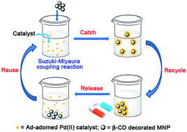
The general methodology is shown in Scheme 1. The Ad-L-Pd(II) complex reported previously is used to promote Suzuki–Miyaura coupling and at the end of the reaction is captured by magnetically recoverable Fe3O4 magnetic nanoparticles (MNPs) garlanded with β-CD hosts. Organic solvent extraction then facilitates release and reuse of the catalyst.
 |
| Scheme 1 Schematic representation of the catch and release of an Ad adorned Pd(II) catalyst by employing iron oxide MNPs decorated with β-CD moieties. | |
Materials and methods
Materials
β-Cyclodextrin (β-CD) (98%) was purchased from Sigma and used after recrystallization from water and drying at 100 °C under vacuum overnight. Succinic anhydride, succinic acid, FeCl3·6H2O, FeCl2·4H2O and PdCl2 were purchased from Sigma-Aldrich. All substrates for Suzuki–Miyaura coupling reactions were purchased from Sigma-Aldrich. The palladium(II) complex bearing an adamantyl (Ad) molecular recognition moiety (Ad-L-PdCl2) was synthesized according to our previously reported method.17 All other reagents and solvents were used as received without purification.
Synthesis of carboxyl-β-CD (β-CD-COOH)
Dry β-CD (5.11 g, 4.5 mmol) was dissolved in 30 mL of anhydrous N,N-dimethylacetamide (DMAc) with stirring at 50 °C. Powdered succinic anhydride (1.80 g, 18 mmol) was added with rapid stirring. The reaction temperature was maintained at 50 °C under a N2 atmosphere for another 2 h and then at 90 °C for one day. The reaction mixture was precipitated with 500 mL of diethyl ether and the resulting light brown powder collected by centrifugation and washed with ethyl acetate (100 mL). The crude product was redissolved in 6 mL of deionized water (DI water) and precipitated with 500 mL of acetone. The white powder was again collected by centrifugation, washed well with acetone (2 × 30 mL), and then dried under vacuum at 40 °C. Yield 5.20 g (74.0%, based on an average substitution degree of 3 as determined by elemental analysis and 1H NMR spectroscopy). FTIR (KBr): ν = 3380 (br, ν(O–H)), 2931 (m, ν(C–H)), 1734 (s, ν(C
O)), 1408 (m), 1365 (m), 1158 (s), 1078 (s), 1029 (s, ν(C–O–C)), 949 (w), 758 (w), 706 (w), 579 (m) cm−1; 1H NMR (300 MHz, D2O, δ): 2.40–2.80 (d, 12H, –OOC–CH2–CH2–COOH), 3.30–4.30 (m, 42H, –CH– of β-CD), 4.90–5.40 (m, 7H, C(1)H of β-CD); 13C NMR (75.5 MHz, DMSO-d6, δ): 173.7 (C
O), 102.1 (C(1) of β-CD), 81.8 (C(4) of β-CD), 74.0–71.0 (C(3), C(2) and C(5) of β-CD), 60.0 (C(6) of β-CD), 28.8, 28.7 (CH2 of succinyl group). Anal. calcd for C54H82O44·7H2O: C 41.54, H 6.20; found: C 40.91, H 5.87.
Synthesis of β-CD-coated Fe3O4 magnetic nanoparticles (β-CD-MNP)
Powdered FeCl3·6H2O (300 mg, 1.11 mmol) and FeCl2·4H2O (115 mg, 0.58 mmol) were dissolved in 30 mL of deionized water in a 2-necked round bottom flask equipped with a condenser. The flask was evacuated, filled with N2 at room temperature and then, 1.0 M NaOH (5.0 mL) added dropwise with stirring at 1000 rpm. The reaction mixture turned from golden to black, and was stirred at room temperature for an additional 30 min. Carboxyl-β-CD (874 mg, 0.56 mmol) in water (10 mL) was added in one portion followed by another 0.4 mL of 1.0 M NaOH solution added to keep the pH at 6. The mixture was slowly heated to 80 °C for 1 h and then cooled to yield a black precipitate that was rinsed thoroughly with water and collected from the supernatant using a permanent magnet.
Synthesis of succinic acid-coated Fe3O4 MNP (succinate MNP)
The procedure was identical to that described above except that succinic acid (66 mg, 0.56 mmol) was used in place of carboxyl-β-CD.
Synthesis of bare Fe3O4 MNP
The procedure was identical to that above except that no ligand was used during the synthesis.
Measurements and characterization methods
General methods
Proton and carbon nuclear magnetic resonance (1H and 13C NMR) spectra were recorded at room temperature on a Bruker Avance DRX 300 MHz NMR spectrometer operating at 300.1 and 75.5 MHz, respectively; or on a Bruker Avance DRX 500 MHz NMR spectrometer operating at 500.1 and 125.8 MHz, respectively. Chemical shifts were reported in parts per million (ppm) on the δ scale, and were referenced to residual protonated solvent peaks: CDCl3 spectra were referenced to CHCl3 at δH 7.26 and CDCl3 at δC 77.36; DMSO-d6 spectra were referenced to (CHD2)(CD3)SO at δH 2.50 and (CD3)2SO at δC 39.6; D2O spectra were referenced to HDO at δH 4.70; acetone-d6 spectra were referenced to (CHD2)(CD3)CO at δH 2.09. X-ray diffraction (XRD) patterns were recorded on a PANalytical's X'Pert PRO diffractometer with Cu Kα radiation. Fourier transform infrared (FTIR) spectra of samples in KBr pellet were recorded on a Perkin-Elmer FTIR 2000 spectrometer in the region of 4000–400 cm−1; 32 scans were signal-averaged with a resolution of 4 cm−1 at room temperature. The transmission electron micrographs were taken on a JEOL JEM-2010F FasTEM field emission transmission electron microscope operated at 100 kV. The hydrodynamic diameter and zeta-potential were determined using a Zetasizer Nano ZS (Malvern Instruments, Southborough, MA). All measurements were taken after the sample solutions were ultrasonicated for 1 min and equilibrated at room temperature for 20 min. The amount of organic component coated onto the MNP was determined by a thermogravimetric analyzer (TGA) using a Mettler Toledo Star System TGA/SDTA 851e in the presence of N2 gas with a heating rate of 20 °C min−1. The scanning electron micrographs were taken with a JEOL JSM-7600F, which is a high resolution thermal field emission scanning electron microscope (FEG SEM) equipped with Oxford AZtec energy system for energy dispersive spectroscopy (EDS). The elemental analyses of dried MNPs with Pd on the surface were carried out using the Oxford AZtec energy system. Inductively Coupled Plasma Mass Spectroscopy (ICP-MS) analysis was carried out using a Perkin Elmer Optima 5300 DV.
Catch and release of catalyst Ad-L-PdCl2 by β-CD-MNP
Catch experiment. β-CD-MNP dispersion in DI water (4.0 mg mL−1, 0.5 mL) was mixed with Ad-L-PdCl2 solution in H2O–CH3OH (1/1, v/v, 0.35 mg mL−1, 1 mL) with stirring at room temperature. The β-CD/Ad molar ratio was 1.0 and the amount of β-CD-MNP adjusted as required (see Results and discussion). The mixture was stirred (500 rpm) for 2 h at room temperature. The β-CD-MNP with captured catalyst was separated using a permanent magnet. The amount of residual catalyst in the clear supernatant was determined by UV-Vis spectroscopy. The catch experiments using bare MNP and succinate MNP were conducted using the same procedure.
Release experiment. The separated β-CD-MNP with captured catalyst was washed twice with 2 mL DI water, separated by centrifugation (8000 rpm, r.t.) or a permanent magnet and freeze-dried. The MNP powder was stirred (500 rpm) with 1.5 mL methanol for 1 h at room temperature. The clear solution was separated by centrifugation (8000 rpm, r.t.) or magnetically and the amount of extracted catalyst in the methanol solution determined by UV-Vis spectroscopy.
Evaluation of catch–release performance with Suzuki–Miyaura coupling reaction
Step 1 Suzuki–Miyaura coupling reaction before catch–release. Ad-L-PdCl2 (0.35 mg, 6.25 × 10⁻⁴ mmol) was dissolved in 0.5 mL MeOH. 4-Bromobenzoic acid (25.1 mg, 0.125 mmol), phenylboronic acid (18.3 mg, 0.15 mmol), and Na2CO3 (31.8 mg, 0.3 mmol) were dissolved in 0.5 mL DI water. The catalyst and substrate solutions were stirred (500 rpm) together at room temperature for 2 h and then the white coupling product isolated by centrifugation (6000 rpm, r.t.). The clear filtrate was used in step 2.
Step 2 catch–release experiment. To the clear filtrate from step 1 (1 mL) was added a β-CD-MNP dispersion in DI water (4.0 mg in 0.5 mL) with stirring (500 rpm) for 2 h at room temperature. Centrifugation (8000 rpm, r.t.) or a permanent magnet was used to separate the MNPs which were then washed twice with DI water (2 mL) and freeze dried. The clear filtrate was used in step 3. The dried MNPs were stirred with 1.5 mL methanol for 1 h at r.t. and again removed magnetically or by centrifugation (8000 rpm, r.t.). This clear filtrate was used in step 4.
Step 3 Suzuki–Miyaura coupling catalyzed by filtrate after catch. The clear filtrate from step 2 (1.5 mL) was mixed with powdered substrate as in step 1, but without Na2CO3. Dilute aqueous NaOH was added to adjust the pH to 11. The reaction mixture was stirred at room temperature for 2, 5 or 24 h (Fig. 9).
Step 4 Suzuki–Miyaura coupling catalyzed by released catalyst. The methanol extract from step 2 was concentrated to 0.5 mL and mixed with freshly prepared substrate in 1.0 mL DI water. The amount of each reactant was identical to step 1. The reaction mixture was stirred at room temperature for 2 h, 5 h or 24 h (Fig. 9).
Results and discussion
Synthesis and characterization of carboxyl-β-CD and β-CD coated MNP
Carboxyl groups were introduced at the narrow end of the β-CD toroid by treatment with excess succinic anhydride in anhydrous N,N-dimethylacetamide (DMAc) at 90 °C (Scheme 2). Elemental analysis and 1H NMR spectroscopy suggested that an average of three of the seven primary alcohols of each β-CD were esterified (ESI, Fig. S1†).
 |
| Scheme 2 Synthesis of carboxyl-β-CD and β-CD-coated Fe3O4 MNPs. | |
Fe3O4 MNPs with β-CD moieties on the surface (β-CD-MNP) were prepared using a one-pot co-precipitation18,19 method from aqueous solution (Scheme 2). The β-CD carboxylic acids are anchored to the Fe3O4 surface through carboxyl chelation to the Fe2+/Fe3+ sites. Ester hydrolysis was minimized by maintaining the pH of reaction at 6. The β-CD-MNPs formed a stable dispersion in water (Fig. 1, left). This dispersion could be aggregated by exposure to a magnetic field (Fig. 1, right) and then re-dispersed by removing the magnet and shaking the vial gently.
 |
| Fig. 1 (Left) Photo of the β-CD-MNPs dispersed in water (0.5 mg mL−1); (right) magnetically induced aggregation of β-CD-MNPs from water. | |
The β-CD-MNPs had a diameter of around 10 nm with a narrow size distribution as observed by TEM (Fig. 2, left). Fringes can be clearly seen in high resolution TEM image (Fig. 2, right) because the electron beam is diffracted by the Fe3O4 crystalline lattice.
 |
| Fig. 2 (Left) TEM image of β-CD-MNPs; (right) expanded section (left). | |
The powder XRD diffraction pattern of β-CD-MNP (Fig. 3) shows the characteristic peaks of standard Fe3O4 crystals (isometric-hexoctahedral crystal system),18,20 matching well with those from the JCPDS card (19-0629) for pure magnetite.21 Characteristic peaks of β-CD are not in the Bragg diffraction range (2θ) 30° to 70° and so do not interfere with the Fe3O4 diffraction peaks.22,23
 |
| Fig. 3 pXRD pattern of β-CD-MNP. | |
The crystallite size of β-CD-MNP was calculated using the Debye–Scherrer equation:24–26
where
D is the average crystallite size (Å),
θ is the Bragg diffraction angle,
β is the full width at half maximum (FWHM) (in radians) and
λ is the X-ray wavelength of Cu Kα radiation (1.5418 Å). The diffraction peak at 2
θ = 35.6° (
Fig. 3) has a FWHM of
ca. 0.01538 rad which corresponds to a crystallite size of 9.5 nm, consistent with the TEM result (
Fig. 2). Dynamic Light Scattering (DLS) experiments were also carried out to measure the hydrodynamic diameter of β-CD-MNP in water (0.1 mg mL
−1). This technique returned an average hydrodynamic diameter of around 100 nm. The discrepancy with the TEM and pXRD determined crystallite size is presumably because of some aggregation in water. The zeta potential of β-CD-MNP was found to be around −20 mV indicating that the MNP surface is negatively charged, which is attributed to uncoordinated carboxyl groups on the β-CD moieties (ESI, Fig. S2
†). Bare Fe
3O
4 MNPs with no organic ligand on the surface and succinic acid-coated Fe
3O
4 MNPs were also synthesized for comparison using a modification of the method outlined in
Scheme 2. Powder XRD measurements indicated that both MNPs have similar diameters to that of β-CD-MNP (ESI, Fig. S3
†). Similarly, DLS measurements showed that these MNPs also had average hydrodynamic diameters of around 70 nm with narrow size distributions. The surfaces of bare Fe
3O
4 MNPs and succinic acid-coated Fe
3O
4 MNPs are positively charged with the mean zeta potential of around 50 mV and 26 mV, respectively.
Characterization of β-CD-MNP surface
FTIR experiments were performed on β-CD-MNP, succinate-MNP, bare MNP and β-CD-succinate (Fig. 4). Strong absorption at 590 cm−1, characteristic of Fe–O bond stretching in nano-sized Fe3O4 particles19,27 was identified for all three MNPs. An absorption band at around 1410 cm−1 was observed for succinate MNP which is due to the bidentate carboxyl group on the MNP surface.19,28–30 Three strong absorption peaks at around 1730, 1160 and 1030 cm−1 were observed in the spectrum of β-CD-MNP, which we attribute to the ester (–C
O stretch) and β-CD moieties (–C–O stretch), respectively.
 |
| Fig. 4 (Top) FTIR spectra for the three Fe3O4 MNPs (β-CD-MNP, bare MNP and succinate MNP); (bottom) FTIR spectrum of β-CD-succinate. | |
The amount of β-CD on the MNPs was estimated using thermogravimetric analysis (TGA) (Fig. 5).
 |
| Fig. 5 TGA of β-CD-MNP and bare MNP (20 °C min−1, N2). | |
Weight losses were observed for β-CD-MNP from room temperature to 900 °C under N2. The first (6%) from room temperature to approximately 200 °C is ascribed to water adsorbed on the MNP surface. The β-CD-MNP subsequently experiences significant weight loss (45%) over the range 200–860 °C due to organic ligand decomposition leaving a residue of Fe3O4 (49%). By comparison, the TGA curve of the bare MNP shows the first weight loss of 4% from room temperature to approximately 100 °C due to water physically adsorbed onto the MNP surface, and then another 4% over the range 100–400 °C due to coordinated water. These results indicate a molar content of 3.1 × 10⁻⁴ mmol of β-CD per milligram of β-CD-MNP.
Evaluation of catch–release catalysis system
A visual comparison was made of the three MNPs with added Ad-L-PdCl2 in H2O–CH3OH (2/1, v/v). All three MNPs dispersed in deionized water to give clear suspensions. Upon addition of the catalyst in methanol, a dark brown precipitate formed immediately in the β-CD-MNP dispersion (Fig. 6, left). By comparison, there was no change for the mixtures of bare MNP and succinate MNP even after 2 days of exposure to the Ad-L-PdCl2 at room temperature (Fig. 6, middle and right). This comparison demonstrates that β-CD-MNP strongly interacts with the Ad-containing catalyst. Upon host–guest complexation, the originally hydrophilic MNP surface becomes hydrophobic with a coating of hydrophobic catalyst molecules that accelerate aggregation. This interesting phenomenon aids the separation of β-CD-MNP after catching the catalyst.
 |
| Fig. 6 Mixtures of Ad-L-PdCl2 (0.35 mg) with different MNPs in 2 mL of H2O–CH3OH (2/1, v/v); (left) β-CD-MNP (2.8 mg), (middle) bare MNP (1.3 mg) and (right) succinate MNP (1.4 mg). | |
The interaction between Ad-L-PdCl2 and each of the three MNPs in aqueous methanol was further investigated by UV-Vis spectroscopy (Fig. 7, top). The MNPs and any associated catalyst were removed magnetically prior to analysis. The concentration of catalyst in the supernatant was determined using a calibration curve (R2 = 0.997, ESI, Fig. S4†).
 |
| Fig. 7 (Top) Variation of UV-Vis absorbance for catalyst solutions in H2O–CH3OH (2/1, v/v) before and after addition of different MNPs (β-CD-MNP 1.4 mg, bare MNP 0.65 mg and succinate MNP 0.7 mg); (bottom) UV-Vis spectra of catalyst in H2O–CH3OH (2/1, v/v) before and after treatment by varying amounts of β-CD-MNP. Initial catalyst amount before catch: 0.35 mg in 1.5 mL H2O–CH3OH (2/1, v/v); all sample solutions were diluted to 1/10 prior to UV-Vis measurement; all tests were carried out at room temperature with slit width 1.0 nm. | |
It can be seen from Fig. 7 (top), that the catalyst solution shows an obvious drop in absorbance after treatment with β-CD-MNP. In this experiment, the molar ratio of β-CD/Ad was ca. 0.7. By comparison, addition of a similar amount of bare and succinate MNPs resulted in only a slight drop in absorbance. In both cases, the catalyst molecules have no specific interactions with the MNP surfaces. However weak and non-specific interactions may cause a slight loss of catalyst by adsorption.
The catch efficiency for the catalyst was studied by varying the proportion of β-CD-MNP (Fig. 7, bottom). About 34% of catalyst was removed by the MNP at a β-CD/Ad molar ratio of 0.7. Increasing the β-CD/Ad molar ratio increases the catch efficiency, but not proportionally. At β-CD/Ad molar ratios of 1.5, 2.0 and 4.0, the catch efficiencies were 57%, 64% and 73%, respectively. As expected, when the catalyst solution was treated twice with 2.0 equiv. of β-CD-MNP, the catch efficiency was higher (82%) than for a single treatment of 4.0 equiv. of β-CD-MNP.
The ability of β-CD-MNP to catch the catalyst from aqueous solution was confirmed by the presence of Pd in the separated β-CD-MNP as determined by SEM-EDS analysis (ESI, Fig. S5†).
Release of the catalyst was achieved by washing the separated β-CD-MNP with methanol for 1 hour at room temperature (Fig. 8). The mixture was separated, and the clear supernatant analyzed by UV-Vis spectroscopy. The concentration of catalyst was determined using a calibration curve (R2 = 0.998, ESI, Fig. S6†).
 |
| Fig. 8 UV-Vis spectra of methanol extracts of separated β-CD-MNP with captured catalyst. Initial catalyst before catch: 0.35 mg in 1.5 mL H2O–CH3OH (2/1, v/v); β-CD/Ad molar ratio: 2.0; extraction with 1.5 mL methanol for 1 hour at room temperature. All solutions were diluted to 1/10 prior to UV-Vis analysis. | |
The reference spectrum (Fig. 8, black) is equivalent to 100% recovery of catalyst bound to the separated β-CD-MNP. The UV-Vis spectrum of a single methanol extraction indicates an 80% recovery of catalyst bound to the MNP. A second extraction yielded an additional 6%.
One complete cycle of catch and release with catalyst solution (1.5 mL) at an initial concentration of 0.233 mg mL−1 in H2O–CH3OH (2/1, v/v) and 2.0 equivalent of β-CD-MNP at room temperature yielded 51% of recovered catalyst as determined by UV-Vis spectroscopy and confirmed by ICP-MS analysis of Pd content in the methanol extract. Higher recovery would be expected from additional treatment with β-CD-MNP and/or additional extraction steps. The β-CD-MNP could be recycled and reused at least 3 times without obvious loss of ability to catch and release the catalyst.
Evaluation of catch–release catalysis with a Suzuki–Miyaura coupling reaction
The practicability of our catch–release catalysis system was evaluated using the Suzuki–Miyaura cross coupling of 4-bromobenzoic acid and phenylboronic acid in aqueous media (Scheme 3).
 |
| Scheme 3 Suzuki–Miyaura coupling reaction of 4-bromobenzoic acid and phenylboronic acid in aqueous media catalyzed by Ad-L-PdCl2. | |
This reaction was carried out in aqueous methanol (1/1, v/v) under an air atmosphere at room temperature with a catalyst loading of 0.5 mol% and proceeded in a typical yield of 96% in 2 hours.
After reaction, the insoluble product was removed by centrifugation and the supernatant treated with 2.0 equivalent of β-CD-MNP. This β-CD-MNP with captured catalyst on the surface was separated, vacuum dried and then dissociated with methanol. The extracted catalyst solution was concentrated and reused with fresh substrate solution in water (Scheme 4).
 |
| Scheme 4 Catch–release cycle. | |
The β-CD-MNP separated from the catch step was vacuum dried and characterized by SEM-EDS. As expected, the elemental analysis of the dried sample indicated the presence of Pd (ESI, Fig. S7†).
The activity of residual catalyst was determined for the various reaction fractions (Fig. 9). Immediately obvious from this graph is the relatively low activity of the filtrate of a Suzuki–Miyaura reaction mixture after addition and removal of β-CD-MNP (A). Almost homeopathic levels of Pd are reported to catalyze this reaction31 and so this low level of activity is testament to the catching ability of the β-CD-MNP. By comparison, a suspension of recovered catalyst still attached to β-CD-MNP (C) and the corresponding methanol solution of released catalyst (B) exhibited similar activity to the recycled filtrate of a Suzuki–Miyaura reaction mixture without catching by β-CD-MNP (D).
 |
| Fig. 9 Comparison of reaction yields for Suzuki–Miyaura coupling catalyzed by (A) filtrate of a Suzuki–Miyaura reaction mixture after catching by β-CD-MNP (step 3, above), (B) the methanol extract of the separated β-CD-MNP from the catch step (step 4, above), (C) the separated β-CD-MNP from the catch step, and (D) recycled filtrate of a Suzuki–Miyaura reaction mixture without catching by β-CD-MNP. Each point represents the mean value ± SD of 2 runs. | |
At a reaction time of 2 h (green bar), the filtrate of a Suzuki–Miyaura reaction mixture after catching by β-CD-MNP (A) showed the lowest ability to catalyse a new Suzuki–Miyaura reaction. The yield at 2 h is only ca. 15%. By comparison, the yields at 2 h were ca. 55%, 64%, 82% for the other three solutions (B–D). Moreover, the yield with solution A did not show a significant increase even after prolonging the reaction time (around 30% yield after 24 h), while with increasing reaction time, all of the other three reaction mixtures could achieve high yields (>90% after 24 h).
The yield of Suzuki–Miyaura coupling with the released catalyst in methanol decreased to 50% (reaction time 5 h) after three catch–release cycles. We attributed this to (i) some decomposition of the Ad-L-Pd(II) catalyst at the relatively high pH of the reaction mixture (pH 11) and (ii) hydrolysis of the succinic ester bonds causing loss of β-CD moieties from the recycled MNPs.
Conclusions
A new catch–release catalysis system was established based on the specific interaction between host β-cyclodextrin torroids tethered to magnetic nanoparticles and a palladium(II) catalyst adorned with a guest adamantane. This system enjoys both tight, non-covalent binding and the possibility of quick and complete release on addition of an appropriate competing organic solvent molecule. The host and guest however are handicapped by their slow degradation under the test reaction conditions, thus limiting the times that the catalyst can be recycled and reused. The potential advantages of this relatively simple system in green chemical synthesis are however notable. Current investigations are directed at seeking for solutions in practical applications.
Acknowledgements
This work was supported by EDB (Singapore)-GSK (GlaxoSmithKline) (Grant ID: R143-000492-592) and the Institute of Materials Research and Engineering (IMRE), A*STAR (Agency for Science, Technology and Research), Singapore.
Notes and references
- R. A. Sheldon, Green Chem., 2005, 7, 267–278 RSC.
- M. Lamblin, L. Nassar-Hardy, J. C. Hierso, E. Fouquet and F. X. Felpin, Adv. Synth. Catal., 2010, 352, 33–79 CrossRef CAS.
- D. J. Cole-Hamilton, Science, 2003, 299, 1702–1706 CrossRef CAS PubMed.
- C. Baleizao, A. Corma, H. García and A. Leyva, J. Org. Chem., 2004, 69, 439–446 CrossRef CAS PubMed.
- Q. M. Kainz and O. Reiser, Acc. Chem. Res., 2014, 47, 667 CrossRef CAS PubMed.
- S. Wittmann, A. Schaetz, R. N. Grass, W. J. Stark and O. Reiser, Angew. Chem., Int. Ed., 2010, 49, 1867–1870 CrossRef CAS PubMed.
- W. Saenger, Angew. Chem., Int. Ed. Engl., 1980, 19, 344–362 CrossRef.
- J. Szejtli, Chem. Rev., 1998, 98, 1743–1753 CrossRef CAS PubMed.
- S. Gould and R. C. Scott, Food Chem. Toxicol., 2005, 43, 1451–1459 CrossRef CAS PubMed.
- R. I. Gelb, L. M. Schwartz and D. A. Laufer, J. Chem. Soc., Perkin Trans. 2, 1984, 15–21 RSC.
- B. L. Zhang and R. Breslow, J. Am. Chem. Soc., 1993, 115, 9353–9354 CrossRef CAS.
- R. I. Gelb and L. M. Schwartz, J. Inclusion Phenom., 1989, 7, 537–543 CrossRef CAS.
- Z.-X. Zhang, K. L. Liu and J. Li, Angew. Chem., Int. Ed., 2013, 52, 6180–6184 CrossRef CAS PubMed.
- J.-L. Zhu, K. L. Liu, Z. Zhang, X.-Z. Zhang and J. Li, Chem. Commun., 2011, 47, 12849–12851 RSC.
- Z.-X. Zhang, K. L. Liu and J. Li, Macromolecules, 2011, 44, 1182–1193 CrossRef CAS.
- Z.-X. Zhang, X. Liu, F. J. Xu, X. J. Loh, E.-T. Kang, K.-G. Neoh and J. Li, Macromolecules, 2008, 41, 5967–5970 CrossRef CAS.
- M. Qi, P. Z. Tan, F. Xue, H. S. Malhi, Z.-X. Zhang, D. J. Young and T. S. A. Hor, RSC Adv., 2015, 5, 3590–3596 RSC.
- L. Gu, Z. Shen, C. Feng, Y. Li, G. Lu, X. Huang, G. Wang and J. Huang, J. Mater. Chem., 2008, 18, 4332–4340 RSC.
- C. L. Lin, C. F. Lee and W. Y. Chiu, J. Colloid Interface Sci., 2005, 291, 411–420 CrossRef CAS PubMed.
- S. Singh, K. Barick and D. Bahadur, J. Hazard. Mater., 2011, 192, 1539–1547 CrossRef CAS PubMed.
- Z. Li, L. Wei, M. Gao and H. Lei, Adv. Mater., 2005, 17, 1001–1005 CrossRef CAS.
- K. Srinivasan and T. Stalin, Spectrochim. Acta, Part A, 2014, 130, 105–115 CrossRef CAS PubMed.
- V. Nikolic, M. Stankovic, A. Kapor, L. Nikolic, D. Cvetkovic and J. Stamenkovic, Pharmazie, 2004, 59, 845–848 CAS.
- R. Massart and V. Cabuil, J. Chim. Phys. Phys.-Chim. Biol., 1987, 84, 967–973 CrossRef CAS.
- B. D. Cullity, Elements of X-ray Diffraction, Addison-Wesley Reading, MA, 1978 Search PubMed.
- A. Taylor, X-ray metallography, Wiley, New York, 1961 Search PubMed.
- S. Singh, K. C. Barick and D. Bahadur, J. Hazard. Mater., 2011, 192, 1539–1547 CrossRef CAS PubMed.
- S. Si, A. Kotal, T. K. Mandal, S. Giri, H. Nakamura and T. Kohara, Chem. Mater., 2004, 16, 3489–3496 CrossRef CAS.
- N. Shukla, C. Liu, P. M. Jones and D. Weller, J. Magn. Magn. Mater., 2003, 266, 178–184 CrossRef CAS.
- Q. X. Liu and Z. H. Xu, Langmuir, 1995, 11, 4617–4622 CrossRef CAS.
- C. Deraedt and D. Astruc, Acc. Chem. Res., 2014, 47, 494–503 CrossRef CAS PubMed.
Footnotes |
† Electronic supplementary information (ESI) available: 1H and 13C NMR spectra of β-CD-COOH; characterization results by pXRD and DLS for β-CD-MNP, bare MNP and succinate MNP; calibration curves of absorbance versus concentration of catalyst in H2O–CH3OH (2/1, v/v) and CH3OH; SEM-EDS analysis of β-CD-MNP with Pd bound the particle surface. See DOI: 10.1039/c6ra01846g |
‡ The first and second authors have made equal contributions. |
|
This journal is © The Royal Society of Chemistry 2016 |
Click here to see how this site uses Cookies. View our privacy policy here.