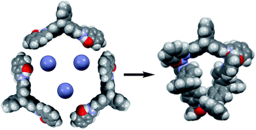
Zinc-mediated diastereoselective assembly of a trinuclear circular helicate

Jesus Sanmartín-Matalobos*a, Matilde Fondoa, Ana M. García-Deibea, Martín Amozaa, Pilar Bermejob, M. Raquel Domínguezb, Antonio J. Motac, J. Luis Pérez-Lustresd, Sourav Bhowmicke and Neeladri Das*e
aDpto. de Química Inorgánica, Facultad de Química, Universidad de Santiago de Compostela, Avenida de las Ciencias s/n, 15782 Santiago de Compostela, Spain. E-mail: jesus.sanmartin@usc.es; Fax: +34 981 528073; Tel: +34 981 563100
bDpto. de Química Analítica, Facultad de Química, Universidad de Santiago de Compostela, Avenida de las Ciencias s/n, 15782 Santiago de Compostela, Spain
cDepartamento de Química Inorgánica, Facultad de Ciencias, Universidad de Granada, 18002 Granada, Spain
dDepartamento de Química Física, CIQUS, Universidad de Santiago de Compostela, Jenaro de la Fuente s/n, 15782 Santiago de Compostela, Spain
eDepartment of Chemistry, Indian Institute of Technology Patna, Bihta 801 103, Bihar, India. E-mail: neeladri2002@yahoo.co.in; neeladri@iitp.ac.in

Received 20th January 2016 , Accepted 15th February 2016

First published on 17th February 2016


Abstract

Coordination of a triptycene-based ditopic Schiff base ligand (H2L) with Zn2+ leads to the formation of a novel trinuclear circular species. A combination of 1H NMR spectroscopy, infrared spectroscopy, mass spectrometry and elemental analysis allow the unambiguous characterization of H2L and Zn3L3·8H2O. DFT molecular modelling has been used to study the possible isomers of the cyclic trinuclear Zn(II) complex that results from the combination of conformational and optical isomers of the ligand. A NOESY experiment demonstrated that in the trinuclear circular zinc(II) helicate the three isomers of the ligand have an anti configuration of N,O-donor domains and an s-cis configuration of their imine bonds. Besides, a UV-VIS absorption and fluorescence emission study of H2L and Zn3L3·8H2O has been performed.

Introduction

In recent years, iptycenes (9,10-dihydro-9,10-[1,2]benzenoanthracene derivatives) have attracted considerable research attention because of their application as porous materials.1–7 Triptycene is the simplest member of the iptycene family, where three benzene rings are held together by a [2.2.2] bicyclic bridge. The rigid three-bladed geometry of triptycene generates a 120° angle between the arene rings and this is attributed to their inefficient packing in the solid state. As a result, void spaces thus generated in the clefts between the arenes8,9 are responsible for the porosity in materials derived from triptycene. This characteristic structural feature of triptycenes has been also utilized in the design of potential fluorescent chemosensors with enhanced solubility in organic solvents, wherein the presence of bulky structural motif minimizes interchain π–π interactions.10,11 This results in lesser extent of self-quenching and consequently fluorescence quantum yield of the triptycene derivative increases considerably.12 In contrast, electron-deficient guest species can penetrate in host clefts and interact with triptycene derivatives leading to fluorescence quenching.12–14

Recently, we have reported fluorescence quenching of triptycene-based polymers by host–guest interaction with fullerene15 and the application of triptycene based tripods as chemosensors for selective detection of metal ions in aqueous media.16 We have also reported the utility of organometallic acceptors containing the triptycene motif as building blocks in the construction of metallasupramolecular cages.17 Herein, we have extended our studies to explore the use of a triptycene-based Schiff base ligand (H2L, Scheme 1) as a supramolecular synthon in coordination driven self-assembly processes towards the design of trinuclear circular helicates. These latter compounds are rather rare and their formation remains challenging, comprising the vast majority of ligands employed for the preparation of such species aromatic diimine derivatives.18,19 Nevertheless, several circular helicates with ligands other than aromatic diimine derivatives have been recently published.20 DFT molecular modelling has been used to determine the most probable diastereoisomer from all possible ones of the obtained cyclic trinuclear zinc(II) complex.


image file: c6ra01783e-s1.tif
Scheme 1 The two possible enantiomers of the triptycene-based ditopic Schiff base ligand, obtained by condensation of 2,6-diaminotriptycene and 2-hydroxybenzaldehyde. The numbering scheme for NMR studies on (9R,10R)-H2L and (9S,10S)-H2L are shown.

According to the best of our knowledge, the use of triptycene-based ditopic Schiff base ligands for the preparation of complexes in which the metal ions define a triangle with the ligands wrapping over and under has been unexplored. We have paid attention to imines because they are able to stabilize many different metals in various oxidation states, controlling the performance of metals in a large variety of useful applications21 in biological, clinical, analytical and industrial fields, in addition to their important roles in catalysis and organic synthesis.

Results and discussion

Schiff base ditopic ligand

The investigation was initiated to design the helicand H2L that can be able to adopt the adequate topology required for optimal binding to naked metal ions leading to trinuclear circular helicates. The extended conjugation between the rigid three-bladed triptycene hub and both salicylaldimine pendants should lead to an Y-shaped spatial arrangement of H2L, with a ca. 120° angle between the two pendant arms. In light of the orientation of the two N,O-donor sets of H2L, either to the same or to opposite sides, respect to the triptycene residue, its conformational isomers have been qualified as syn or anti. Besides, two configurations of the imine bond, s-cis and s-trans,22 are possible for the anti-isomer, as Fig. 1 shows. We speculated that the assembly of three naked metal ions and three anti-isomers of the bideprotonated ligand could lead to trinuclear circular helicates (M3L3), while the participation of syn conformers only can lead to circular, but non-helical, trinuclear species.
image file: c6ra01783e-f1.tif
Fig. 1 Capped sticks (left) and spacefill (right) representations of anti-s-cis (top), syn (centre) and anti-s-trans (bottom) conformational isomers of H2L obtained by DFT energy-minimisation. Only (R,R) isomers are represented for simplicity. Black arrows are indicating the orientation of the N,O-donor sets for chelation (syn or anti). Configurations of the C[double bond, length as m-dash]N bond, s-cis and s-trans, are highlighted in green. Relative energy (ΔE, ΔH and ΔG in kcal mol−1) for the three conformers studied for H2L (anti-s-trans, anti-s-cis and syn) from DFT energy-minimisations is shown at bottom.

Condensation reaction of 2-hydroxybenzaldehyde and 2,6-diaminotriptycene (2[thin space (1/6-em)]:[thin space (1/6-em)]1 molar ratio) occurs efficiently in ethanol solution, leading to the triptycene-based Schiff base ditopic ligand H2L. Although we have not been able to obtain single crystals adequate for X-ray diffraction studies. H2L has been isolated and unambiguously characterized by elemental analysis, mass spectrometry, infrared and NMR spectroscopies. Two-dimensional proton–proton (COSY) in combination with proton–carbon (HMQC and HMBC) correlation spectra have been used to achieve a full assignment of NMR signals.

To gain insight into the structure of the conformational isomer of H2L in solution, we have undertaken a 1D NOE experiment (Fig. 2) in combination with molecular modelling (Fig. 1). The iminic proton H-17, resonating at δ 8.89 (selective band), showed correlations with the proton signals at δ 7.58 (H-21, 3.8%), δ 7.54 (H-1, 3.6%) and δ 7.03 ppm (H-7, 3.5%). These data are only consistent with one of the three modelled structures, the syn conformer of H2L. This shows H-17⋯H-21, H-17⋯H-1 and H-17⋯H-7 distances of about 2.37, 2.10, and 2.13 Å, respectively.


image file: c6ra01783e-f2.tif
Fig. 2 1D 1H NMR spectrum (in black) and selective 1D NOE spectrum (in red) of H2L in dmso-d6. The signal at 8.89 ppm for selective NOE irradiation with a shape pulse was omitted for clarity.

With respect to the energy differences between the three calculated conformational isomers of H2L, DFT studies indicate that the syn-isomer has the most favourable ΔG (Fig. 1), but in practice, all they are isoenergetic. Furthermore, all of them are kinetically accessible, since the transition states for their interconversion are very low, giving rise to a rather flat potential energy surface. As a consequence, H2L can easily adopt an adequate spatial arrangement to behave as helicand, with both anti conformations that can lead to neutral trinuclear circular helicates. Furthermore, the syn conformer would lead to trinuclear circular species, but non-helical.

Cyclic trinuclear Zn(II) complex

To explore the application of this triptycene-based N,O-donor ligand as a potential building block for constructing novel supramolecular architectures, we studied the reaction of L2− with naked Zn2+ ions. This self-assembly reaction yielded the cyclic trinuclear complex Zn3L3·8H2O, which has been isolated and unambiguously characterized by elemental analysis, mass spectrometry (Fig. 3), infrared and NMR spectroscopies (ESI). TLC was used to ensure purity of the compound.
image file: c6ra01783e-f3.tif
Fig. 3 Comparison of experimental (in grey) and theoretically calculated (in red) peak at m/z about 1667 for Zn3L3·8H2O.

Mass-spectrometric analysis was used to confirm the M3L3 composition of the neutral complex obtained via self-assembly of three units of the metal acceptor (Zn2+) and three units of the helicand triptycene based ditopic ligand (L2−). The only possible shape that can be assigned to the M3L3 composition of the complex is that of a neutral discrete cyclic trinuclear species. CHNO microanalysis supported the formation of the octahydrated metal complex with stoichiometry in equimolar ratio (M[thin space (1/6-em)]:[thin space (1/6-em)]L). The infrared stretching frequencies of C[double bond, length as m-dash]N and C–O groups are consistent with coordination of both imine-N and phenol-O atoms to the metal ions.23,24 The 1H NMR spectrum of Zn3L3·8H2O showed the absence of phenol protons, which is indicative of double deprotonation of the ligand (L2−), and subsequent coordination to Zn2+ metal centres. Besides, a typical high-field chemical shift (about 0.4 ppm) of the imino protons of the arms in 2 and 6-positions clearly shows the participation of the azomethine groups in the coordination. Two-dimensional proton–proton (COSY) in combination with proton–carbon (HMQC and HMBC) correlation spectra have been used to achieve a full assignment of NMR signals.

We have explored the possible isomers of the cyclic trinuclear Zn(II) complex using DFT molecular modelling. Fig. 4 shows the six most stable diastereoisomers that have been considered for the theoretical calculations. Other combinations were dismissed because it was not possible the assembly of cyclic trinuclear complexes. Four of the six possible diastereoisomers are circular helicates, as they result from the assembly of three naked metal ions and three anti conformers (as s-cis or s-trans forms) of the racemic ligand. One might note that helicity is not possible when the trinuclear complex contains syn conformers of the ligand. The handedness for these four helicates is determined by the configuration of the C[double bond, length as m-dash]N bond. Thus, the Δ helicate with three anti-s-cis conformers contains three (R,R) ligands, while the Δ helicate with three anti-s-trans conformers contains three (S,S) ligands. A change from s-cis to s-trans or vice versa, forces a change of chirality as well. Therefore, helicity for these trinuclear circular species is conditioned by both conformation and chirality.


image file: c6ra01783e-f4.tif
Fig. 4 Six possible diastereoisomers of the cyclic trinuclear zinc(II) complex (four circular helicates and two circular non-helical species) obtained by DFT energy-minimization. Only Δ isomers of helicates have been considered and minimized for simplicity. To make it easier to see, schematic representations have been included.

Fig. 5 shows the relative energy (from DFT energy-minimisations in phase gas) for the six possible isomers studied in the cyclic trinuclear zinc(II) complex formation. These studies indicate that the most favourable ΔG value corresponds to the circular non-helical species that combines one R,R-anti-s-trans form and two R,R-syn forms of the ligand. In contrast, the less favourable ΔG value corresponds to the circular helicate that combines two R,R-anti-s-cis forms and one S,S-anti-s-trans form of the ligand. However, the energy difference between the species having the least favourable and the most favourable ΔG values is less than 3 kcal mol−1, and therefore anyone of six isomers can be expected in solution.


image file: c6ra01783e-f5.tif
Fig. 5 Relative energy (from DFT energy-minimisations) for the six possible isomers studied in the cyclic trinuclear zinc(II) complex formation.

For the complete characterization of the cyclic trinuclear zinc(II) complex formed, its three-dimensional structure in solution was investigated using a 2D NOESY experiment (Fig. 6) in combination with molecular modelling (Fig. 4). This allows us to deduce which of the six isomers of the cyclic trinuclear zinc(II) complex is in solution (Fig. 6). The iminic proton H-17, resonating at δ 8.98 ppm, showed correlations with H-21 (at δ 7.53 ppm) and H-1/H-5 (at δ 7.71 ppm), which in the modelled structure are separated by about 2.2 Å. These data are only consistent with one of the six possibilities, the circular helicate that combines three anti-s-cis forms of the ligand. This result reflects the free rotation around the carbon–nitrogen single bond of the ligand, which in free form adopted a syn relative disposition, but coordinated is adopting the anti-s-cis form. Thus, the isomer experimentally observed in solution results from the zinc-assisted diastereoselective assembly of three units of the anti-s-cis helicand H2L, as (R,R)- or (S,S)-enantiomers.


image file: c6ra01783e-f6.tif
Fig. 6 NOESY spectrum of Zn3L3·8H2O. Cross peaks corresponding to the correlation of H-17 with H-1/H-5 and H-21 is highlighted.

Optical studies

A study on the optical properties of H2L and Zn3L3·8H2O showed that both compounds have yellow fluorescence emission (near to 570 nm under UV excitation, λexc = 350 nm), although with a significant difference in their intensities, as zinc complexation to H2L leads to significant fluorescence quenching. Thus, the noise observed in the fluorescence spectrum of Zn3L3·8H2O (Fig. 7) is apparently caused by its low quantum yield.
image file: c6ra01783e-f7.tif
Fig. 7 Emission (red line) and excitation (blue line) spectra of Zn3L3·8H2O (in THF, 3.2 × 10⁻⁵ M). The absorption spectrum is also shown (black line).

The excitation spectrum is overlapped with the absorption spectrum of Zn3L3·8H2O in its band of lower energy. Due to the strong absorption of the solvent used (THF), it was not possible to record, in a trustworthy way, the excitation spectrum beyond 280 nm. Anyway, the good agreement between the absorption and excitation spectra demonstrates the presence of an only fluorescent species in the ground electronic state. The fluorescence emission spectrum shows a large Stokes shift, about 12[thin space (1/6-em)]500 cm−1, that corresponds to significant reorganization energy of ca. 6250 cm−1. This implies a strong intramolecular structural reorganization in the excited electronic state or conversion from the Franck–Condon state to a dark state. Reorganization of the weakly polar solvent THF could also have a minor contribution to the full Stokes shift.

A study of the stability of the fluorescence signal of an acetonitrile solution of H2L in alkaline medium shows that its intensity decreases vs. time (Fig. 8). An NMR monitoring of a CD3CN solution of H2L in the presence of NMe4OH during 3 h has thrown some light on the observed instability of the fluorescence intensity. H2L without UV excitation remains unchanged for about 2 h and 30 min, but after 3 h small signals of 2-hydroxybenzaldehyde and 2,6-diaminotriptycene appear in the 1H NMR spectrum as result of the Schiff base hydrolysis, which can be promoted by UV light.25


image file: c6ra01783e-f8.tif
Fig. 8 Variation of the fluorescence emission spectrum with time for λ = 525 nm at λexc = 350 nm of an acetonitrile solution of H2L in the presence of NMe4OH (a), and after addition of Zn2+ (50 μL, 1 g L−1) and SDBS (100 μL, 1.5 mM) (b).

To retard the hydrolysis of H2L in alkaline medium under UV light, we have studied the use of the following surfactants: sodium dodecylbenzenesulfonate (SDBS), sodium dodecylsulfate (SDS) and saponin at concentration levels of 1.5, 1.1 and 0.9 mM, respectively.26–28 The best results were found for SDBS added after the addition of Zn2+ (coefficient of variation about 1.5%). In these conditions fluorescence intensity of H2L was stable during at least 60 min (Fig. 8).

Fluorescence emission was measured at different concentration levels of Zn2+ (0–50 μg L−1) to know if there is a correlation between analytical signal and concentration of Zn2+ added to a solution of H2L in alkaline medium. The reproducibility within each level was acceptable (coefficients of variation of fluorescence intensities were generally below 10% for each measurement), but no correlation between analytical signal and concentration of Zn2+ was found.

Then, we have studied whether the UV-vis spectroscopy data (absorbance) correlate with the concentration of Zn2+ (0–2.4 μg L−1) added to a solution of H2L in alkaline medium (Fig. 9). The obtained results have shown coefficients of variation in the range 0.1–0.3%. The limits of detection (LOD) and quantification (LOQ) obtained at 348 nm were 4.2 and 14.1 μg L−1, respectively.


image file: c6ra01783e-f9.tif
Fig. 9 Variation of the UV-vis absorbance spectrum with the addition of Zn2+ at 267 and 348 nm.

Conclusions

The new triptycene-based ditopic Schiff base ligand H2L was synthesized and satisfactorily characterized. H2L is able to adopt the adequate spatial arrangement required for optimal binding to naked Zn2+ ions leading to a trinuclear circular helicate, or a trinuclear circular non-helical species. The assembly of three naked metal ions and three anti conformers of the ligand would result in a helicate, while the assembly of three metal ions and three syn conformers of H2L would lead to a non-helical species.

A novel trinuclear circular species has been isolated and unambiguously characterized by elemental analysis, mass spectrometry, infrared and NMR spectroscopies. We have synthesized the circular helicate Zn3L3·8H2O, which results from the zinc-assisted diastereoselective assembly of three units of the anti-s-cis helicand H2L, as (R,R)- or (S,S)-enantiomers.

H2L and Zn3L3·8H2O have yellow fluorescence emission, although with a significant difference in their intensities, as zinc complexation to H2L leads to significant fluorescence quenching. We have demonstrated that absorbance at 267 and 348 nm correlate with the concentration of Zn2+ added to a solution of L2−.

Experimental

Synthesis and characterization of the compounds

H2L. A solution of 2,6-diaminotriptycene (100 mg, 0.352 mmol) and 2-hydroxybenzaldehyde (0.1 mL, 0.703 mmol) in ethanol (10 mL) with 4–5 drops of acetic acid was stirred at room temperature for overnight. Reaction progress was monitored by TLC. Then, it was obtained a yellow solid, which was washed with methanol to get the desired product. Further purification was not required. Yield: above 90%. 1H NMR (400 MHz, dmso-d6), ppm 13.05 (s, 2H, 2×OH), 8.89 (s, 2H, 2×H-17), 7.57 (d, 2H, J = 7.7, J = 1.7, 2×H-21), 7.53 (d, 2H, J = 2.1, H-1 and H-5), 7.49 (d, 2H, J = 7.8, H-4 and H-8), 7.44 (d, 2H, J = 8.6, J = 2.1, H-13 and H-16), 7.35 (t, 2H, J = 7.5, J = 1.7, 2×H-19), 7.03 (d, 2H, J = 7.8, J = 2.1, H-3 and H-7), 7.00 (d, 2H, J = 8.5, J = 2.3, H-14 and H-15), 6.92 (t, 2H, J = 7.5, J = 0.9, 2×H-20), 6.90 (d, 2H, J = 8.3, 2×H-18), 5.71 (s, 2H, H-9 and H-10). 13C NMR (125 MHz, dmso-d6): δ/ppm 163.5 (2×C-17), 160.6 (2×C-18a), 147.0 (2×C-10a), 145.6 (C-2, C-6), 145.2 (C-11, C-12), 144.2 (2×C-8a), 133.6 (2×C-19), 132.9 (2×C-21), 125.7 (C-14, C-15), 124.9 (C-4, C-8), 124.2 (C-13, C-16), 119.7 (2×C-17a), 119.6 (2×C-20), 118.8 (C-3, C-7), 117.1 (C-1, C-5), 117.0 (2×C-18), 52.6 (C-9, C-10). IR (ATR, ν/cm−1): 3441 (OHphenol), 1623, 1609 (C[double bond, length as m-dash]N), 1190 (C–O). MS (MALDI-TOF+) m/z (%): 493.7 (100) [H2L + H]+. UV-vis (acetonitrile, nm) λmax = 267, 348. Elemental analysis: C, 82.6; H, 4.6; N, 5.6%. Calcd for C34H24N2O2 (492.0) C, 82.9; H, 4.9; N, 5.7%.
Zn3L3·8H2O. To an acetonitrile (10 mL) suspension of H2L (37 mg, 0.075 mmol), 1.5 mL of a 0.1 M methanol solution of NMe4OH·5H2O (27.5 mg, 0.15 mmol) were added. The resultant yellow solution was mixed with a methanol (2.5 mL) solution of Zn(ClO4)2·6H2O (28.5 mg, 0.075 mmol). The mixture was stirred at room temperature for 30 min and concentrated under vacuum. The yellow solid that precipitated was filtered off, washed with 15 mL of methanol during 30 min and dried in air. TLC was used to ensure purity of the compound. Alternatively, Zn3L3·8H2O was obtained from a methanol solution (2 mL) of H2L (37 mg, 0.075 mmol) and Zn(OAc)2·H2O (30 mg, 0.075 mmol), which was heated under reflux for 2 h. Yield: 21 mg, 50%. 1H NMR (400 MHz, dmso-d6), ppm 8.94 (s, 2H, 2×H-17), 7.67 (d, 2H, J = 2.0, H-1 and H-5), 7.49 (d, 2H, J = 8.1, J = 1.5, 2×H-21), 7.40 (d, 2H, J = 8.5, J = 2.0, H-13 and H-16), 7.31 (t, 2H, J = 8.6, J = 1.6, 2×H-19), 7.22 (d, 2H, J = 7.9, H-4 and H-8), 7.00 (d, 2H, J = 8.5, J = 2.2, H-14 and H-15), 6.66 (d, 2H, J = 7.7, J = 2.1, H-3 and H-7), 6.65 (d, 2H, J = 8.0, 2×H-18), 6.63 (t, 2H, J = 7.2, J = 0.9, 2×H-20), 5.52 (s, 2H, H-9 and H-10). 13C NMR (100 MHz, dmso-d6): δ/ppm 171.32 (C-18a); 169.35 (2×C17); 147.15 (C-4a + C-8a); 144.59 (C-9a + C-10a); 144.33 (C-11 + C-12); 143.85 (C-2 + C-6); 137.95 (2×C21); 136.42 (2×C19); 125.84 (C-14 + C-15); 124.96 (C-4 + C-8); 124.36 (C-13 + C-16); 122.98 (C-20); 120.02 (C-3 + C-7); 119.16 (C-17a); 115.24 (C-18); 114.84 (C-1 + C-5); 52.44 (C-9 + C-10). IR (KBr, ν/cm−1): 3435 (OHwater), 1607 (C[double bond, length as m-dash]N), 1184 (C–O). MS (MALDI-TOF+) m/z (%): 1667.1 (36%) [Zn3L3 + H]+, 1605.2 (7%) [Zn2L(H2L)2 + H]+, 1111.1 (36%) [Zn2L2 + H]+, 1048.1 (100%) [Zn(H2L)2 + H]+. UV-vis (THF, nm) λmax = 270, 350. Elemental analysis: C, 67.7; H, 4.5; N, 4.6; O, 12.0%. Calcd for C102H82N6O14Zn3 (1809.2) C, 67.7; H, 3.5; N, 4.6; O, 12.4%.

Physical measurements

H2L and its complexes were characterized by a combination of elemental analysis, infrared spectroscopy, mass spectrometry and 1H NMR spectroscopy (see ESI).

Elemental analyses of C, H, N and O were performed on a Thermo Finnigan, FLASH EA 1112 series analyzer. IR spectra were recorded as KBr pellets on a FT-IR Mattson Instruments 202Q spectrophotometer in the range 4000–600 cm−1. MALDI-TOF mass spectra were recorded on a Bruker Ultraflex III TOF/TOF using methanol as solvent and DCTB as matrix. 1H NMR spectra were recorded on a Bruker DPX-250 spectrometer using DMSO-d6 as solvent.

Computational details

The calculations were carried out at the DFT-B3LYP level with the Gaussian 09 program.29 The geometries were fully optimized by the gradient technique with the following basis: for Zn, the LANL2DZ basis set; for C, H, N and O were described by the standard polarized 6-31G* basis set. The nature of the optimized structures was assessed through a frequency calculation and the changes of Gibbs free reaction energies (ΔG values) were obtained by taking into account zero-point energies, thermal motion, and entropy contribution at standard conditions (temperature of 298.15 K, pressure of 1 atm).

Although H2L displays two chiral centres at C-9 and C-10, and consequently, there are (R,R) and (S,S) enantiomers, only the (R,R) enantiomer was considered for simplicity. It was assumed that the azomethine group presents an E-configuration, since this is typically stabilized by an intramolecular Ophenol–H⋯Nimine bonding.30 Therefore, for all these conformers, the imine proton (H-17) is always in close proximity to H-21. One might note that in the anti-s-cis conformer, one of the iminic H atoms is close to H-1, while the other one is near to H-5, while in the case of the anti-s-trans conformer the iminic H atoms are close to H-3 and H-7, respectively. In the third case, the syn conformer, the closest protons are H-1 and H-7. These latter couplings are those experimentally observed in the selective NOE spectrum of H2L.

Optical studies

We have observed two intense absorption bands for H2L and Zn3L3·8H2O at wavelengths about 270 and 350 nm, using the UV-visible spectroscopy. Thus, we have done excitation at wavelength 350 nm for fluorescence spectroscopic analysis of H2L and Zn3L3·8H2O. In order to study the fluorescence emission of H2L in alkaline medium used to the obtaining of Zn3L3·8H2O, we have prepared an acetonitrile solution of H2L in the presence of NMe4OH.

Both fluorescence emission and UV-vis absorption of H2L in alkaline medium has been studied dissolving 37 mg of H2L in 7.5 mL acetonitrile with 1.6 mL of a methanol solution of tetramethylammonium hydroxide (0.1 M). Since the absorbance was very high, it was necessary use 20 μl of the solution of H2L in 2 mL of MeOH to perform the UV-VIS studies. Instrumental parameters of both UV-vis absorption and fluorescence emission measurements are shown in ESI. The limits of detection (LOD) and quantification (LOQ) were calculated according to LOD = 3SD/m and LOQ = 10SD/m, were SD is the standard deviation of eleven measurements of a blank and m is the slope of the calibration graph. Coefficients of variation were calculated from the averages and standard deviations as follows: 100 × SD/average.

Absorption spectra were performed using a Varian Cary 3 UV-vis spectrophotometer. Fluorescence emission and excitation spectra were performed using a SPEX Fluorolog-2 spectrofluorometer. The photometric correction were obtained using the Gardecki and Maroncelli method,31 giving spectral deviations of less than 5% in the range 300–750 nm. All the spectra have been corrected by the base line of the solvent. Results correspond to the mean value of five spectra measured independently and consecutively. Fluorescence spectra have been represented as a fluorescence quantum distribution (FQD) vs. wavenumber.

Acknowledgements

N. D. thanks the Indian Institute of Technology (IIT) Patna, for financial support. S. B. thanks IIT Patna for an Institute Research Fellowship.

Notes and references

  1. J. H. Chong and M. J. MacLachlan, Chem. Soc. Rev., 2009, 38, 3301 RSC.
  2. J. H. Chong, S. J. Ardakani, K. J. Smith and M. J. MacLachlan, Chem.–Eur. J., 2009, 15, 11824 CrossRef CAS PubMed.
  3. M. Mastalerz, H.-J. S. Hauswald and R. Stoll, Chem. Commun., 2012, 48, 130 RSC.
  4. M. Mastalerz and I. M. Oppel, Eur. J. Org. Chem., 2011, 5971 CrossRef CAS.
  5. J. H. Chong and M. J. MacLachlan, J. Org. Chem., 2007, 72, 8693 CrossRef PubMed.
  6. M. Mastalerz and I. M. Oppel, Angew. Chem., Int. Ed., 2012, 51, 5252 CrossRef CAS PubMed.
  7. M. W. Schneider, H.-J. Siegfried Hauswald, R. Stoll and M. Mastalerz, Chem. Commun., 2012, 48, 9861 RSC.
  8. T. M. Long and T. M. Swager, Adv. Mater., 2001, 13, 601 CrossRef CAS.
  9. N. T. Tsui, A. J. Paraskos, L. Torun, T. M. Swager and E. L. Thomas, Macromolecules, 2006, 39, 3350 CrossRef CAS.
  10. C.-F. Chen and Y.-X. Ma, Iptycene Chemistry: from Synthesis to Applications, Springer-Verlag, Berlin, 2013 Search PubMed.
  11. J.-S. Yang and T. M. Swager, J. Am. Chem. Soc., 1998, 120, 5321 CrossRef CAS.
  12. J.-S. Yang and T. M. Swager, J. Am. Chem. Soc., 1998, 120, 11864 CrossRef CAS.
  13. H. Nie, Y. Zhao, M. Zhang, Y. Ma, M. Baumgarten and K. Mullen, Chem. Commun., 2011, 47, 1234 RSC.
  14. (a) Z. Zhu and T. M. Swager, Org. Lett., 2001, 3, 3471 CrossRef CAS PubMed; (b) D. Zhao and T. M. Swager, Org. Lett., 2005, 7, 4357 CrossRef CAS PubMed; (c) A. J. McNeil, P. Müller, J. E. Whitten and T. M. Swager, J. Am. Chem. Soc., 2006, 128, 12426 CrossRef CAS PubMed; (d) A. Satrijo, S. E. Kooi and T. M. Swager, Macromolecules, 2007, 40, 8833 CrossRef CAS PubMed; (e) J. Bouffard and T. M. Swager, Macromolecules, 2008, 41, 5559 CrossRef CAS; (f) E. L. Dane, S. B. King and T. M. Swager, J. Am. Chem. Soc., 2010, 132, 7758 CrossRef CAS PubMed.
  15. S. Mondal, S. Chakraborty, S. Bhowmick and N. Das, Macromolecules, 2013, 46, 6824 CrossRef CAS.
  16. S. Chakraborty, S. Mondal and N. Das, Inorg. Chim. Acta, 2014, 413, 214 CrossRef CAS.
  17. (a) S. Chakraborty, S. Mondal, Q. Li and N. Das, Tetrahedron Lett., 2013, 54, 1681 CrossRef CAS; (b) S. Chakraborty, S. Mondal, S. Bhowmick, J. Ma, H. Tan, S. Neogi and N. Das, Dalton Trans., 2014, 43, 13270 RSC.
  18. (a) B. Hasenknopf, J.-M. Lehn, B. O. Kneisel, G. Baum and D. Fenske, Angew. Chem., Int. Ed. Engl., 1996, 35, 1838 CrossRef CAS; (b) C. Provent, S. Hewage, G. Brand, G. Bernardinelli, L. J. Charbonnière and A. F. Williams, Angew. Chem., Int. Ed. Engl., 1997, 36, 1287 CrossRef CAS; (c) K. E. Allen, R. A. Faulkner, L. P. Harding, C. R. Rice, T. Riis-Johannessen, M. L. Voos and M. Whitehead, Angew. Chem., Int. Ed., 2010, 49, 6655 CrossRef CAS PubMed; (d) L. J. Childs, N. W. Alcock and M. J. Hannon, Angew. Chem., Int. Ed., 2012, 51, 4244 CrossRef PubMed; (e) J.-F. Ayme, J. E. Beves, D. A. Leigh, R. T. McBurney, K. Rissanen and D. Schultz, Nat. Chem., 2012, 4, 15 CrossRef CAS PubMed.
  19. (a) D. A. Leigh, R. G. Pritchard and A. J. Stephens, Nat. Chem., 2014, 6, 978–982 CrossRef CAS PubMed; (b) O. R. Clegg, R. V. Fennessy, L. P. Harding, C. R. Rice, T. Riis-Johannessen and N. C. Fletcher, Dalton Trans., 2011, 40, 12381 RSC; (c) J.-F. Ayme, J. E. Beves, D. A. Leigh, R. T. McBurney, K. Rissanen and D. Schultz, J. Am. Chem. Soc., 2012, 134, 9488 CrossRef CAS PubMed; (d) J.-F. Ayme, J. E. Beves, C. J. Campbell and D. A. Leigh, Angew. Chem., Int. Ed., 2014, 53, 7823 CrossRef CAS PubMed; (e) L. Bain, S. Bullock, L. Harding, T. Riis-Johannessen, G. Midgley, C. R. Rice and M. Whitehead, Chem. Commun., 2010, 46, 3496 RSC.
  20. (a) S. A. Baudron, H. Ruffin and M. W. Hosseini, Chem. Commun., 2015, 51, 5906 RSC; (b) L. Zhang, P. Zhang, L. Zhao, J. Wu, M. Guo and J. Tang, Inorg. Chem., 2015, 54, 5571 CrossRef CAS PubMed; (c) C. S. Wood, T. K. Ronson, A. M. Belenguer, J. J. Holstein and J. R. Nitschke, Nat. Chem., 2015, 7, 354 CrossRef CAS PubMed; (d) S.-Y. Lin, L. Zhao, Y.-N. Guo, P. Zhang, Y. Guo and J. Tang, Inorg. Chem., 2012, 51, 10522 CrossRef CAS PubMed.
  21. (a) R. Kumari, S. Bhowmick, N. Das and P. Das, J. Biol. Inorg. Chem., 2014, 19, 1221 CrossRef CAS PubMed; (b) A. K. Bhendkar, K. Vijay and A. W. Rant, Acta Cienc. Indica, Ser. Chem., 2004, 30, 29 CAS.
  22. IUPAC, Compendium of Chemical Terminology (the "Gold Book"), Compiled by A. D. McNaught and A. Wilkinson, Blackwell Scientific Publications, Oxford, 2nd edn, 1997, XML on-line corrected version: http://goldbook.iupac.org (2006-) created by M. Nic, J. Jirat, B. Kosata; updates compiled by A. Jenkins,  DOI:b10.1351/goldbook.
  23. Encyclopedia of Analytical Chemistry, ed. R. A. Meyers, John Wiley & Sons Ltd, Chichester, 2000, pp. 10815–10837 Search PubMed.
  24. (a) J. Sanmartín, M. R. Bermejo, A. M. García-Deibe, I. M. Rivas and A. R. Fernández, J. Chem. Soc., Dalton Trans., 2000, 22, 4174 RSC; (b) J. Sanmartín, M. R. Bermejo, A. M. García-Deibe, O. R. Nascimento, L. Lezama and T. Rojo, J. Chem. Soc., Dalton Trans., 2002, 22, 1030 RSC.
  25. Z. Huang, D. Wan and J. Huang, Chem. Lett., 2001, 708 CrossRef CAS.
  26. A. C. Dash, B. Dash and D. Panda, J. Org. Chem., 1985, 50, 2905–2910 CrossRef CAS.
  27. A. N. Colina, M. S. Diaz and M. I. Gutierrez, J. Lumin., 2013, 144, 198 CrossRef CAS.
  28. A. Dutta, B. Boruah, A. K. Manna, B. Gohain, P. M. Saikia and R. K. Dutta, Spectrochim. Acta, Part A, 2013, 104, 150 CrossRef CAS PubMed.
  29. M. J. Frisch, G. W. Trucks, H. B. Schlegel, G. E. Scuseria, M. A. Robb, J. R. Cheeseman, G. Scalmani, V. Barone, B. Mennucci, G. A. Petersson, H. Nakatsuji, M. Caricato, X. Li, H. P. Hratchian, A. F. Izmaylov, J. Bloino, G. Zheng, J. L. Sonnenberg, M. Hada, M. Ehara, K. Toyota, R. Fukuda, J. Hasegawa, M. Ishida, T. Nakajima, Y. Honda, O. Kitao, H. Nakai, T. Vreven, J. A. Montgomery Jr, J. E. Peralta, F. Ogliaro, M. Bearpark, J. J. Heyd, E. Brothers, K. N. Kudin, V. N. Staroverov, R. Kobayashi, J. Normand, K. Raghavachari, A. Rendell, J. C. Burant, S. S. Iyengar, J. Tomasi, M. Cossi, N. Rega, N. J. Millam, M. Klene, J. E. Knox, J. B. Cross, V. Bakken, C. Adamo, J. Jaramillo, R. Gomperts, R. E. Stratmann, O. Yazyev, A. J. Austin, R. Cammi, C. Pomelli, J. W. Ochterski, R. L. Martin, K. Morokuma, V. G. Zakrzewski, G. A. Voth, P. Salvador, J. J. Dannenberg, S. Dapprich, A. D. Daniels, Ö. Farkas, J. B. Foresman, J. V. Ortiz, J. Cioslowski and D. J. Fox, Gaussian 09, Revision A.2., Gaussian, Inc., Wallingford CT, 2009 Search PubMed.
  30. (a) C. J. Whiteoak, G. Salassa and A. W. Kleij, Chem. Soc. Rev., 2012, 41, 622 RSC; (b) K. C. Gupta and A. K. Sutar, Coord. Chem. Rev., 2008, 252, 1420 CrossRef CAS.
  31. J. A. Gardecki and M. Maroncelli, Appl. Spectrosc., 1998, 52, 1179 CrossRef CAS.

Footnote

Electronic supplementary information (ESI) available: Spectroscopic studies on the compounds. See DOI: 10.1039/c6ra01783e

This journal is © The Royal Society of Chemistry 2016