
Discovery of anti-Ebola drugs: a computational drug repositioning case study†
Prashant S. Kharkar*a,
Ponnadurai Ramasamib,
Yee Siew Choongc,
Lydia Rhymanb and
Sona Warriera
aSPP School of Pharmacy and Technology Management, SVKM's NMIMS, V. L. Mehta Road, Vile Parle (West), Mumbai-400 056, India. E-mail: prashant.kharkar@nmims.edu; Fax: +91 22 2618 5422; Tel: +91 22 4233 2016
bComputational Chemistry Group, Department of Chemistry, Faculty of Science, University of Mauritius, Réduit 80837, Mauritius
cInstitute for Research in Molecular Medicine (INFORMM), Universiti Sains Malaysia, Malaysia
First published on 3rd March 2016
Abstract
Computational drug repositioning has complemented and guided the experimental drug repositioning assignments in the recent past. Structure- and ligand-based strategies have been tried for the generation of novel repositioning ideas. In the present computational work, an attempt has been made to reposition a set of approved/existing drugs as novel anti-Ebola agents, targeting VP35 viral protein. One of the crystal structure ligands bound to VP35 protein was used as a query for the initial shape- and electrostatics-based virtual screening using OpenEye Scientific Software suite. The top-ranking hits from the ligand-based virtual screening were further docked in the ligand-binding pocket of the VP35 protein. The virtual hits from both the ligand- and structure-based screening were examined carefully in terms of tightness of their binding interactions with the macromolecular target. Several top hits belonged to therapeutic categories like antidiabetics (e.g., nateglinide), antihypertensives (e.g., telmisartan) and antibiotics (e.g., ticarcillin), etc., totally unrelated to the proposed antiviral indication. The computational repositioning hypotheses generated at the end of this study are likely to interest several researchers around the globe that are desperately looking for a breakthrough treatment to curb the recent menace created by a relatively older virus. The proposed hits that are approved/existing drugs may serve as starting points for the clinical repositioning for anti-Ebola indication or as lead structures in fast-track anti-Ebola drug discovery campaigns.
Introduction
The most recent outbreak of Ebola hemorrhagic fever (EHF), also known as Ebola virus disease (EVD) in Sierra Leone, was one of the many dreadful microbial attacks on us, humans, in the 21st century. Several infections such as chikungunya and swine flu that were never previously heard of, have become commonplace. In addition, increased resistance to the current antibiotics has created a situation where we are left with very few therapeutic options for the treatment of microbial infections. The world has seen the emergence of Mycobacterium tuberculosis (Mtb) strains causing totally drug-resistant tuberculosis (TDRTB). We do not have a drug to treat this disease. This only emphasizes that ‘microbes have become more powerful and deadly’. The situation will only become worse with the rampant use of powerful antibiotics. Ultimately we need newer, potent antibiotics with novel modes of action so that we are a step ahead of so-called smart microbes.1Coming back to EVD, this often fatal illness associated with the viral infection is known to be caused by Ebola filovirus and the prognosis varies in severity with the virus species. At present, EVD epidemic appears to be widespread and irrepressible because of a delay in the therapeutic response and ineffective public health care delivery system.2 Ebola virus is not new. It's been more than 40 years that we know Ebola. It was localized to some parts of the African continent. But now it has spread to the developing and developed nations, which were caught unaware. There is no specific treatment available as on today. We are in dire need of a drug/vaccine which can put brakes on this deadly infection. Therefore, in response to Ebola virus outbreak, advanced steps are being taken by the scientific community to gain momentum for development of drugs against EVD. Drug discovery and development through conventional approaches fail to meet the need for anti-Ebola drugs. Several vaccine and small-molecule drug discovery programs across the globe are dedicated to develop an anti-Ebola treatment.3
Drug repositioning (finding new uses for the existing/approved drugs) could overcome some of these obstacles and help in the rapid discovery and development of therapeutics for EVD.4 Many of the small-molecule drugs, e.g., atorvastatin, are currently under evaluation for the treatment of Ebola virus. Drug repositioning perfectly fits in the script since it can be one of the shortest routes to develop a much-needed anti-Ebola drug(s) quickly. This approach has a potential to deliver a solution to the problem rapidly due to the shortened clinical development time and cost.3,4
In this era of reduced pharmaceutical productivity, development of drugs for the concerned virus can be facilitated by in silico methods, including but not limited to, computational drug repositioning. A database of FDA-approved drugs was recently used for computational screening for finding anti-Ebola agents.4 The pharmacophores developed using potential anti-Ebola drugs, e.g., chloroquine and amodiaquine,5 clomiphene and toremiphene,6 exhibiting in vitro and in vivo activity, was used for the virtual screening against an array of approved/existing/experimental drugs.7
Numerous small molecules have actually been tested in small numbers of humans for activity against the Ebola virus.8 One such drug under investigation is favipiravir, which acts by inhibiting viral RNA-dependent RNA polymerase selectively and has demonstrated activity against a number of other RNA viruses. In the records, one Ebola patient, who has since recovered, was given favipiravir, and hence the drug was supplied to the World Health Organization (WHO).8 Of the several targets for anti-Ebola drugs, viral protein 35 (VP35), a protein that facilitates immune evasion by antagonizing antiviral signaling pathways, is much discussed and researched till date.9 The structural and functional studies of VP35 also elucidate the potential of multifunctional Ebola VP35 as a therapeutic target.
VP35 facilitates the viral replication and pathogenesis of Ebola virus. It is known to perform multiple functions, by suppressing host interferon (IFN) responses as well as by being an innate part of the EBOV viral RNA polymerase complex.10 Although the mechanism by which VP35 functions is not completely understood, but by far it has been reported that VP35 inhibits production of IFN-α/β by impairing the RIG-I pathway (retinoic acid-inducible gene 1).11,12 One of the reports that describe this inhibition refers to the binding of VP35 to dsRNA, which is subsequently associated with VP35 IFN-antagonist function.13,14 Structurally, the VP35 carboxy-terminal domain consists of many functionally important regions referred to as the interferon-inhibitory domain (IID). It shows the presence of a central basic area which interacts with the phosphodiester backbone of dsRNA. These interactions lead to certain mutations that attenuate VP35 inhibition of IFN-α/β production.
Another role of VP35 is to be a key component of the EBOV RNA polymerase complex. The structure of this viral complex is composed of the EBOV nucleoprotein (NP), VP35, VP30 and the large protein (L), the catalytic subunit of the polymerase.15 In this complex, VP35 interacts with both L and NP, and these interactions facilitate viral transcription and replication.16 Therefore it can be concluded that inhibition of VP35 leads to reduced viral amplification. Overall, VP35 represents a novel target for the design and development of potent inhibitors leading to effective anti-Ebola treatment.
To our delight, the Protein Data Bank (PDB) search (August 23, 2015) for the crystal structure of VP35 yielded 40 hits out of which 30 were released in last five years (2010–2015). A total of nine VP35 crystal structures contain small-molecule ligands. This observation emphasizes the appreciation of the critical role played by the Ebola VP35 protein in host immune suppression and its utility as a potential drug target.
In the present study, we have performed a combined shape- and electrostatics-based virtual screening of a dataset of small-molecule drugs in DrugBank17 using one of the VP35 crystal structure ligands 1 (5-[(2R)-3-benzoyl-2-(4-bromothiophen-2-yl)-4-hydroxy-5-oxo-2,5-dihydro-1H-pyrrol-1-yl]-2-methylbenzoic acid) (Fig. 1)18 as a query. The top 500 hits from the ligand-based screening were further subjected to the structure-based virtual screening using molecular docking. The aim of these exercises was to generate repositioning hypotheses for the small-molecule drugs in DrugBank. Interested researchers may use the ‘interesting hits’ from this study for further experimental in vitro and in vivo studies involving Ebola virus. There are reports in the literature on computational drug repositioning.19,20
 | ||
| Fig. 1 Structures of crystal structure ligands 1 (PDB ID 4IBD) (used as query for ROCS and EON screening) and 2 (PDB ID 4IBG) (used for validation of docking protocol). | ||
Materials and methods
Hardware and software
All the computational analyses described here were done on Lenovo UltraBook Laptop (Intel® Core™ i5-3317U CPU@1.70 GHz, RAM 4 GB) running Windows 7 Home Basic Operating System. OpenEye Scientific Software suite and the products therein21 (trial version licensed to PSK for six months) were used for performing various molecular modeling studies. Structure building and related operations were performed using Vida version 4.2.1. The ionization constants (pKa) were normalized to physiological pH (pH 7.4) and AM1BCC partial charges were calculated in QUACPAC version 1.5.0. The multiconformer database was generated for every molecule with the help of Omega version 2.4.6.Query and drugs database
Molecule 1 (Fig. 1) was used for generation of query needed for shape- and electrostatics screening in Rapid Overlay of Chemical Structures (ROCS) version 3.1.2 and EON version 2.1.0, respectively. The query was generated in ROCS with default settings (Fig. 2). The screening collection comprised of 1135 approved/existing small-molecule drugs downloaded from DrugBank.17 The original set of 1541 drugs from DrugBank was further modified by deleting metals (e.g., lithium), inorganic and drugs with molecular weight >700 daltons. The drugs database was further subjected to 2D to 3D conversion, pKa normalization, calculation of AM1BCC partial charges and generation of multiconformer database in the end. These databases were then used for shape and electrostatics screening. | ||
| Fig. 2 ROCS query generated using 1 (VP35 crystal structure ligand, PDB ID 4IBD). The chemical features used in the alignment features are shown in as spheres. | ||
ROCS screening
In this shape-based alignment method, a solid-body optimization process is utilized for maximizing the volume overlap between the molecules to be aligned. Only non-hydrogen (heavy atoms) is used. Since shape and volume are closely related, maximization of volume overlap is considered analogous to the shape similarity.22 Even though ROCS is primarily a shape-based method, user-specified chemical features can be included in the alignment and similarity analyses, thereby considering both shape and chemistry during the alignment process. Following query generation, a simple ROCS run was performed with the multiconformer database of drugs generated previously. All the default settings were used [Best Hits: 500; Rank by: Tanimoto Combo; Color Optimize: Yes; Full Optimization: Yes]. The output of ROCS simple run served as input for the electrostatics-based screening in EON.Electrostatics screening
EON, unlike ROCS, calculates electrostatic similarity (expressed as Electrostatic Tanimoto (ET) score) between them.23 ROCS-aligned molecules with AM1BCC partial charges were used for EON screening. A due care was taken during the initial modeling operations such as pKa normalization and the subsequent partial charge calculations using AM1BCC method since the ionization state(s) and the formal charges affect electrostatics significantly. Electrostatic similarity calculation is also dependent on the quality of alignment; ROCS-aligned molecules provide the best option. All the default settings in EON were used for electrostatic similarity calculation between the query and the drugs database. The screening was performed with the query molecule 1 (Fig. 1) listed in the ROCS output as the first molecule. The molecules were ranked according to ET_combo score (sum of shape Tanimoto and ET_pb). ET_pb uses outer dielectric of 80 in the Poisson–Boltzmann (PB) electrostatics calculation. Table 1 lists top-ranking hits (#25) from EON screening. The electrostatic potential maps generated in EON for the query 1 and one of the hits, ceftriaxone, are shown in Fig. 3.| Rank | Drug | Category | Structure | ET_pb | ET_coul | ET_combo | EON_shape Tanimoto | Estimated free energy of binding (kcal mol−1) |
|---|---|---|---|---|---|---|---|---|
| a Approved and experimental drugs selected as candidate for treatment of EVD.4 | ||||||||
| 1 | — | 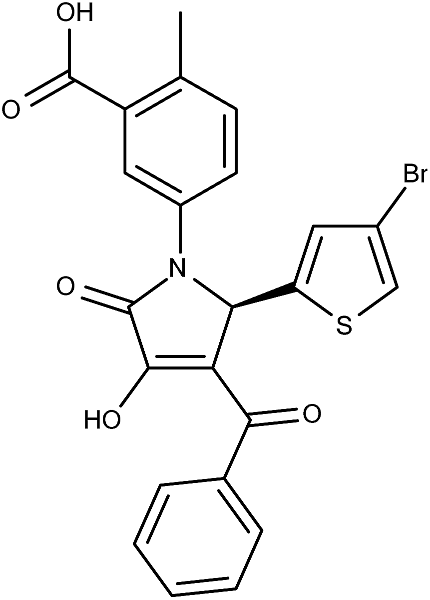 |
— | — | — | — | −9.88 | |
| 1 | Sitaxentan | Antihypertensive (pulmonary) |  |
0.633 | 0.914 | 0.926 | 0.924 | −14.37 |
| 2 | Alitretinoina | Treatment of cutaneous lesions |  |
0.66 | 0.91 | 0.906 | 0.246 | −3.91 |
| 3 | Ceftriaxone | Antibiotic |  |
0.606 | 0.937 | 0.823 | 0.217 | −12.33 |
| 4 | Acitretina | Antipsoriatic |  |
0.637 | 0.936 | 0.809 | 0.172 | −3.71 |
| 5 | Cidofovir | Antiviral |  |
0.473 | 0.633 | 0.783 | 0.31 | −4.21 |
| 6 | Telmisartan | Antihypertensive |  |
0.601 | 0.908 | 0.775 | 0.174 | −6.46 |
| 7 | Nateglinidea | Antidiabetic |  |
0.54 | 0.874 | 0.745 | 0.205 | −3.78 |
| 8 | Ceftizoxime | Antibiotic |  |
0.557 | 0.88 | 0.735 | 0.178 | −11.35 |
| 9 | Treprostinil | Antihypertensive (pulmonary) |  |
0.432 | 0.846 | 0.732 | 0.301 | −3.41 |
| 10 | Tenoxicam | Anti-inflammatory |  |
0.367 | 0.817 | 0.71 | 0.343 | −7.86 |
| 11 | Dicoumarol | Anticoagulant |  |
0.368 | 0.615 | 0.663 | 0.295 | −6.06 |
| 12 | Ethacrynic acid | Diuretic | 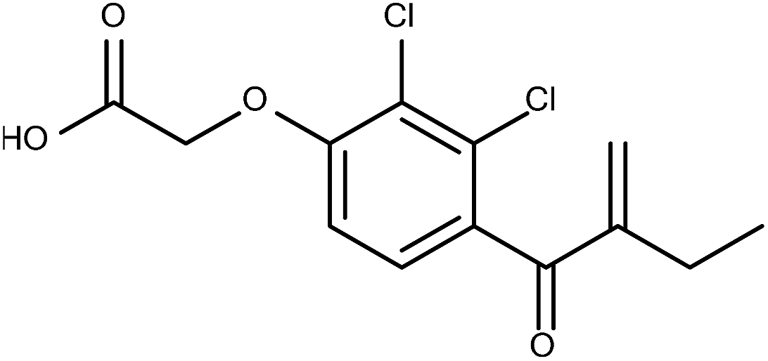 |
0.478 | 0.847 | 0.656 | 0.177 | −4.13 |
| 13 | Chlorambucila | Anticancer |  |
0.466 | 0.867 | 0.646 | 0.18 | −3.14 |
| 14 | Ticarcillin | Antibiotic |  |
0.52 | 0.664 | 0.643 | 0.123 | −17.89 |
| 15 | Nitroxoline | Antibacterial (urinary) |  |
0.41 | 0.824 | 0.641 | 0.232 | −5.31 |
| 16 | Nedocromil | Anti-inflammatory |  |
0.321 | 0.603 | 0.637 | 0.316 | −4.2 |
| 17 | Aztreonam | Antibiotic |  |
0.417 | 0.609 | 0.636 | 0.219 | −5.13 |
| 18 | Bexarotenea | Treatment of cutaneous T-cell lymphoma |  |
0.381 | 0.862 | 0.616 | 0.235 | −4.58 |
| 19 | Olmesartan | Antihypertensive |  |
0.375 | 0.593 | 0.611 | 0.237 | −6.28 |
| 20 | Aspartame | Neutraceutical |  |
0.284 | 0.118 | 0.605 | 0.32 | −3.59 |
| 21 | Cinoxacin | Antibiotic |  |
0.313 | 0.748 | 0.604 | 0.29 | −5.66 |
| 22 | Minocycline | Antibiotic |  |
0.346 | 0.602 | 0.594 | 0.248 | −4.65 |
| 23 | Sparfloxacin | Antibiotic |  |
0.156 | 0.061 | 0.591 | 0.435 | −5.06 |
| 24 | Ceftibuten | Antibiotic |  |
0.26 | 0.533 | 0.589 | 0.329 | −14.71 |
| 25 | Pemetrexed | Anticancer |  |
0.538 | 0.609 | 0.588 | 0.051 | −4.95 |
 | ||
| Fig. 3 Electrostatic potential maps generated in EON for query 1 (green capped-stick model) (line map) and one of the top-scoring hits, ceftriaxone (capped-stick model) (line map). The acidic moiety is surrounded by large red contour (level +1.9) in both the query and the hit. | ||
Docking analyses
The hits obtained from EON screening were further subjected to docking analyses in order to study their binding modes (Fig. 4).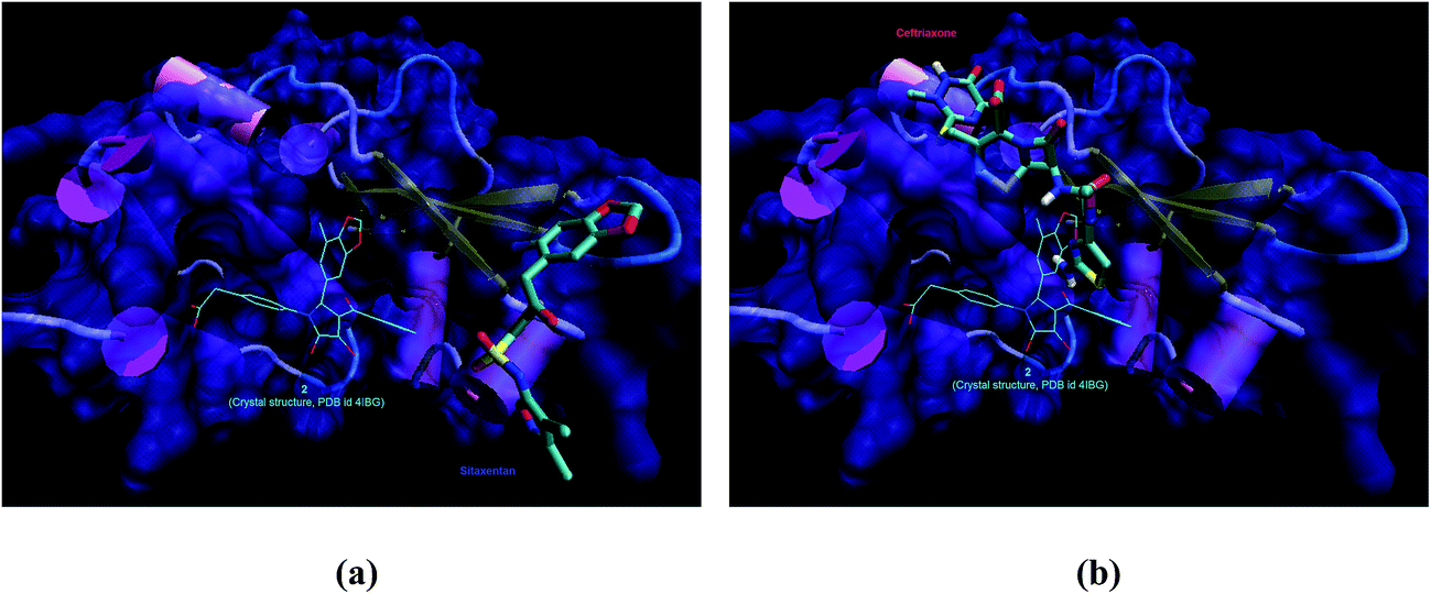 | ||
| Fig. 4 Binding modes of (a) sitaxentan (stick representation) and (b) ceftriaxone (stick representation) in the active site of VP35 eIID (PDB ID 4IBG; ribbon representation). Crystal structure ligand 2 (line representation) is shown for reference. | ||
![[thin space (1/6-em)]](https://www.rsc.org/images/entities/char_2009.gif) 500000; root mean square tolerance of 1.0 Å and 100 docking runs by AutoDock3.0.24 The ligand conformation with lowest free energy of binding in the most populated cluster was selected for comparison. The results of docking analyses are summarized in Table 1.
500000; root mean square tolerance of 1.0 Å and 100 docking runs by AutoDock3.0.24 The ligand conformation with lowest free energy of binding in the most populated cluster was selected for comparison. The results of docking analyses are summarized in Table 1.Results and discussion
In the present investigation, combined ligand- and structure-based virtual screening was performed for generating drug repositioning hypotheses for Ebola virus VP35 protein. One of the VP35 crystal structure ligands, 1, was used as a query molecule. The ligand was located in a binding pocket within a region of Ebola VP35 interferon inhibitory domain (eIID) crucial for viral replication complex formation.18 Ligand 1 belonged to benzoic acid series (B-ring) of the pyrrole-based inhibitors. The choice of 1 for ligand-based virtual screening was arbitrary.EON screening
A single run based on the query molecule 1 yielded a total of 500 hits. Careful inspection of this list revealed that a variety of drug belonging to therapeutic classes such as antibiotics, antihypertensives, anti-inflammatory, anti-hyperlipidaemic, antidiabetics, etc. were included. We focused our attention on the top 100 hits from this list. Table 1 lists top-ranking hits (#25) from EON screening with relevant parameters measuring shape and electrostatics similarities between the hits and the query 1. All the top hits (#100) along with the relevant parameters can be found in Table S1 (see ESI† section). These hits were then inspected for their molecular and physicochemical property distributions. The scatter plots are given in Fig. 1S to 6S (see ESI† section). The striking difference was seen with the scatter plot of polar surface area (PSA) versus #RotlBonds (RB) (Fig. 3S, ESI† section). A total of 36% hits possessed PSA >140 Å2, normally allowed higher limit for oral drugs. Majority of these drugs were from β-lactam and tetracycline antibiotics, in addition to others. In addition, predicted CNS permeation was found to be quite low (0.0 for 70% of the drugs) (Fig. 5S, ESI† section). This is particularly interesting since the query has acid moiety present in its structure. The EON screening would likely to pick up molecules with acidic functionality. It is a well-known fact that acids are prevented from crossing blood–brain barrier (BBB) and gaining entry into the CNS.25 Interested readers may access these property distribution plots for more information and understanding.Majority of the hits (Table 1) belonged to the antibiotics category, particularly from the β-lactam, tetracyclines and quinolone antibacterials. None of the top-ranking hits were earlier reported anti-Ebola drugs – chloroquine and amodiaquine,5 clomiphene and toremiphene.6 The obvious reason could be the chemotype of the query. While 1 belonged to acid class, it would prefer acidic drugs to match the electrostatics. The above mentioned anti-Ebola drugs are basic in nature. Hence, the query did not pick up such drugs from the DrugBank screening collection. In our opinion, it would be worthwhile to try out different query molecules in future, once some experimental proof establishes them as VP35 ligands. Fig. 3 shows the electrostatic similarity between the query molecule 1 and the top ranked drug, ceftriaxone (rank 3, Table 1). A large red contour near the cefem ring –COOH group is seen in the hit similar to 1. Other hits containing –SO2NH–, –COOH and bioisosteric groups represented antibiotics, antihyperlipidaemic, diuretic, antihypertensive, etc., categories.
Literature search of the top-ranked hits in the list was further carried out to see if any of these were ever proposed or evaluated as anti-Ebola therapeutics. Recent drug repositioning campaigns for EVD clearly list FDA-approved drugs known to possess anti-Ebola activity and FDA-approved and experimental drugs proposed as potential anti-Ebola agents which await experimental confirmation.4 None of the drugs proposed as anti-Ebola agents in the present study (Tables 1 and 1S†) were present in the first set of known anti-Ebola drugs. We could find few of the hits from the present study in the proposed anti-Ebola drug dataset selected by another method4 (Tables 1 and 1S†). Relevant references have been included for easy access to this information. In addition, some other reports (news articles, patents and research papers, etc.) also indicated the usefulness of few approved drugs as anti-Ebola agents (see Table 1S, ESI† section). Such literature evidence increased our confidence in the present study. Among the hits were drugs such as fluvastatin, rosuvastatin, atovaquone and danazol reported for the treatment of diseases caused by the filoviruses.26 These drugs have shown moderate to potent activity as inhibitors of Ebola virus replication in Vero cells. There are several news reports claiming some drugs such as high-dose vitamin C useful for the treatment of Ebola virus (not discussed here; references included in Table 1S ESI† section). The validity of such reports can be quickly tested in relevant experiments. Nonetheless, the literature is full of several interesting reports related to approved drugs and their anti-Ebola activity.
Further to the ligand-based analyses, we have screened the top 100 EON hits by docking simulation into the binding pocket of VP35. Sitaxentan and ceftriaxone were further investigated on their binding mode and interactions with VP35. From the docking studies, results showed that sitaxentan was docked above the β-sheets of Val237-Gln331 and Val294-Arg298. On the other hand, ceftriaxone was docked in the cavity of α-helix Ala238-Asn254, β-sheet Val294-Arg298 and coil/turn Val284-Pro293 of VP35. The crystal ligand was located between α-helix Ala238-Asn254 and β-sheets of Val237-Gln331 and Val294-Arg298. Sitaxentan has a higher binding affinity towards VP35 than that of ceftriaxone but the amine tail of ceftriaxone occupied the similar binding side with the crystal structure ligand 2. It would be therefore a similar ligand in term of binding pocket occupancy. Application of a more stringent calculation on the simulation can be applied to see details picture of the interactions.
Conclusions
In the present study, to motivate Ebola researchers interested in finding a new drug treatment, several repositioning hypotheses were generated for approved/existing small molecular drugs. The crucial Ebola VP35 protein, a potential molecular target against which anti-Ebola therapeutics can be developed, crystal structures and associated ligands were used as a starting point for the combined ligand- and structure-based approaches. The ligand-based virtual screening (shape- and electrostatics-based) yielded several interesting hits such as ceftriaxone, ceftizoxime (antibiotics), telmisartan (antihypertensives), nateglinide (antidiabetics), cidofovir (antiviral), etc. The binding modes of these potential repositioning candidates were established using molecular docking using the eII domain of VP35. The top-ranking hits such as ceftriaxone, were found to dock ‘well’ in the ligand-binding pocket of Ebola protein. Since the query molecule contained an acidic functional group, several of these hits contain carboxylic acid group or its biosostere. This information can be crucial for the design or rather redesign of the novel ligand for the VP35 protein from Ebola. The present study and the associated repositioning hypotheses may potentially prove thought-provoking to the curious researchers to undertake experimental evaluations in the related assays. The experimental results are likely to validate the hypotheses and observations which will help us in developing anti-Ebola drugs.Conflict of interest
The authors declare no financial involvement or conflict.Acknowledgements
PSK would like to acknowledge the support given by Dr R. S. Gaud, Dean, SPP School of Pharmacy and Technology Management, SVKM's NMIMS, Mumbai during execution of this work. PR and LR acknowledge the facilities from the University of Mauritius. YSC would like to thank the computational facilities provided from the Fundamental Research Grant Scheme (FRGS; 203/CIPPM/6711439) from the Malaysia Ministry of Education.References
- R. Hammami and I. Fliss, Drug Discovery Today, 2010, 15, 540–546 CrossRef CAS PubMed.
- S. Shrivastava, P. Shrivastava and J. Ramasamy, Asian Pac. J. Trop. Dis., 2015, 5, 253–262 CrossRef.
- Ebola vaccines, therapies, and diagnostics, http://www.who.int/medicines/emp_ebola_q_as/en/, accessed on August 22 2015.
- V. Veljkovic, P. Loiseau and B. Figadere, F1000Research, 2015, 4, 34 Search PubMed.
- P. B. Madrid, S. Chopra, I. D. Manger, L. Gilfillan, T. R. Keepers, A. C. Shurtleff, C. E. Green, L. V. Iyer, H. H. Dilks, R. A. Davey, A. A. Kolokoltsov, R. Carrion Jr, J. L. Patterson, S. Bavari, R. G. Panchal, T. K. Warren, J. B. Wells, W. H. Moos, R. L. Burke and M. J. Tanga, PLoS One, 2013, 8, e60579 CAS.
- L. M. Johansen, J. M. Brannan, S. E. Delos, C. J. Shoemaker, A. Stossel, C. Lear, B. G. Hoffstrom, L. E. Dewald, K. L. Schornberg, C. Scully, J. Lehár, L. E. Hensley, J. M. White and G. G. Olinger, Sci. Transl. Med., 2013, 5, 190ra79 CrossRef PubMed.
- S. Ekins and M. Coffee, F1000Research, 2015, 4, 48 Search PubMed.
- N. Litterman, C. Lipinski and C. Ekins, F1000Research, 2015, 4, 38 Search PubMed.
- D. Leung, K. Prins, C. Basler and G. Amarasinghe, Virulence, 2010, 1, 526–531 CrossRef PubMed.
- C. A. Zampieri, N. J. Sullivan and G. J. Nabel, Nat. Immunol., 2007, 8, 1159–1164 CrossRef CAS PubMed.
- C. F. Basler, A. Mikulasova and L. Martinez-Sobrido, J. Virol., 2003, 77, 7945–7956 CrossRef CAS PubMed.
- W. B. Cardenas, Y. M. Loo and M. Gale, J. Virol., 2006, 80, 5168–5178 CrossRef CAS PubMed.
- K. C. Prins, S. Delpeut and D. W. Leung, J. Virol., 2010, 84, 3004–3015 CrossRef CAS PubMed.
- D. W. Leung, K. C. Prins and D. M. Borek, Nat. Struct. Mol. Biol., 2010, 17, 165–172 CAS.
- P. Moller, N. Pariente, H. D. Klenk and S. Becker, J. Virol., 2005, 79, 14876–14886 CrossRef PubMed.
- K. C. Prins, J. M. Binning, R. S. Shabman, D. W. Leung, G. K. Amarasinghe and C. F. Basler, J. Virol., 2010, 84, 10581–10591 CrossRef CAS PubMed.
- C. Knox, V. Law, T. Jewison, P. Liu, S. Ly, A. Frolkis, A. Pon, K. Banco, C. Mak and V. Neveu, Nucleic Acids Res., 2011, 39, D1035–D1041 CrossRef CAS PubMed.
- C. S. Brown, M. S. Lee and D. W. Leung, J. Mol. Biol., 2014, 426, 2045–2058 CrossRef CAS PubMed.
- M. K. Teli and G. K. Rajanikant, J. Chem. Inf. Model., 2013, 53, 1818–1824 CrossRef CAS PubMed.
- P. S. Kharkar, S. Borhade, A. Dangi and S. Warrier, J. Comput. Sci., 2015, 10, 217–224 CrossRef.
- OpenEye Scientific Software Products - Vida version 4.2.1, QUACPAC version 1.5.0, Omega version 2.4.6, Make Receptor version 3.0.0 and OEDOCKING version 3.0.0-are available from OpenEye Scientific Software, 9 Bisbee Court, Suite D, Santa Fe, NM 87508 Search PubMed.
- T. S. Rush, J. A. Grant, L. Mosyak and A. Nicholls, J. Med. Chem., 2005, 48, 1489–1495 CrossRef CAS PubMed.
- P. Markt, R. K. Petersen, E. N. Flindt, K. Kristiansen, J. Kirchmair, G. Spitzer, S. Distinito, D. Schuster, G. Wolber, C. Laggner and T. Langer, J. Med. Chem., 2008, 51, 6303–6317 CrossRef CAS PubMed.
- G. M. Morris, D. S. Goodsell, R. S. Halliday, R. Huey, W. E. Hart, R. K. Belew and A. J. Olson, J. Comput. Chem., 1998, 19, 1639–1662 CrossRef CAS.
- P. S. Kharkar, F1000Research, 2014, 3, 40 Search PubMed.
- L. M. Johansen, J. Lehár, B. G. Hoffstrom, G. G. Olinger and A. R. Stossel, US Pat., 8475804, 2013.
Footnote |
| † Electronic supplementary information (ESI) available: The list of all the hits from EON screening (#100) with relevant parameters from EON and docking runs along with the molecular and physicochemical property distribution plots in comparison with the query molecule are given. See DOI: 10.1039/c6ra01704e |
| This journal is © The Royal Society of Chemistry 2016 |