Yu Zhu* and
Yousheng Tao
Key Laboratory of Design and Assembly of Functional Nanostructures, Chinese Academy of Sciences, Fujian Provincial Key Laboratory of Nanomaterials, Fujian Institute of Research on the Structure of Matter, University of Chinese Academy of Sciences, Fuzhou, Fujian 350002, China. E-mail: zhuyu@fjirsm.ac.cn
First published on 4th March 2016
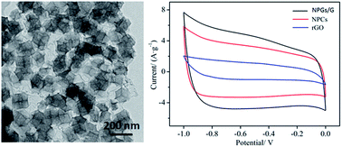
Nitrogen-doped nanoporous carbon/graphene networks (NPC/G) have been prepared by zeolitic imidazolate frameworks (ZIF-8) homogeneously encapsulated in graphene oxide (GO) networks, which were carbonized at high-temperature under a N2 atmosphere and washed to get rid of impurities. The obtained NPC/G composites are highly graphitizing and exhibit a high specific surface area up to 703 m2 g−1. By optimizing the annealing conditions, the nanoporous frameworks derived from ZIF-8 can be effectively maintained, which are beneficial for electrolyte ion adsorption and transportation. Furthermore, the NPC/G composites are used as electrode materials for supercapacitive energy storage and show a high specific capacitance of 235 F g−1 at a current density of 1 A g−1, which is larger than that of NPCs and rGO. This provides a new route for designing novel structures to improve electrochemical performance. More interestingly, these electrode materials also present excellent rate capability and 85% retention of its initial capacitance after 1000 cycles in 1 M KOH aqueous solution.
In the last decade, graphene has shown promising applications in high-performance electrochemical energy storage devices owing to its excellent electronic properties, high specific surface area (2630 m2 g−1), and chemical and mechanical stability.10–14 However, it was repeatedly demonstrated that the face-to-face restacking behavior of graphene nanosheets hindered the charge transport and thereby limited its application for supercapacitors. To prevent restacking of graphene nanosheets and increase their efficient electrical conducting paths, different kinds of spacer materials were reported for improving the performance of supercapacitors.15–17 For example, incorporating pseudocapacitive nanostructured transition metal oxides or hydroxides (MnO2, Ni(OH)2, etc.)18–22 into graphene sheets was adopted to avoid the stacking of nanosheets for improving the capacitance. Unfortunately, the so-called pseudocapacitors, even though it is possible to store more energy than EDLC, suffer from drawbacks such as a lower power density and lower stability during cycling.1,10
Recently, nanoporous carbons have been shown as promising electrode candidates applied in EDLCs.23–28 In order to prepare highly nanoporous carbons with excellent features for supercapacitors, metal-organic frameworks (MOFs) are often considered as precursor or sacrificial templates owing to their highly specific surface areas, good thermal stability, tailoring pore structures and controlled channels.29–33 Therefore, it is anticipated that constructing uniform MOF derived nanoporous carbons embedded into graphene nanosheets to obtain nanoporous carbon/graphene networks is highly desirable for supercapacitors owing to the combination of nanopore structures and electron transfer. Moreover, the imidazole-based ligands of ZIF-8 will yield N-doped porous carbons, which can enhance the properties of the porous carbons. For example, N-doped carbons may afford pseudocapacitance and improve the electronic conductivity of carbon-based materials.34–36
In this study, the NPC/G networks were prepared by ZIF-8 nanocrystals homogeneously encapsulated in GO nanosheets, followed by calcining and washing processes. The obtained NPC/G networks have a high specific surface area, hierarchical pore structure and high nitrogen content, which are beneficial for electrolyte ion adsorption and transportation. Furthermore, the NPC/G as electrode materials were applied in supercapacitive energy storage for the first time and exhibited a high specific capacitance, excellent rate capability and 85% retention of its initial capacitance after 1000 cycles in 1 M KOH aqueous solution.
Electrochemical experiments were performed on a CHI 660e electrochemical workstation (Chenhua, Shanghai, China) with a standard three-electrode cell in 1 M KOH aqueous solution. A platinum foil (1.0 cm2) and mercuric oxide electrode were used as counter and reference electrode, respectively. The working electrode was prepared by coating a slurry of sample onto a Ni-foam electrode (1 × 1 cm2) and drying in a vacuum oven at 333 K overnight. Typically, the NPC/G was uniformly mixed with polyvinylidene difluoride (PVDF) binders in a weight ratio of 95
:
5 and made as a slurry using N-methyl-2-pyrrolidone. To evaluate the electrochemical characteristics of the materials as electrodes in aqueous systems, cyclic voltammetric (CV) and galvanostatic charge–discharge (GC) measurements were performed. Cyclic stabilities were characterized using galvanostatic charge–discharge measurements over 1000 cycles at a charge–discharge rate of 3 A g−1. The specific capacitance Cg was calculated based on the following equation:
| Cg = I × Δt/(m × ΔV) |
Graphene oxide (GO) was prepared by an improved Hummers’ method37–40 (for the detailed synthetic procedure, see the ESI†). The as-prepared GO was characterized by XRD and FT-IR spectroscopy (see Fig. S1 and S2, in the ESI†). The XRD result showed that there was a larger interlayer spacing of d002 (9.5 Å) than that of graphite (3.35 Å), attributed to the successful integration of a large amount of hydroxyl and epoxide functional groups in the GO nanosheets. The IR spectrum of GO prepared by this routine was essentially consistent with that reported in the literature,37,41,42 and the following functional groups were identified: O–H stretching vibrations (3432 cm−1), C
O stretching vibration (1737 cm−1), C
C from sp2 CC bonds (1632 cm−1), C–OH vibrations (1400 cm−1), and C–O vibrations (1227 cm−1). Further, the surface morphology and texture of the GO samples were investigated by TEM and AFM measurements. As shown in Fig. S3,† a lamellar nanostructured architecture is observed. The obtained nanosheets are further examined by AFM analyses in Fig. S4† which reveal an average thickness of approximately 0.8 nm for GO.
ZIF-8 was chosen as the precursor in the presence of GO for preparing ZIF-8/GO nanocomposites. The evolution of the structure during the synthesis of ZIF-8/GO and NPC/G was demonstrated using XRD. As revealed by the X-ray diffraction (XRD) pattern shown in Fig. 2, all diffraction peaks with a strong intensity for ZIF-8/GO match well with the simulated ZIF-8, indicating a pure phase and high crystallinity. After the heat treatment, the diffraction peak of NPC/G at around 25° was assigned to the interplane (002) reflection of graphite carbon, and there were no diffraction peaks of impurities (Zn species), indicating thorough carbonization of ZIF-8 and that the zinc species were fully removed by acid washing.
The SEM and TEM observations shown in Fig. 3a and c demonstrate that the ZIF-8 nano-polyhedrons, with a size of around 150 nm, were successfully embedded into the GO networks. To our knowledge, this is the first example of a novel architecture consisting of nanoporous ZIF-8 particles embedded in a microsized graphene network. Such a type of structure could effectively prevent the agglomeration of graphene during high-temperature treatment. In addition, in the absence of GO, Zn salts in methanol were added to imidazole solution for preparing ZIF-8. As revealed by the SEM image shown in Fig. 3b, ZIF-8 exhibited a nano-polyhedron structure with a size of around 500 nm, which is larger than that of those in ZIF-8/GO. This result suggests that GO can confine the growth of ZIF-8 nanoparticles, which is similar to the report by Wang’s group.43
![]() | ||
| Fig. 3 SEM images of (a) ZIF-8/GO and (b) ZIF-8. (c) TEM image of ZIF-8/GO. (d) Schematic illustration for ZIF-8/GO. | ||
After high-temperature calcination, the ZIF-8/GO nanocomposites can be transformed to derivative NPC/G with size and morphological retention as revealed by the SEM images shown in Fig. 4a and b. To further investigate the morphology and structure of NPC/G, TEM characterization has been carried out. As presented in Fig. 4c, the NPC particles homogeneously encapsulated in the graphene nanosheets maintained the structural integrity and prevented the agglomeration of graphene nanosheet. Moreover, the high-resolution TEM image in Fig. 4d further confirms that NPC/G has a nanoporous structure, suggesting that the optimized annealing conditions can prevent the collapse of the structure. In the Raman spectra of NPC/G before and after calcination, as shown in Fig. S5,† there are two dominant peaks at 1350 and 1584 cm−1, corresponding to the D band and G band. Notably, the ID/IG value of NPC/G (ID/IG = 0.99) was higher than that of ZIF-8/GO (ID/IG = 0.91), indicating the formation of abundant defects and disordered carbon during the heat-treated process.
The surface area and pore size distribution of NPC/G were characterized by N2 adsorption–desorption measurements. As shown in Fig. 5a, the NPC/G displayed a type-IV isotherm with a distinct hysteresis loop in the relative pressure region of 0.43 < P/P0 < 1.0, indicating a hierarchically porous structure with micropores and mesopores.44,45 The BET surface area of NPC/G was up to 703 m2 g−1. The pore size distribution, calculated from the density functional theory (DFT) method, revealed that the NPC/G possessed micro- (0.47 nm, 1.3 nm), meso- (2–50 nm) and macropores (50–100 nm), as shown in Fig. 5b. Compared with pore size distribution of NPC/G, the NPCs exhibited a similar distribution in micropores, and an obvious difference in mesopores and macropores (Fig. S6†). This result revealed that the micropores might originate from the carbon framework by calcination, and the mesopores and macropores might be mainly attributed to the stacking of carbon frameworks and graphene.44 The proportion of hierarchical pore distribution endowed NPC/G with a large electrode/electrolyte contact area and facilitated diffusion of electrolyte ions.
![]() | ||
| Fig. 5 (a) Nitrogen adsorption (○)–desorption (●) isotherm of the NPC/G networks at 77 K. (b) Pore size distribution of NPC/G. | ||
Nowadays, various synthesis methods and characterization techniques for N-graphene have been explored. For example, Pumera’s group presented an optimized, scalable technique for fabrication of large quantities of highly nitrogen doped (>7 at%) graphene.46 The charge distribution and electron doping in nitrogen-doped graphene were investigated by Joucken et al.,47 and individual nitrogen dopants in monolayer graphene were characterized by Pasupathy et al.48 The nitrogen doping effects on the structure of graphene were studied by Sun’s group.42 In addition, different synthesis and characterization methods for nitrogen-substituted graphene were reviewed by Wang et al.49 Different from the above strategy for nitrogen directly doping into graphene frameworks, in this study, zeolitic imidazolate frameworks (ZIF-8) were adopted to embed into GO nanosheets for yielding N-doped porous carbon/graphene composites. Nitrogen doping of porous carbons can enhance the capacitive property by affording pseudocapacitance and improving the electronic conductivity of carbon-based materials.34 Therefore, X-ray photoelectron spectroscopy (XPS) was used to determine the nitrogen content and the nitrogen type in NPC/G. As shown in Fig. 6, the N 1s peak of NPC/G was deconvoluted to four different peaks located at 398.4, 400.0, 401.1, and 405.4 eV, corresponding to pyridinic nitrogen (N-1), pyrrolic nitrogen (N-2), quaternary nitrogen (N-3) and pyridinic N+–O−, respectively.36,43 N-1 and N-2 can provide faradaic pseudocapacitance, whereas N-3 can effectively improve the conductivity of porous carbons. The N-1, N-2 and N-3 content was 4.7%, 4.2% and 4.0%, respectively, indicating that the NPC/G may have an excellent capacitive behavior. In addition, elemental analysis showed N content of 11.17% (mass percent), which was close to the result of XPS.
Motivated by the advantages of a hierarchically porous structure and high content of nitrogen,50–54 the capacitance behavior has been investigated by cyclic voltammetric (CV) and galvanostatic charge–discharge (GC) measurements. Compared with NPCs and rGO, as shown in Fig. 7, the NPC/G has a larger covered area in the CV curve at a scan rate of 25 mV s−1, indicating that NPC/G has a larger specific capacitance than that of NPCs and rGO, suggesting that this novel nanoporous architecture would be beneficial for improving the performance of supercapacitors.
As displayed in Fig. 8a, comparing the CV curves of materials obtained at different calcinating temperatures (700, 800, and 900 °C), it was found that the NPC/G networks carbonized at 800 °C exhibit the largest CV area, which may be attributed to that the NPC/G can effectively maintain the nanoporous framework derived from ZIF-8, and possess a high surface area.44 The electrochemical performance of NPC/G was further studied carefully. The CV curves of NPC/G showed quasi-rectangular shapes at different scan rates as presented in Fig. 8b. With increasing scan rates from 5 mV s−1 to 100 mV s−1, the specific capacitances decreased from 200 F g−1 to 136 F g−1. The more detailed data are listed in Table S1.† In general, the current density tends to increase with the increase of the scanning rate.27,30,32 The CV curves retained their shape even at high scan rates, indicating a good high-rate performance of the active material. As presented in Fig. 8c, the GC curves of NPC/G displayed almost symmetrical triangular shapes at different current densities ranging from 1 to 10 A g−1, implying the excellent electrochemical reversibility of NPC/G. The specific capacitances based on the GC curves are exhibited in Fig. 8c. NPC/G has specific capacitances of 235, 201, 186, 176 and 168 F g−1 at current densities of 1, 2, 3, 4 and 5 A g−1, respectively. For comparison, the CV, GC curves and specific capacitance of NPCs are shown in Fig. S7† and 9. The NPCs hold the specific capacitances of 171, 141, 131, 124 and 119 F g−1 at current densities of 1, 2, 3, 4 and 5 A g−1, respectively. Obviously, the NPC/G networks showed a higher specific capacitance than that of the NPCs. This result indicated that the construction of NPC/G networks was beneficial for improving the specific capacitance. Meanwhile, this would also provide a new route for designing novel structures to improve the electrochemical performance.
To our knowledge, some nanoporous carbons derived from MOFs have been used as electrode materials for supercapacitors with high performances, as listed in Table 1. The NPC/G networks show a high specific capacitance of 235 F g−1 at current density of 1 A g−1, which is comparable to some materials derived from MOFs reported previously.
| Material | Specific capacitance (F g−1) | Reference |
|---|---|---|
| NPC/G | 235 (1 A g−1) or 200 (5 mV s−1) | This work |
| Z-800 | 130 (50 mV s−1) | 27 |
| NPC-800 | 238 (20 mV s−1) | 24 |
| C800 | 188 (5 mV s−1) | 55 |
| CZIF69a | 168 (5 mV s−1) | 56 |
| AS-ZC-800 | 211 (10 mV s−1) | 32 |
| NPC800 | 151 (0.05 A g−1) | 57 |
| MC-A | 222 (0.25 A g−1) | 58 |
| HPC | 166 (0.1 A g−1) | 59 |
| C-S700 | 182 (2 mV s−1) | 60 |
| C-MOF-2 | 170 (10 mV s−1) | 61 |
| C-Zn-BTC | 134 (10 mV s−1) | 61 |
Moreover, the cyclic stability of the NPC/G composites was measured at a current density of 3 A g−1, as shown in Fig. 10. The specific capacitance retention was 85% after 1000 cycles. The degradation of 15% of the specific capacitance may be caused by two factors as follows. One is that the hetero-atoms (N or O) in the framework or as functional groups on the surface are not stable during long cycling and a gradual decrease of capacitance is observed.62,63 The other is structural breakdown and decay of electrical conductivity during the charging/discharging process.64
Footnote |
| † Electronic supplementary information (ESI) available: Synthetic procedure, XRD, FT-IR spectroscopy, TEM image of GO nanosheet, AFM scan of GO sheet, pore size distribution of NPCs, Raman spectra of NPC/G before and after calcination, CV curves of NCPs at different scan rates, and specific capacitance at different scan rates for NCP/G. See DOI: 10.1039/c6ra01623e |
| This journal is © The Royal Society of Chemistry 2016 |