Photophysical, G-quadruplex DNA binding and cytotoxic properties of terpyridine complexes with a naphthalimide ligand

Zhize Ou*a, Yimeng Qiana, Yunyan Gao*a, Yunqing Wanga, Guoqiang Yangb, Yi Lic, Kaiyue Jianga and Xin Wanga
aThe Key Laboratory of Space Applied Physics and Chemistry, Ministry of Education, Department of Applied Chemistry, School of Science, Northwestern Polytechnical University, Xi'an, 710072, People's Republic of China. E-mail: ouzhize@nwpu.edu.cn; gaoyunyan@nwpu.edu.cn; Fax: +86 29 88431677; Tel: +86 29 88431677
bCAS Key Laboratory of Photochemistry, Institute of Chemistry, Chinese Academy of Sciences, Beijing 100190, People's Republic of China
cKey Laboratory of Photochemical Convesion and Optoelectronic Material, Technical Institute of Physics and Chemistry, Chinese Academy of Sciences, Beijing, 100190, People's Republic of China

Received 17th January 2016 , Accepted 6th April 2016

First published on 7th April 2016


Abstract

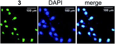
Two novel metal complexes 2–3 (metal = PdII, PtII) have been synthesized by reacting the corresponding tolylterpyridine complexes and the 4-aminonaphthalimide derivative 1. The interactions of the complexes with duplex DNA and telomeric G-quadruplex DNA have been investigated by UV-Vis spectroscopy and fluorescence spectroscopy. The studies reveal that the complexes 2–3 possess high affinity and reasonable selectivity for telomeric G-quadruplex DNA over duplex DNA. Spectroscopic and molecular docking studies suggest that the complexes 2–3 interact with telomeric G-quadruplex DNA mainly through groove binding. The compounds 1–3 are emissive (Φem > 0.22), making it possible to study the localization of 1–3 in A549 using fluorescence microscopy. The complexes 2–3 are mainly localized in nuclei, while 1 is localized in the nuclei and cytoplasmic region after 0.5 h incubation. The complex 3 inhibits A549 cells selectively over non-cancerous NIH3T3 cells, with higher antitumor activity than 1 and cisplatin.


1. Introduction

The study of the sequence specific binding of small molecules with DNA has been an important topic in the design of new drugs.1 G-rich sequences of DNA can fold into four-strand helical structures known as G-quadruplexes, which are prevalent in human genes, especially in telomere ends and promoter regions of oncogenes (for example c-myc, c-kit, and bcl-2).2 Human telomeres possess a single-stranded DNA (ssDNA) overhang of TTAGGG repeats.3 In normal cells, telomeres shorten with each cell division until they become critically short and dysfunctional. Telomerase, which is over-expressed in approximately 85% of cancer cell, extends the length of human telomeric DNA by adding d(TTAGGG) repeats to the 3′-terminus and is crucial for the immortalization of cancerous cells. Considerable circumstantial evidence suggests that telomerase activity can be directly inhibited if single-stranded telomeric DNA is folded into a quadruplex. Thus, G-quadruplex binding molecules have recently emerged as a new class of potential selective antitumor therapeutics.4 Apart from the purely organic heteroaromatic compounds reported as G-quadruplex binders, the metal complexes can also interact strongly and selectively with quadruplexes, due to their specific geometries and ligand orientation for optimal G-quadruplex binding.5 Organometallic complexes with biologically active ligands, such as acridine,6 porphyrin,7 erlotinib,8 quindoline,9 perylene,10 exhibit enhanced G-quadruplex affinity, in some cases, display promising anticancer properties.

Naphthalimide derivatives, as a class of DNA-binding agents, have been extensively explored as antitumor agents.11 Several naphthalimide derivatives (such as amonafide, mitonafide, bisnafide and elinafide) have reached clinical trials as antitumor agents.12 Structure–activity relationship studies have pointed out that some crucial parameters can influence the anticancer activities of naphthalimides, including fused aromatic ring,13 position and size of side chains,14 and the nature of the imide substituent.15 In recent years, research on targeted and multifunctional naphthalimide–metal complex conjugates has received increasing attention, mainly due to their enhanced anticancer activity.16 It has been noted that the presence of naphthalimide ligand can enhance the cellular uptake and accumulation of metal complex in the nuclei of tumour cells compared to the naphthalimide free analogue.17

Given the fact that telomeric DNA is localized in nucleus, investigation of the cellular uptake and localization of G-quadruplex targeting drugs will provide valuable clues for the rational design of more efficient anticancer drugs.18 Naphthalimide derivatives display high affinity for the G-quadruplex.19 It can be expected that selective recognition of G-quadruplex may be achieved by conjugation of napthalimides with terpyridine complex.20–22 On the other hand, the modification of the complex with naphthalimide makes it possible for fluorescence imaging of the intracellular distribution of metal complex.23 In this work, the 4-aminonaphthalimide derivative 1 is synthesized and further reacts with terpyridine metal complexes to give the complexes 2–3 (Scheme 1). The photophysical properties, G-quadruplex binding ability, cellular location and cytotoxicity of 1–3 are investigated, which can provide useful information for the design of novel G-quadruplex binding naphthalimide–metal complex conjugates.


image file: c6ra01441k-s1.tif
Scheme 1 The synthetic route of N-dimethylaminoethyl-4-aminoethylamino-1,8-naphthalimide 1 and its terpyridine complexes. Reagents and conditions: (a) dioxiane, 80 °C, 24 h; (b) acetonitrile, reflux, 12 h; (c) CF3COOAg, DMF, RT, 24 h; (d) compound 1, DMF, RT, 24 h.

2. Results and discussion

2.1 Synthesis and characterization

In literature, 4-aminonaphthalimide derivatives have shown anticancer activity against a variety of human tumour cells.24 Use of naphthalimide derivatives to enhance the cytotoxic effect of metal complexes has been recently reported.16 The synthetic route of the 4-aminonaphthalimide 1 and its complexes is shown in Scheme 1. 4-Bromine-1,8-naphthalanhydride is used as the raw material and the intermediate product further reacts with ethylenediamine to give 4-aminonaphthalimide 1. The tertiary amine side chain in 1 would facilitate the internalization of naphthalimides.25 The tolylterpyridine (ttpy) complexes 2–3 are obtained in a single-step reaction from the naphthalimide ligand 1, by reacting it with [Pd(ttpy)Cl]Cl or [Pt(ttpy)Cl]Cl following the literature procedure.26 All the terpyridine complexes can be obtained in good yields (69–83%) and are characterized with 1H NMR, 13C NMR, MALDI-TOF and elemental analysis.

2.2 Photophysical properties

In the family of naphthalimides, 4-aminonaphthalimides show strong absorption and emission in the visible region, with high photostability and large Stokes' shift.27 The UV-Vis absorption of naphthalimide 1 shows two intense absorption bands at 279 and 437 nm, respectively (Fig. 1 and Table 1). The longer-wavelength absorption of 1 is characterized by a broad band which can be attributed to the intramolecular charge transfer (ICT) between the 4-amino group and the naphthalimide ring system.28 The absorption spectra of complexes 2–3 show a band at around 435 nm characteristic of 4-aminonaphthalimide, and a band at around 280 nm, which can be assigned to the intraligand (IL) transition of naphthalimide and terpyridine ligands.29 The bands in the range of 300–400 nm can be observed in the absorption spectra of complex 2–3, which can be attributed to metal-to-ligand charge transfer (MLCT) transitions because of their high intensity and their dependence on metal ion.30
image file: c6ra01441k-f1.tif
Fig. 1 UV-Vis absorption spectra of 1–3 (30 μM) in HEPES (10 mM, pH 7.4) containing 0.1 M KCl.
Table 1 Spectroscopic and excited-state properties of the ligand 1 and its complexes 2–3 (λex = 435 nm)
Compound Absorption λAbs/nm (ε/×104 L mol−1 cm−1) Emission (298 K)
λem (nm) Φem τf (ns) kf × 107/(s−1) knr × 107/(s−1)
1 279 (1.40), 437 (1.28) 536 0.31 5.78 5.36 1.19 × 10⁸
2 280 (1.98), 349 (0.77), 365 (0.79), 437 (0.99) 537 0.34 5.40 6.30 1.22
3 279 (2.21), 322 (0.85), 434 (1.26) 538 0.22 4.55 4.65 1.65


The complexes 2–3 are stable in organic solvents, such as DMF, DMSO and methanol for several weeks. The absorption spectra of 2–3 in the buffer solution containing 0.15 M KCl do not change in several hours, indicating that they are stable in aqueous solution (Fig. S1a). However, addition of glutathione (GSH) brings about some changes in the absorption spectra of 2–3 (Fig. S1b), indicating the occurrence of substitution reactions of 2–3 with GSH.31

Fluorescence spectra and fluorescence quantum yields of 1–3 are studied in buffer solution. The excited state parameters of 1 and its complexes are summarized in Table 1. It is found that there is no significant shift in the position of fluorescence maxima, but the fluorescence quantum yields (Φem) follows the trend of 12 > 3, suggesting that the metal ions would affect the excited state of 1. Since the decay profiles are fitted satisfactorily by a single exponential function (Fig. S2), the excited-state life time (τf), radiative decay rate constant (kf) and nonradiative rate constant (knr) could be determined accurately (eqn (1) and (2)).32 The rate constant for nonradiative decay (knr), increases obviously when 1 coordinated with platinum, which can be attributed to the strong heavy atom effect of platinum.33

 
kr = Φem/τf (1)
 
knr = (1 − Φem)/τf (2)

2.3 DNA binding titration

DNA interaction studies of 1–3 have been evaluated by UV-Vis and fluorescence titration experiments. Addition of CT DNA to a solution of 1 leads to a 13.0% hypochromism without significant spectral shift (Fig. S3a). By using a plot of Dεap versus D (Fig. S3a inset) according to the Scatchard equation,34 the binding constant (Ka) of 1 with CT DNA is calculated to be 7.35 × 10⁴ M−1. On the other hand, when addition of Htelo G-quadruplex to the solution of 1, the decrease in the absorbance of 1 is within 5%, indicating weak interaction between 1 and Htelo G-quadruplex (Fig. S3b).

In contrast, addition of Htelo quadruplex to a solution of 2–3 leads to a 20–37% hypochromism at λ = 279 nm in the UV-Vis spectrum without significant red shift (Fig. 2a), suggesting that 2–3 interact with G-quadruplexes through external binding mode.35 The DNA binding constants derived from Scatchard equation are given in Table 2. It seems that the binding constants of the complexes 2 and 3 for Htelo G-quadruplex are monovalent cation-dependent, maybe due to the structural polymorphism of G-quadruplexes under different ionic conditions.36 Both 2 and 3 show high affinity for telomeric G-quadruplex DNA (Ka > 5 × 10⁶ M−1), with binding constants comparable to those of previously reported good quadruplex DNA binders (106 to 108 M−1).37 To examine the binding specificity, the interaction between the complexes and duplex DNA is also investigated by UV-Vis titration (Fig. 2b). Both 2 and 3 bind to CT DNA with Ka values of 104 to 105 M−1, exhibiting reasonable selectivity for telomeric G-quadruplex versus duplexes, similar to that of dimetallic complex.38


image file: c6ra01441k-f2.tif
Fig. 2 UV-Vis titration of 3 (30 μM) with (a) Htelo and (b) CT DNA in HEPES (10 mM, pH 7.4) containing 0.1 M NaCl. The arrow indicates the changes upon addition of DNA. Inset: plot of Dεap versus D. D is the concentration of DNA, Δεap = |εAεF|. εA = Aobs/[complex], εF corresponds to the extinction coefficients of the unbound complex.
Table 2 Association constants (Ka, M−1) of the compounds 1–3 with CT DNA and Htelo G-quadruplex DNA determined by UV-Vis spectroscopy
Compound 0.1 M Na+ 0.1 M K+
CT DNA Htelo Selectivitya CT DNA Htelo Selectivity
a HteloKa/CT DNAKa.b Means not detected.
1 7.35 × 10⁴ N.D.b N.D.b 6.2 × 10⁴ N.D.b N.D.b
2 8.1 × 10⁴ 1.43 × 10⁷ 177 9.5 × 10⁴ 2.39 × 10⁷ 252
3 9.47 × 10⁴ 3.74 × 10⁷ 394 1.74 × 10⁵ 6.83 × 10⁶ 39.2


Along with the UV-Vis spectral changes, a decrease in the fluorescence intensity of 3 is observed upon addition of CT DNA and Htelo G-quadruplex by up to ca. 25%, without any shift of the emission peak (Fig. 3). Similar changes can also be observed for 1 and 2. It is generally accepted that insignificant shift in emission peak is the most probable consequence of groove binding.39 Thus, these results primarily indicate that the compounds 1–3 interact with DNA mainly through groove binding. It is also likely that the metal-terpyridine fragments in 2–3 interact electrostatically with the negatively charged phosphate backbone of the DNA.40


image file: c6ra01441k-f3.tif
Fig. 3 Emission spectra of 3 (30 μM) in HEPES (10 mM, pH 7.4) containing 0.1 M NaCl upon addition of (a) CT DNA and (b) Htelo G-quadruplex (λex = 435 nm).

2.4 Circular dichroism (CD) studies

CD spectroscopic studies are carried out to investigate the interaction of 1–3 with Htelo. In the absence of potassium or sodium, Htelo displays two positive absorption bands at 255 and 295 nm, indicating the coexistence of unfolded sequence and quadruplexes structure. After addition of the complexes 2–3, the overall shape of the spectrum is retained (Fig. S4), indicating that the complexes do not template quadruplex formation from the unfolded Htelo sequence.41

As shown in Fig. 4a, in the presence of Na+, the CD spectrum of Htelo shows a strong positive band at 290 nm and a negative band at 265 nm, which indicates the formation of an anti-parallel G-quadruplex structure. After addition of the complexes 2–3, the intensity of both the positive and negative peaks increases, suggesting that the antiparallel structure is stabilized by the complexes 2–3.


image file: c6ra01441k-f4.tif
Fig. 4 CD spectra of Htelo (10 μM) in 10 mM Tris–HCl (pH 7.4) in the presence of (a) 0.1 M Na+ and (b) 0.1 M K+ upon addition of the complexes 2–3 (20 μM).

In the presence of K+, the CD spectrum of Htelo shows two positive maxima at about 265 and 295 nm and a negative band at 235 nm, indicative of a hybrid of anti-parallel and parallel conformation (Fig. 4b). After addition of 2 mol equiv. of 2–3 into this DNA, the band at 290 nm increases while a negative band at around 260 nm appears. The intensification of the band at 295 nm characterizes the stabilization of antiparallel conformation of quadruplex. These results suggest that in the presence of K+, the complexes 2–3 could induce structural changes favouring the antiparallel conformation of Htelo quadruplex DNA.38,42 No induced CD spectra (ICD) can be observed in the MLCT region (300–400 nm) of 2–3, indicating that they may interact with G-quadruplexes through external binding mode.43

2.5 FRET melting assay

The ability of 1–3 to stabilize the telomeric G-quadruplex is further evaluated by FRET experiments. As shown in Fig. S5 and Table 3, the melting temperature of FHtelo decreases slightly in the presence of 1, indicating weak interaction between 1 and telomeric G-quadruplex. In contrast, the complexes 2–3 appear as remarkably strong quadruplex stabilizers. Indeed, ΔTm values are significantly higher with 2 and 3Tm = 29.7 and 25.1 °C, respectively) than that with [Pt(ttpy)Cl] (ΔTm = 11.3 °C) and [Pd(ttpy)Cl] (ΔTm = 20.3 °C).44
Table 3 ΔTm (°C) data for 1 and its complexes 2–3 binding to FHtelo G-quadruplex using FRET methoda,b
Complex Tm ΔTm Competitive FRET
1 μM ds26c 5 μM ds26c 50 μM CT DNAd
a The concentrations of FHtelo and the complexes are 0.2 and 1.0 μM, respectively.b ΔTm = [Tm(FHtelo + compound) − Tm(FHtelo)].c Expressed in terms of strand concentration.d Expressed in terms of base pair concentration.e Means not detected.f ΔTm values reported in ref. 44.
1 50.4 −2.3 N.D.e N.D. N.D.e
2 82.4 29.7 21.5 16.3 14.9
3 77.8 25.1 17.1 17.3 23.3
[Pd(ttpy)Cl]f   20.3      
[Pt(ttpy)Cl]f   12.9      


To verify the G-quadruplex selectivity over duplex, a competitive FRET melting assay is performed in the presence of 1–5 μM ds26 or 50 μM CT DNA (Fig. S5 and Table 3). It can be seen that in the presence of duplex DNA, the stabilization of G-quadruplex is still maintained to some extent for the complexes 2 and 3 (between 50.2 and 92.8%). At high concentration of ds26 and CT DNA, 3 appears clearly the most resistant compound.

2.6 Molecular docking analysis

To gain further insight into the binding mode of the complexes 2–3 with quadruplex DNA, docking studies are carried out. The X-ray crystal structure of Htelo quadruplex DNA (PDB ID:1KF1), is docked with the DFT optimized molecular structure of metal complex using Autodock 4.2.45 The docking results show that the naphthalimide moieties in complexes 2–3 mainly bind in the groove region of G-quadruplex (Fig. 5). Some aromatic rings in 2 interact with the loop formed by DT17, DT18 and DA19, while the ttpy moiety in 3 is stacked on the quadruplex, with its planar core overlapping on the guanine bases. The groove-binding mode is critical for the selectivity of 2–3 for G-quadruplex versus duplex DNA (Table 2).46 A phosphate group from the DNA backbone point to the metal ion of the complexes (∼3 Å) suggesting that electrostatic interactions also play an important role (Fig. 5c and f).
image file: c6ra01441k-f5.tif
Fig. 5 Different views of the docked model of 2 (a–c) and 3 (d–f) with Htelo quadruplex DNA (PDB code of the telomeric G-quadruplex structure used for the docking: 1KF1). The red dotted line represents the distance from DNA backbone point to the platinum ion.

2.7 The cellular localization of naphthalimide and its complexes

By linking with 4-aminonaphthalimide, the cellular localization of bioactive molecules can be conveniently tracked by fluorescence imaging.47 Fluorescence microscopy is used to study the localization of 1–3 in A549 lung carcinoma cells. A popular cell permeant nuclear stain, DAPI is chosen for co-staining. After incubation at 37 °C for 30 min, both 3 and DAPI can enter into the living cells (Fig. 6a–c). The strong green fluorescence of 3 is observed in nuclei, and some weak green fluorescent spots are scattered in the other regions, suggesting that 3 is concentrated in nuclei. The blue fluorescence of DAPI was mainly found in nuclei, which may be due to the fluorescence turn-on property of DAPI upon binding to DNA. After incubation at 37 °C for 2.0 h, the fluorescence of 3 is mainly observed in nuclei (Fig. 6d–f). These results suggest that 3 has similar cell permeability with DAPI and can localize in nuclei. Similar results can be observed for 2 (Fig. S6).
image file: c6ra01441k-f6.tif
Fig. 6 Double staining of A549 cells with 3 (5 μM) and DAPI (5 μM) at 37 °C for 0.5 h (a–c) and 2.0 h (d–f), respectively. Images from left to right: fluorescence image of 3 (laser 488 nm); laser images of DAPI (laser 405 nm); merged image.

The fluorescence of 1 can also be observed in cells after incubation for 0.5 h (Fig. 7a–c). Among the stained live cells, the contrast between nuclei and cytoplasm stained by 1 is much weaker than that stained by 2–3. The enhancement of the nuclear localization of 2–3 may be due to the positive charge in 2–3 and their higher DNA affinity.48 In addition, the further stronger fluorescence in nuclei than in other regions can be observed after 2.0 h incubation, suggesting that 1 can be localized in nuclei (Fig. 7d–f).


image file: c6ra01441k-f7.tif
Fig. 7 Double staining of A549 cells with 1 (5 μM) and DAPI (5 μM) at 37 °C for 0.5 h (a–c) and 2.0 h (d–f). Images from left to right: fluorescence image of 1 (laser 488 nm); laser images of DAPI (laser 405 nm); merged image.

The absorption spectral results indicate that incubation of 3 with excess amount of GSH could lead to part dissociation the complex (Fig. S1). However, the fluorescence imaging results of 2–3 are clearly different from those of 1, indicating that most of the complexes 2–3 are intact under our experimental conditions. Structural integrity and metabolism mechanism of 2–3 for longer time need further study.

2.8 Cytotoxicity of 1–3

It has been revealed that small molecules, which can stabilize the G-quadruplex, can inhibit the cancer cell-immortalizing enzyme telomerase by sequestration of the telomere substrate and regulate the overexpression of oncogenes.49 Therefore, a good G-quadruplex stabilizer may show antitumor effects. The cytotoxicity of 1–3 against cancer cell lines A549 in vitro are assessed by MTT assay (Fig. S7 and Table 4). For comparison, cisplatin is used as control. After a 48 h incubation, the compounds 1–3 show different antitumor activity. The IC50 values of complexes 2–3 (4.53 and 2.41 μM, respectively) are much lower than that of 1 (IC50 = 12.4 μM). Notably, the cytotoxicity of 2–3 against A549 cancer cell line is ca. 3-fold and 6-fold higher than that of cisplatin, respectively. For comparation, the compounds 1–3 are also tested against NIH3T3 normal fibroblasts (Table 4). The complex 3 inhibits A549 cells selectively over non-cancerous NIH3T3 cells, suggesting that 3 may be promising anti-proliferative agent.
Table 4 Data for partition coefficients and cell growth inhibition against A549 and NIH3T3 cells (μM) of cisplatin, 1 and its complexes 2–3
Compound log[thin space (1/6-em)]P A549IC50 NIH3T3IC50 NIH3T3IC50/A549IC50
a Data from ref. 50.
1 −0.83 12.4 2.31 0.19
2 −0.56 4.53 3.41 0.75
3 −0.37 2.41 6.22 2.58
Cisplatin −2.53a 17.6 26.1 1.48


It is well known that lipophilicity correlates with DNA binding and cytotoxic potency for some reported platinum–anticancer complexes.51 The octanol/water partition coefficients (log[thin space (1/6-em)]P) for 1 and its complexes 2–3 are determined. The log[thin space (1/6-em)]P values increase in the order of 1 (−0.83) < 2 (−0.56) < 3 (−0.37), which is consistent with that of the cytotoxicity of these compounds. The selective cytotoxicity of 3 against cancer cell A549 may correlate with its selective G-quadruplex binding and nuclear location.52

3. Conclusion

In conclusion, we have synthesized a 4-aminonaphthalimide derivative 1 and its terpyridine complexes 2–3. These compounds all have moderate fluorescence quantum yields in buffer solution. The complexes 2–3 show high affinity and selectivity to telomeric G-quadruplex versus duplex DNA. Beside the effective permeability, the positively charged terpyridine complexes 2–3 display preferential localization in nuclei. The cytotoxicity of the complexes 2–3 is much higher than that of 1 against A549 cell, suggesting that enhanced cytotoxicity can be achieved by cooperation of terpyridine complex and naphthalimide. These results provide useful information for the design of bifunctional naphthalimides which possess sequence-specific DNA binding and anticancer activities.

4. Experimental

4.1 Materials and methods

1-(4,5-Dimethyl-2-thiazolyl)-3,5-diphenylformazan (MTT), K2PtCl4, CF3COOAg, dichloro(1,5-cyclooctadiene)palladium, 4,6-diamino-2-phenyl indole (DAPI), 4-(4-methylphenyl)-2,2′:6′,2′′-terpyridine (tolylterpyridine, ttpy), glutathione, calf thymus DNA (CT DNA) and 4-bromo-1,8-naphthalic anhydride were purchased from Sigma-Aldrich. The Htelo strand (5′-GGG-TTA-GGG-TTA-GGG-TTA-GGG-3′) and FHtelo (5′-FAM-GGG-TTA-GGG-TTA-GGG-TTA-GGG-TAMRA-3′) were used for the human telomeric studies. For the duplex studies a ds26 strand was used (5′-CAA-TCG-GAT-CGA-ATT-CGA-TCC-GAT-TG-3′). All DNA oligomers were purchased from Sangon (Shanghai, China) and purified by Waters 2695 Alliance HPLC (U.S.A).

The organic solvents were distilled prior to use. [Pt(ttpy)Cl]Cl and [Pd(ttpy)Cl]Cl was prepared by a literature method.44

Unless otherwise stated, spectroscopic titration experiments were performed in 10 mM HEPES (pH 7.4). Stocking solutions of 1–3 (1 mM) were made in dimethyl sulfoxide (DMSO).

4.2 Synthesis

4.2.1 Preparation of N-dimethylaminoethyl-4-bromo-1,8-naphthalimide. N,N-Dimethyl diamine (0.5 mL, 4.6 mmol) was added dropwise to a stirring suspension of 4-bromo-1,8-naphthalic anhydride (1 g, 3.6 mmol) in dioxane (20 mL). The resulting mixture was heated at 80 °C for 24 h under nitrogen, and then allowed to cool to room temperature. The solvents were removed under vacuum. The solid was dissolved in chloroform and purified by column chromatography on silica gel eluting with petroleum ether/tetrahydrofuran (3[thin space (1/6-em)]:[thin space (1/6-em)]2) to give 1.11 g pale yellow solid (yield 89%). 1H NMR(CDCl3, 400 MHz) (ppm): 8.65 (m, 1H), 8.55 (m, 1H), 8.40 (d, 1H, J = 8.0 Hz), 8.03 (d, 1H, J = 8.0 Hz), 7.85 (m, 1H), 4.34 (m, 2H), 2.68 (m, 2H), 3.37 (s, 6H).
4.2.2 Preparation of N-dimethylaminoethyl-4-aminoethylamino-1,8-naphthalimide (1). A mixture of N-dimethylaminoethyl-4-bromo-1,8-naphthalimide (0.19 g, 0.55 mmol) and ethylenediamine (2.5 mL, 37.5 mmol) was refluxed for 12 h in the presence of catalytic amount of CuSO4·5H2O (0.04 g, 0.16 mmol) in acetonitrile (10 mL). After completion of the reaction, the solvents were removed under vacuum. The solid was dissolved in chloroform and purified by column chromatography on silica gel eluting with chloroform/methanol (10[thin space (1/6-em)]:[thin space (1/6-em)]1) to give 0.174 g yellow solid (yield 97.1%). 1H NMR (DMSO-d6, 600 MHz) (ppm): 8.68 (d, 1H, J = 8.4 Hz), 8.40 (d, 1H, J = 7.2 Hz), 8.23 (d, 1H, J = 9.0 Hz), 8.04 (m, 1H), 7.65 (m, 1H), 6.79 (d, 1H, J = 8.4 Hz), 4.11 (m, 2H), 3.37 (m, 2H), 3.17 (m, 2H), 2.88 (m, 2H), 2.47 (m, 2H), 2.22 (s, 6H). 13C NMR (DMSO-d6, 150 MHz) (ppm): 163.87, 162.82, 161.29, 150.81, 134.17, 130.58, 129.37, 128.55, 124.09, 121.74, 119.99, 107.49, 103.78, 56.65, 46.49, 45.38, 37.15, 36.85.
4.2.3 Preparation of terpyridine palladium complex of 1 (2). A solution of CF3COOAg (22.0 mg, 0.100 mmol) in 1 mL of DMFwas added gradually to a suspension of [Pd(ttpy)Cl]Cl (25 mg, 0.050 mmol) in 5 mL of DMF and the mixture was stirred at room temperature in the dark for 24 h. The resulting mixture was then filtered through celite to remove AgCl. To the resulted solution, a solution of 2 (19 mg, 0.058 mmol) in 2 mL of DMF was added and stirred at room temperature for 24 h. The product was precipitated from diethyl ether and recrystallized twice from DMF-ether to give 34 mg brown solid (yield 69.2%). 1H NMR (DMSO-d6, 600 MHz) (ppm): 8.87 (s, 2H), 8.81 (d, 2H, J = 7.8 Hz), 8.64 (m, 2H), 8.45 (m, 2H), 8.29 (d, 1H, J = 8.4 Hz), 8.10 (d, 2H, J = 8.4 Hz), 7.95 (s, 1H), 7.85 (m, 2H), 7.80 (m, 1H), 7.71 (m, 1H), 7.47 (d, 2H, J = 7.8 Hz), 6.86 (d, 1H, J = 8.4 Hz), 4.98 (s, 3H), 4.23 (m, 2H), 3.64 (m, 2H), 3.16 (m, 2H), 2.89 (s, 1H), 2.73 (s, 1H), 2.60 (s, 6H), 2.45 (s, 2H). 13C NMR (DMSO-d6, 150 MHz) (ppm): 164.7, 163.78, 158.75, 158.50, 151.47, 150.84, 142.69, 134.74, 134.63, 131.40, 130.44, 129.96, 129.96, 128.18, 126.30, 124.97, 122.36, 120.83, 108.97, 104.47, 43.57, 40.86, 37.74, 35.64, 21.05. MS (MALDI-TOF): [M − H]+980.7, calcd for C44H38F6N7O6Pd, 980.2, elemental analysis calcd (%), C44H39F6N7O6Pd: C 53.80, H 4.00, N 9.98, found: C 53.74, H 4.06, N 9.93.
4.2.4 Preparation of terpyridine platinum complex of 1 (3). The procedure was similar to that for complex 2, except that the terpyridine platinum complex [Pt(ttpy)Cl]Cl (29.4 mg, 0.050 mmol) was used in place of [Pd(ttpy)Cl]Cl. Yield: 83.0%. 1H NMR (DMSO-d6, 600 MHz) (ppm): 8.77 (s, 2H), 8.71 (d, 2H, J = 7.8 Hz), 8.66 (m, 2H), 8.64 (m, 1H), 8.44 (m, 2H), 8.26 (d, 1H, J = 8.4 Hz), 8.05 (d, 2H, J = 7.8 Hz), 7.84 (m, 2H), 7.71 (m, 2H), 7.46 (d, 2H, J = 7.8 Hz), 6.83 (d, 1H, J = 9 Hz), 5.42 (s, 1H), 4.14 (m, 2H), 3.81 (s, 1H), 3.60 (m, 2H), 3.14 (m, 2H), 2.68 (m, 2H), 2.45 (s, 3H), 2.36 (s, 6H). 13C NMR (DMSO-d6, 150 MHz) (ppm): 164.57, 163.67, 158.41, 158.02, 155.02, 153.51, 152.41, 150.76, 142.83, 142.49, 134.70, 131.40, 130.54, 129.91, 129.42, 129.28, 128.30, 125.98, 124.99, 122.38, 121.12, 120.83, 56.15, 44.18, 40.85, 37.77, 36.18, 21.49. MS (MALDI-TOF): [M − H]+1069.5, calcd for C44H38F6N7O6Pt, 1070.2; elemental analysis calcd (%), C44H39F6N7O6Pt: C 49.35, H 3.67, N 9.16, found (%): C 49.31, H 3.71, N 9.10.

4.3 UV-Vis absorption titration of DNA with 1–3

Solution of 1–3 (30 μM) were prepared in HEPES buffer (10 mM HEPES, pH 7.4) containing 0.1 M KCl or 0.1 M NaCl, and aliquots of stock duplex or G-quadruplex DNA solution were added, and incubated at 25 °C for 15 min. For the absorption spectra, equal volume of DNA was added to both complex solution and reference solution to eliminate the absorbance of DNA itself. The binding constant Ka was determined from a Dεap vs. D plot according to the following equation:34
 
Dεap = Dε + 1/[(Δε)Ka] (3)
where D is the concentration of DNA, Δεap = |εAεF|, εA = Aobs/[compound], Δε = |εBεF|, and εB and εF correspond to the extinction coefficients of the DNA-compound adduct and unbound compound, respectively.

4.4 Circular dichroism (CD) studies

The Htelo strand was used for the human telomeric studies. The oligonucleotides were dissolved in water to yield a 100 μM stock solution, and then diluted using 10 mM Tris–HCl (pH 7.4) with or without specific cation to be tested. Prior to use in the CD assay, the DNA solution was annealed by heating to 95 °C for 5 min and slowly cooled to room temperature. After that, 400 μL of 40 μM complex was prepared in corresponding Tris–HCl buffer and mixed with the annealed DNA. The final concentration of DNA and the complex was 10 and 20 μM, respectively. The solution was incubated at room temperature for 2 h before examination.

4.5 Fluorescence resonance energy transfer (FRET) melting assay

The FRET experiments were carried out in 10 mM lithium cacodylate buffer (pH 7.2) containing 10 mM KCl and 90 mM LiCl. The fluorescent labelled oligonucleotide FHtelo strand (5′-FAM-GGGTTAGGGTTAGGGTTAGGG-TAMRA-3′) was used as the FRET probe, in which FAM and TAMRA was 6-carboxyfluorescein and 6-carboxytetramethylrhodamine, respectively. FHtelo was diluted from 20 μM stock solution to 0.2 μM, and then annealed by heating to 90 °C for 5 min, followed by cooling to room temperature. After that, 700 μL of 1–3 were prepared in buffer solution and mixed with the annealed FHtelo. The final concentrations of FHtelo and 1–3 were 0.2 and 1.5 μM, respectively. Measurements were performed on a Hitachi F-4600 with excitation at 492 nm and detection at 516 nm. The temperature of the solution was increased from 27 to 96 °C at intervals of 0.5 °C min−1, maintaining a constant temperature of 3 min before each measurement. The ds26 and CT DNA were used as double strand competitor and added into the solution containing FHtelo and 1–3 before FRET assay. Final analysis of the data was carried out using Origin 8.0 (OriginLab Corp.). The melting results were the average of two replicates.

4.6 Molecular docking studies

Docking studies were carried out using AutoDock (Version 4.2).52 The metal complexes 2–3 were optimized by using density functional theory (DFT) at the B3LPY/6-31G(d) level with a LanL2MB basis set.53 The optimized structures were used to do the docking. The crystal structure of the quadruplex was obtained from the Protein Data Bank (PDB code 1KF1). AutoDock Tools 1.5.6 was used to establish the Autogrid points and to visualize the docked ligand G-quadruplex structures. For the docking modeling, the DNA structure was kept rigid during the docking while the metal complex was allow to have rotatable bonds. DNA was enclosed in the grid box defined by Auto Grid (dimensions 60′′ × 60′′ × 60′′ Å) used to recognize the binding site of complexes in G-quadruplex. One hundred independent docking runs were performed.

4.7 Fluorescence imaging

A549 cells were incubated in Dulbecco's Modified Eagle Medium (DMEM, Gibco) supplemented with 10% fetal bovine serum (FBS, Gibco) and 1% penicillin/streptomycin (Hyclone). Cells were cultured in a humidified incubator at 37 °C with 5% CO2. For the purpose of staining and imaging, cells were seeded in a 35 mm × 12 mm style cell culture dish (Φ 20 mm glass bottom) and cultured for 24 h (cell confluence must be over 80%). The cells were incubated with naphthalimides and/or DAPI for different time. After washing with PBS for three times, cells were imaged using an Olympus IX70 fluorescence microscope (Olympus Corporation, Japan). The fluorescence of DAPI was excited by a 405 nm laser and collected from 425 to 475 nm; the fluorescence of naphthalimides was excited by a 488 nm laser and collected from 500 to 600 nm. Florescent images were processed using the Olympus FV10-ASW 1.6 viewer software.

4.8 Cytotoxicity assay

All tissue culture media and reagents were obtained from Hy-Clone, Thermo Scientific A549 and NIH3T3 cells were maintained in bulbecco's modified eagle medium (DMEM) containing 10% fetal bovine serum. A549 and NIH3T3 cells were plated at 2 × 10⁵ per well in a Nunc 48 well plate and allowed to grow for 24 h. The cells were exposed to increasing concentrations of the complexes and incubated for 48 h. The loading medium was then removed and the cells were fed with medium containing MTT from Sigma. Cell viability was then measured by reading the absorbance at 570 nm using a Thermo MK3 Multiscan microplate reader. The signal was normalized to 100% viable (untreated) cells.

4.9 Determination of partition coefficients (log[thin space (1/6-em)]P)54

To obtain octanol-saturated water (OSW) and water-saturated octanol (WSO), 100 mL of water was stirred with 100 mL of octanol for 24 h, followed by centrifugation for 5 min. The compounds were dissolved in 1.0 mL of OSW to a typical concentration of 0.05 mM and then mixed with 1.0 mL WSO. The samples were mixed in a multi-tube vortexer incubator for 24 h at room temperature and then centrifuged for 5 min. The layers were separated carefully, and the concentration of platinum complexes in WSO and OSW was determined by UV-Vis spectroscopy. Partition coefficients of compounds were calculated using the equation log[thin space (1/6-em)]P = lg([compound]WSO/[compound]OSW), where [compound]WSO and [compound]OSW was the concentration of compounds in WSO and OSW, respectively.

Acknowledgements

This research is supported by National Natural Science Foundation of China (21073143), “Chunhui Project” from the Ministry of Education of China (NO. Z2009-1-71002, Z2009-1-71006), NPU Foundation for Graduate Innovation. The measurements of NMR were performed at the Center for Physicochemical Analysis and Measurements in ICCAS.

Notes and references

  1. (a) G. K. Darbha, U. S. Rai, A. K. Singh and P. C. Ray, Chem.–Eur. J., 2008, 14, 3896 CrossRef CAS PubMed; (b) J. W. Szewczyk, E. E. Baird and P. B. Dervan, Angew. Chem., Int. Ed. Engl., 1996, 35, 1487 CrossRef CAS; (c) C. Bailly, S. Echepare, F. Gago and M. J. Waring, Anti-Cancer Drug Des., 1999, 14, 291 CAS.
  2. (a) K. Paeschke, T. Simonsson, J. Postberg, D. Rhodes and H. J. Lipps, Nat. Struct. Mol. Biol., 2005, 12, 847 CrossRef CAS PubMed; (b) A. T. Phan, Y. S. Modi and D. J. Patel, J. Am. Chem. Soc., 2004, 126, 8710 CrossRef CAS PubMed; (c) E. Hiyama, T. Kodama, K. Shinbara, T. Iwao, M. Itoh, K. Hiyama, J. W. Shay, Y. Matsuura and T. Yokoyama, Cancer Res., 1997, 57, 326 CAS; (d) A. M. Zahler, J. R. Williamson, T. R. Cech and D. M. Prescott, Nature, 1991, 350, 718 CrossRef CAS PubMed.
  3. J. L. Munoz-Jordan, G. A. M. Cross, T. De Lange and J. D. Griffith, EMBO J., 2001, 20, 579 CrossRef CAS PubMed.
  4. (a) S. Balasubramanian, L. H. Hurley and S. Neidle, Nat. Rev. Drug Discovery, 2011, 10, 261 CrossRef CAS PubMed; (b) D. Yang and K. Okamoto, Future Med. Chem., 2010, 2, 619 CrossRef CAS PubMed.
  5. S. N. Georgiades, N. H. Abd Karim, K. Suntharalingam and R. Vilar, Angew. Chem., Int. Ed. Engl., 2010, 49, 4020 CrossRef CAS PubMed.
  6. L. Rao and U. Bierbach, J. Am. Chem. Soc., 2007, 129, 15764 CrossRef CAS PubMed.
  7. X. Zheng, Q. Cao, Y. Ding, Y. Zhong, G. Mu, P. Z. Qin, L. Ji and Z. Mao, Dalton Trans., 2015, 44, 50 RSC.
  8. Z. Ou, B. Ju, Y. Gao, Z. Wang, G. Huang and Y. Qian, Acta Phys.-Chim. Sin., 2015, 31, 2386 CAS.
  9. Y. Ma, T.-M. Ou, J.-Q. Hou, Y.-J. Lu, J.-H. Tan, L.-Q. Gu and Z.-S. Huang, Bioorg. Med. Chem., 2008, 16, 7582 CrossRef CAS PubMed.
  10. L. Rao, J. D. Dworkin, W. E. Nell and U. Bierbach, J. Phys. Chem. B, 2011, 115, 13701 CrossRef CAS PubMed.
  11. M. F. Brana and A. Ramos, Curr. Med. Chem.: Anti-Cancer Agents, 2001, 1, 237 CrossRef CAS PubMed.
  12. S. Banerjee, E. B. Veale, C. M. Phelan, S. A. Murphy, G. M. Tocci, L. J. Gillespie, D. O. Frimannsson, J. M. Kelly and T. Gunnlaugsson, Chem. Soc. Rev., 2013, 42, 1601 RSC.
  13. Q. Yang, P. Yang, X. Qian and L. Tong, Bioorg. Med. Chem. Lett., 2008, 18, 6210 CrossRef CAS PubMed.
  14. K. Wang, Y. Wang, X. Yan, H. Chen, G. Ma, P. Zhang, J. Li, X. Li and J. Zhang, Bioorg. Med. Chem. Lett., 2012, 22, 937 CrossRef CAS PubMed.
  15. Z. Li, Q. Yang and X. Qian, Bioorg. Med. Chem. Lett., 2005, 15, 3143 CrossRef CAS PubMed.
  16. (a) K. Ghebreyessus, A. Peralta, M. Katdare, K. Prabhakaran and S. Paranawithana, Inorg. Chim. Acta, 2015, 434, 239 CrossRef CAS; (b) R. B. P. Elmes, M. Erby, S. A. Bright, D. C. Williams and T. Gunnlaugsson, Chem. Commun., 2012, 48, 2588 RSC; (c) K. J. Kilpin, C. M. Clavel, F. Edafe and P. J. Dyson, Organometallics, 2012, 31, 7031 CrossRef CAS; (d) S. Roy, S. Saha, R. Majumdar, R. R. Dighe and A. R. Chakravarty, Inorg. Chem., 2009, 48, 9501 CrossRef CAS PubMed; (e) J. M. Perez, I. Lopez-Solera, E. I. Montero, M. F. Brana, C. Alonso, S. P. Robinson and C. Navarro-Ranninger, J. Med. Chem., 1999, 42, 5482 CrossRef CAS PubMed.
  17. C. P. Bagowski, Y. You, H. Scheffler, D. H. Vlecken, D. J. Schmitz and I. Ott, Dalton Trans., 2009, 10799 RSC.
  18. (a) D. Sun, Y. Liu, Q. Yu, D. Liu, Y. Zhou and J. Liu, J. Inorg. Biochem., 2015, 150, 90 CrossRef CAS PubMed; (b) N. H. Abd Karim, O. Mendoza, A. Shivalingam, A. J. Thompson, S. Ghosh, M. K. Kuimova and R. Vilar, RSC Adv., 2014, 4, 3355 RSC.
  19. A. Peduto, B. Pagano, C. Petronzi, A. Massa, V. Esposito, A. Virgilio, F. Paduano, F. Trapasso, F. Fiorito, S. Florio, C. Giancola, A. Galeone and R. Filosa, Bioorg. Med. Chem., 2011, 19, 6419 CrossRef CAS PubMed.
  20. (a) V. Guelev, J. Lee, J. Ward, S. Sorey, D. W. Hoffman and B. L. Iverson, Chem. Biol., 2001, 8, 415 CrossRef CAS PubMed; (b) R. Gupta, L. Jixiang, X. Guojian and J. W. Lown, Anti-Cancer Drug Des., 1996, 11, 581 CAS.
  21. L. Gonzalez-Bulnes and J. Gallego, J. Am. Chem. Soc., 2009, 131, 7781 CrossRef CAS PubMed.
  22. H. Bertrand, D. Monchaud, A. De Cian, R. Guillot, J.-L. Mergny and M.-P. Teulade-Fichou, Org. Biomol. Chem., 2007, 5, 2555 CAS.
  23. S. Banerjee, J. A. Kitchen, S. A. Bright, J. E. O'Brien, D. C. Williams, J. M. Kelly and T. Gunnlaugsson, Chem. Commun., 2013, 49, 8522 RSC.
  24. M. Verma, V. Luxami and K. Paul, RSC Adv., 2015, 5, 41803 RSC.
  25. J. Zhou, A. Chang, L. Wang, Y. Liu, X. Liu and D. Shangguan, Org. Biomol. Chem., 2014, 12, 9207 CAS.
  26. S. S. Hosseini, M. Bhadbhade, R. J. Clarke, P. J. Rutledge and L. M. Rendina, Dalton Trans., 2011, 40, 506 RSC.
  27. S. Saha and A. Samanta, J. Phys. Chem. A, 2002, 106, 4763 CrossRef CAS.
  28. J. Zhou, H. Y. Liu, B. Jin, X. J. Liu, H. B. Fu and D. H. Shangguan, J. Mater. Chem. C, 2013, 1, 4427 RSC.
  29. Z. Ou, Z. Feng, G. Liu, Y. Chen, Y. Gao, Y. Li and X. Wang, Chem. Lett., 2015, 44, 425 CrossRef CAS.
  30. Q. Yang, L. Wu, Z. Wu, L. Zhang and C. Tung, Inorg. Chem., 2002, 41, 5653 CrossRef CAS PubMed.
  31. Z. D. Bugarcic, F. W. Heinemann and R. van Eldik, Dalton Trans., 2004, 2, 279 RSC.
  32. N. J. Turro, Modern Molecular Photochemistry, Benjamin-Cummings, Menlo Park, CA, 1978, p. 110 Search PubMed.
  33. T. M Cooper, D. M. Krein, A. R. Burke, D. G. McLean, J. E. Haley, J. Slagle, J. Monahan and A. Fratini, J. Phys. Chem. A, 2012, 116, 139 CrossRef PubMed.
  34. C. V. Kumar and E. H. Asuncion, J. Am. Chem. Soc., 1993, 115, 8547 CrossRef CAS.
  35. N. H. Campbell, N. H. Abd Karim, G. N. Parkinson, M. Gunaratnam, V. Petrucci, A. K. Todd, R. Vilar and S. Neidle, J. Med. Chem., 2012, 55, 209 CrossRef CAS PubMed.
  36. B. Sarah, G. N. Parkinson, H. Pascale, A. K. Todd and N. Stephen, Nucleic Acids Res., 2000, 34, 5402 Search PubMed.
  37. R. Kieltyka, J. Fakhoury, N. Moitessier and H. F. Sleiman, Chem.–Eur. J., 2008, 14, 1145 CrossRef CAS PubMed.
  38. K. Suntharalingam, A. J. P. White and R. Vilar, Inorg. Chem., 2010, 49, 8371 CrossRef CAS PubMed.
  39. (a) S. S. Mati, S. S. Roy, S. Chall, S. Bhattacharya and S. C. Bhattacharya, J. Phys. Chem. B, 2013, 117, 14655 CrossRef CAS PubMed; (b) S. Murphy, S. A. Bright, F. E. Poynton, T. McCabe, J. A. Kitchen, E. B. Veale, D. C. Williams and T. Gunnlaugsson, Org. Biomol. Chem., 2014, 12, 6610 RSC.
  40. S. Banerjee, J. A. Kitchen, T. Gunnlaugsson and J. M. Kelly, Org. Biomol. Chem., 2012, 10, 3033 CAS.
  41. (a) J. Choi and T. Majima, Chem. Soc. Rev., 2011, 40, 5893 RSC; (b) J. Kypr and M. Vorlickova, Biopolymers, 2002, 67, 275 CrossRef PubMed.
  42. L. Wang, Y. Wen, J. Liu, J. Zhou, C. Li and C. Wei, Org. Biomol. Chem., 2011, 9, 2648 CAS.
  43. (a) J. Wang, K. Lu, S. Xuan, Z. Toh, D. Zhang and F. Shao, Chem. Commun., 2013, 49, 4758 RSC; (b) K. J. Castor, Z. Liu, J. Fakhoury, M. A. Hancock, A. Mittermaier, N. Moitessier and H. F. Sleiman, Chem.–Eur. J., 2013, 19, 17836 CrossRef CAS PubMed.
  44. E. Largy, F. Hamon, F. Rosu, V. Gabelica, E. De Pauw, A. Guedin, J.-L. Mergny and M.-P. Teulade-Fichou, Chem.–Eur. J., 2011, 17, 13274 CrossRef CAS PubMed.
  45. S. Ghosh, O. Mendoza, L. Cubo, F. Rosu, V. Gabelica, A. J. P. White and R. Vilar, Chem.–Eur. J., 2014, 20, 4772 CrossRef CAS PubMed.
  46. (a) J. Li, X. Jin, L. Hu, J. Wang and Z. Su, Bioorg. Med. Chem. Lett., 2011, 21, 6969 CrossRef CAS PubMed; (b) W. Tan, J. Zhou and G. Yuan, Rapid Commun. Mass Spectrom., 2014, 28, 143 CrossRef CAS PubMed.
  47. (a) S. Huang, R. Han, Q. Zhuang, L. Du, H. Jia, Y. Liu and Y. Liu, Biosens. Bioelectron., 2015, 71, 313 CrossRef CAS PubMed; (b) M. H. Lee, J. H. Han, P. S. Kwon, S. Bhuniya, J. Y. Kim, J. L. Sessler, C. Kang and J. S. Kim, J. Am. Chem. Soc., 2012, 134, 1316 CrossRef CAS PubMed.
  48. (a) M. Mauro, A. Aliprandi, D. Septiadi, N. Seda Kehr and L. De Cola, Chem. Soc. Rev., 2014, 43, 4144 RSC; (b) S. Wu, C. Zhu, C. Zhang, Z. Yu, W. He, Y. He, Y. Li, J. Wang and Z. Guo, Inorg. Chem., 2011, 50, 11847 CrossRef CAS PubMed.
  49. (a) H. R. Nasiri, N. M. Bell, K. I. McLuckie, J. Husby, C. Abell, S. Neidle and S. Balasubramanian, Chem. Commun., 2014, 50, 1704 RSC; (b) C. X. Xu, Y. X. Zheng, X. H. Zheng, H. Qian, Z. Yong, L. N. Ji and Z. W. Mao, Sci. Rep., 2013, 3, 2060 Search PubMed.
  50. L. Martellia, F. D. Mariob, P. Bottic, E. Ragazzia, M. Martellid and L. Kellande, Biochem. Pharmacol., 2007, 74, 20 CrossRef PubMed.
  51. A. Savic, L. Filipovic, S. Arandelovic, B. Dojcinovic, S. Radulovic, T. J. Sabo and S. Grguric-Sipka, Eur. J. Med. Chem., 2014, 82, 372 CrossRef CAS PubMed.
  52. (a) S. M. H. Ali, Y. Yan, P. P. F. Lee, K. Z. X. Khong, M. A. Sk, K. H. Lim, B. Klejevskaja and R. Vilar, Dalton Trans., 2014, 43, 1449 RSC; (b) S. Cogoi, A. E. Shchekotikhin, A. Membrino, Y. B. Sinkevich and L. E. Xodo, J. Med. Chem., 2013, 56, 2764 CrossRef CAS PubMed.
  53. P. J. Hay and W. R. Wadt, J. Chem. Phys., 1985, 82, 270 CrossRef CAS.
  54. S. Ding, A. J. Pickard, G. L. Kucera and U. Bierbach, Chem.–Eur. J., 2014, 20, 16164 CrossRef CAS PubMed.

Footnote

Electronic supplementary information (ESI) available: Additional figures and NMR spectra of compounds. See DOI: 10.1039/c6ra01441k

This journal is © The Royal Society of Chemistry 2016
Click here to see how this site uses Cookies. View our privacy policy here.