Maria V. Vedunovaab,
Tatiana A. Mishchenkoab,
Elena V. Mitroshinaab,
Natalia V. Ponomarevaa,
Andrei V. Yudintseva,
Alla N. Generalovaac,
Sergey M. Deyevac,
Irina V. Mukhinaab,
Alexey V. Semyanova and
Andrei V. Zvyagin*ad
aThe Institute of Biology and Biomedicine, Lobachevsky State University of Nizhny Novgorod, Nizhny Novgorod, Russia
bMolecular and Cell Technologies Group, Nizhny Novgorod State Medical Academy, Nizhny Novgorod, Russia
cM. M. Shemyakin & Yu. A. Ovchinnikov Institute of Bioorganic Chemistry of the Russian Academy of Sciences, Moscow, Russia
dARC Centre of Excellence for Nanoscale BioPhotonics (CNBP), Department of Physics & Astronomy, Faculty of Science and Engineering, Macquarie University, Sydney, NSW 2109, Australia. E-mail: andrei.zvyagin@.mq.edu.au; Fax: +61 2 9850 8115; Tel: +61 2 9850 7760
First published on 29th March 2016
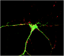
The widespread use of nanomaterials causes public concerns associated with their potential toxicological hazards. New-generation nanomaterials – upconversion nanoparticles (UCNPs) – hold promise for theranostics applications due to their unique optical properties, enabling imaging at the sub-centimetre depth in live biological tissue. In brain tissue, nanoparticle-aided optical imaging and treatment are deemed desirable. To this aim, we carried out cytotoxicity studies of UCNPs in primary hippocampal cultures. The most common core/shell UCNPs (NaYF4:Yb3+:Tm3+/NaYF4) were synthesized using a solvothermal method and hydrophilized with amphiphilic polymaleic anhydride octadecene (PMAO); polyethyleneimine (PEI). Bare UCNPs were produced by using tetramethyl ammonium hydroxide (TMAH). PMAO-, PEI- and TMAH-UCNPs (0.8 mg mL−1) were incubated for 72 hours with primary hippocampal culture and exhibited noticeable cytotoxicity. Our studies showed profound morphological modification of all treated cells with the maximum and minimum uptake observed in PMAO- and TMAH-UCNP-treated cells, respectively. The spontaneous calcium activity in cells treated with TMAH-UCNP, PMAO-UCNP dropped to (17 ± 3)%, (6 ± 3)% of its original level and was completely inhibited in the PEI-UCNP-treated cultures. This study demonstrated that bare and polymer surface-coated upconversion nanoparticles are toxic to dissociated hippocampal cells, evident through aberrant morphological changes, deviant variations of Ca2+ activity, and cell death.
In general, nanoparticle physical and chemical characteristics can be programmed,8 while the large effective surface decorated with reactive functional groups allows attachment of task-specific guiding, therapeutic, or/and contrast-rendering modules.9 These properties allow versatile design of biohybrid photoluminescent nanocomplexes for theranostics applications.10,11 Considering the limited accessibility of brain to surgical intervention and imaging, UCNP-aided visualization of specific molecular structures, diagnostic imaging and therapy of brain are of particular interest. At the same time, challenges of targeted delivery, optical imaging and photosensitization of UCNPs and other nanoparticles and macromolecules in the brain must be addressed. Although the delivery of large-size molecules and nanomaterials is hampered by the brain-blood barrier, this barrier is compromised in several pathological conditions, such as brain tumors.12–14
In view of these reasons, it is important to evaluate safety and biocompatibility of the emerging nanoparticles, including upconversion nanoparticles, for brain cells and tissues. At present, it is generally held that UCNPs comprising rare-earth doped Na+ and Ca2+ oxide15 or fluorides matrixes (e.g. NaYF4,16 NaLuF4,5 etc.) are nontoxic or mildly toxic to cells and tissues.17–20 Mild deteriorative effects of the concentration on the cell survival rate have been reported only for the highest UCNP doses of 500–2000 μm mL−1.21,22 At the same time, UCNPs conjugated with the chemotherapeutic targeting agents (doxorubicin, photoactivatable platinum(IV) prodrug, etc.) demonstrated significant cytotoxicity in the tumor cell lines.19 It is generally consented that the nanoparticle interaction with cellular systems is primarily governed by the surface moieties, although the cytotoxic properties of the core (bare nanoparticle) are also important in case the integrity of the surface coating is compromised.22
Despite a wealth of information published on this topic, evidence of the biosafety of UCNPs is still largely circumstantial due to the considerable disparity of the UCNP characteristics used in the reported cytotoxicity tests. For example, the tested concentrations of the nanoparticles ranged from 5 to 2000 μg mL−1, incubation time ranged from 1 h to 9 days,22,23 the tested cell types varied from normal, tumor, stem and differentiated cells, as well as the composition, size, shape, surface charge, synthesis and surface modification procedures of the UCNPs themselves. It is important to note that all reported studies of the UCNP cytotoxicity were carried out using the established cell lines with the propensity for fast and unlimited proliferation. These cells are less susceptible to adverse conditions and toxic agents owing to the gene activity modification responsible for the cell proliferation and cell cycle progression.24 It is also consented that immortalized cultures of one cellular phenotype reproduce poorly the signaling properties of the parent cells. In particular, neural cell lines exhibit no functional activities, which cells in the central nervous system do.25 The neural network is considered a functionality unit of the central nervous system sustaining the higher nervous system activity, such as transmission and storage of information, learning and memory.26 Hence, the neural system response to nanomaterials needs to be investigated at least at the hierarchical level of the primary hippocampal cultures, which ensures adequate assessment of UCNP toxic effects, including dysfunctional network activity. This activity is conventionally visualized by means of Ca2+-imaging, which reports on the changes of the intracellular calcium concentration in each cell of the neural network, and the contribution of each neuron or astrocyte to the network activity.
In this paper, we address the biocompatibility of upconversion nanomaterial. The most conventional composition of UCNP was chosen as a core/shell beta-phase sodium yttrium fluoride co-doped with 18% (molar ratio) ytterbium; 1.4% erbium and 0.6% thulium (β-NaYF4:Yb3+:Er3+:Tm3+/NaYF4), followed by surface modifications.22 Our selection of the tested surface modifications was guided by the reported studies by Guller et al.,22 where the following three surface modifications were graded from the least to most cytotoxic: (1) bare UCNP produced by the ligand-exchange reaction with a phase-transition agent, tetramethylammonium hydroxide (TMAH-UCNPs); (2) amphiphilic polymaleic anhydride octadecene (PMAO), and; (3) positively-charged polymer, polyethyleneimine (PEI). The functionality of the neural network activity was assessed by labeling, imaging and analyzing the Ca2+ distribution in single neurons and in ensemble.
(1) The ligand exchange reaction was carried out with tetramethylammonium hydroxide (TMAH) to evaluate the intrinsic toxicity of the as-synthesised UCNPs. TMAH is a low molecular phase-transition catalyst that is dissociated in water by producing OH– ions. TMAH is adsorbed on the surface of UCNPs,27 partially displacing oleic acid moieties from the OA-UCNP surface. UCNPs were transferred into an aqueous fraction by preparation of an UCNP micro-emulsion in aqueous solution of TMAH, followed by solvent evaporation.
(2 and 3) Amphiphilic polymers were adsorbed onto the UCNP surface via hydrophobic interactions between the oleate ligand and the hydrocarbon chain of the polymer. The hydrophilic counterpart of the amphiphilic polymers was directed outwards (i.e., into water), rendering OA-UCNP hydrophilic. We used the following amphiphilic polymers: poly(maleic anhydride-alt-1-octadecene) (UCNP-PMAO), polyethyleneimine (UCNP-PEI). We deployed a solvent evaporation method for UCNP surface modification. The solvent evaporation method is usually used for modification of inorganic nanocrystals surface-coated with water-insoluble polymers.28 In accordance with this method, UCNPs were dispersed in volatile solvent (e.g., chloroform), and polymer solution dissolved in chloroform was then added. After ultrasonic treatment, the mixture was stirred at room temperature for 1.5 h to adsorb the polymer chains on the surface of UCNPs. The resulting mixture was then added drop-wise to water or aqueous solution with stirring and sonication to dilute the mixture by at least ten-fold. The intermolecular forces between the polymer chains enabled the formation of particles in water (or aqueous solution) i.e. insoluble polymer particles containing UCNPs. Evaporation of the solvent resulted in an aqueous dispersion of UCNPs surface-modified with the polymer.
We obtained four samples of aqueous colloids of surface-modified UCNPs from these surface modification and complexation procedures. Aqueous colloids of the modified UCNPs were stable over at least two weeks, and stability was immune to electrolytes (0.15 M NaCl and buffer solutions). It is worth noting that there were practically no changes in the photoluminescent properties of UCNPs after their modification.
Phase-contrast images of the cultures were taken weekly to record the culture status using a DMIL HC (Leica, Germany) inverted microscope with a 10×/0.2 Ph1 objective. UCNPs of the concentration of 0.8 mg mL−1 were incubated with the culture medium on the day 14 of the culture development in vitro (DIV). The spontaneous calcium activity was recorded on the 3rd day, following the UCNP incubation.
The immunostained cultures were examined under the confocal laser-scanning microscope (Zeiss LSM 710, Germany), as described above. The laser intensity, gain and offset were fixed for each acquisition. Quantitative evaluation was performed using ImageJ (Research Service Branch, NIH).
The fourteen-days primary hippocampal cultures were incubated for 72 h with the tested colloidal nanomaterial, which represented surface-modified UCNPs in PBS. The choice of the incubation time of 72 h was governed by unusually low metabolic rate of the dissociated cells, especially, neurons in comparison with the established cell lines. Moreover, according to our previous studies, neurons of the hippocampal cultures were most susceptible to unfavorable conditions within the first 72 h after treatment.32
The composition of the core/shell UCNPs (NaYF4:Yb3+:Tm3+/NaYF4) was selected as the most popular3,16, whereas the incubation concentration of 0.8 mg mL−1 was chosen as typical for cytotoxicity tests.22 Three types of the surface-modification were chosen. Small-molecular-weight TMAH was demonstrated to partly displace original oleic acid surface groups used for the coordination of the UCNP during its synthesis.27 As a result, the as-synthesized UCNP surface groups were stripped off making nanoparticles (UCNP-TMAH) bare and miscible with water and cultural medium. This surface-modification procedure was particularly valuable, because it allowed testing cytotoxic properties of the upconversion nanomaterial itself decoupled from the coating material properties, as it was tested in a number of the cytotoxicity tests.20 The UCNP surface modification with amphiphilic polymer represented by polymaleic anhydride octadecene (PMAO) is a preferred approach demonstrated by Pokhrel et al.34 and us.22 PMAO molecules bind to the hydrophobic tails of oleic acid functional groups on the UCNP surface, so that the molecule hydrophilic heads are exposed outwards, thus hydrophilizing the nanoparticle. The cross-linking of PMAO molecules consolidates the coating and shields the UCNP core from environmental exposure, so that the cytotoxicity properties of this biohybrid nanocomplex (UCNP-PMAO) are determined by the PMAO alone. Coating of the nanoparticles with positively-charged polymer, polyethyleneimine (PEI) represents another widely accepted strategy used, for example, in polyelectrolyte coating procedures.22,35 It is well known that positively charged polymers, and specifically PEI, are efficient for cell internalization and transfection, although exhibiting notable cytotoxicity. According to our reported results on the surface-modified cytotoxicity of UCNPs in human skin cells, the least and most cytotoxicity were found in the case of the keratinocytes treatment with UCNP-TMAH and UCNP-PEI, respectively.22
The cytotoxicity test results are shown in a histogram of Fig. 1A. A significant decrease in the number of viable cells 72 h post-treatment was observed for all tested UCNPs. The lowest cytotoxicity was detected in case of the DIV 17 culture incubation with UCNP-TMAH, where the percentage of viable cells dropped to (57 ± 9)%, whereas (97 ± 1)% of viable cells were detected in the control group. The greatest cell loss (22 ± 8)% was noted in the primary cultures treated by UCNP-PEI. The UCNP-PMAO material also caused a decrease in the number of viable cells to (47 ± 9)%. Therefore, the UNCP-PEI material exhibited the greatest toxic effect compared with the other studied nanomaterials.
In order to gain insight into the mechanisms of the observed cytotoxicity of UCNPs, we carried out detailed analysis of the culture responses to the UCNP-induced stress, including morphological and electrophysiological evaluations of the culture status. The primary hippocampal culture is heterogeneous in terms of cell types, with the most abundant populations being neurons and astrocytes. The neurons are more sensitive to toxic agents than the astrocytes. The cell shape changes observed in the bright-field microscopy images of the dissociated hippocampal cells, as shown in Fig. 1B–E (left column), represented a notable manifestation of the UCNP-induced morphological changes of the cells, where the neuron reshaping was particularly obvious (shown by circles). The partial dendrites reduction and rounding bodies of neurons was observed.
We performed immunocytochemical staining of neurons and astrocytes with Microtubule Associated Protein 2 (MAP2) and Glial fibrillary acidic protein (GFAP), respectively to demonstrate the morphological changes of primary hippocampal cultures in more details, with the results shown in Fig. 2. The choice of MAP2 as the neuron marker allow to visualize large-molecular-weight proteins (∼70 kDa) of the cytoskeleton. Only neurons having such proteins are able to demonstrate the normal network activity. The displacement of the large-molecular-weight proteins with small-molecular-weight proteins was a hallmark of the neurodegeneration at the cellular level, and visualized via the depletion of the MAP2 staining. In comparison with the control cell culture exhibiting normal functionality (see Fig. 2A), noticeable disruptions of the neural brunches (dendrites) were clearly observable in the primary cultures incubated with UCNP-TMAH and UCNP-PMAO, as shown, respectively, in Fig. 2B and C. Moreover, the neuron interaction with the UCNP-PEI nanomaterial caused severe shortening of the brunches, eventually leading to their complete disintegration (Fig. 2D). This disintegration was visualized as the profound fragmentation of the initially continuous MAP2-stained neural network (cf. Fig. 2A). The important indicators of the neuron functionality, such as the neurite length, number of branches and the spine density on neurons, were undetectable due to the convoluted neuronal growth pattern typical for the high cell density, which was required to form the functional networks.
Changes in the shape of astrocytes in the cultures treated by UCNP-PEI were also revealed. In the control sample (Fig. 2A), astrocytes were cottony-shaped, distinctly isolated, connecting to each other via brunch terminal tips. They formed a relatively ordered evenly distributed array, as it is schematically presented in Fig. 3A1 and shown in Fig. 2A. The mean separation between the astrocytes was measured to be (44 ± 3) μm (see Fig. 3B). In the UCNP-PEI sample, astrocyte shapes were changed to take aberrant elongated forms. The uniform cell array pattern was also changed to a skewed pattern, with the mean astrocyte separation reduced to (22 ± 7) μm in the plane transverse to the elongation axis, as schematically presented in Fig. 3A2 and discernable in Fig. 2D. The astrocytes displayed a tendency for clumping, forming conglomerates with almost indistinguishable individual cellular elements. These changes of the astrocyte architecture are schematically illustrated in Fig. 3.
The UCNP-TMAH and UCNP-PMAO colloidal nanomaterials induced morphological modifications in the primary cultures. The astrocyte mean separations were markedly reduced in all directions, with l–s measured to be (17 ± 1) μm and (24 ± 3) μm for UCNP-TMAH and UCNP-PMAO, respectively, incomparison with l = (46 ± 2) μm in the control group (see Fig. 3). Note that these changes are typical for the primary cultures undergoing stress exposure, which corroborates the toxic effects of the tested nanoparticles.
Since the cultural medium contained fetal calf serum, protein corona was formed on UCNPs, which, mediated the nanomaterial interaction with cells and hence affected the cytotoxic effects exhibited by the tested surface-functionalized UCNPs.
Our next test related to the important functional activity of neural networks in the primary hippocampal cultures. Investigation of the functional metabolic calcium activity of the neuron-glial networks was conducted with the calcium-sensitive dye Oregon Green 488 (OG) application, which is widely used in studies concerning the dynamics of calcium homeostasis in nervous cells.36,37
Beginning from the days 5–7 of the brain development in vitro, single calcium events are detected in cell networks. Calcium oscillations from different cells are random, asynchronous and rare with the percentage of cells exhibiting the calcium activity less than 12%. Functionally mature neural networks are formed in DIV 14 cell cultures, as reported by us.30,31 There are calcium events characterized by frequency of 2.2 ± 0.3 oscillations per min of the duration of 6.9 ± 0.5 s in cultures. 80% of the cells demonstrate synchronized network activity. The process of the functional network activity formation was demonstrated to be associated with the appearance of complex chemical synapses in the primary hippocampal cultures (detailed in ESI†).
We investigated the spontaneous calcium activity in the primary hippocampal cultures incubated with the surface-modified UCNPs for 72 h. The results are presented in Fig. 4. The percentage number of cells exhibiting spontaneous calcium activity in the control cultures was approximately 90%, the duration of Ca2+ oscillations 7.9 ± 0.2 s, Ca2+ oscillation frequency 1.82 ± 0.07 min−1. The introduction of the tested nanomaterial to the culture caused significant changes in the calcium activity. We observed a decrease in the number of cells exhibiting spontaneous calcium activity in all experimental groups (UCNP-TMAH, 17 ± 3%; UCNP-PMAO, 6 ± 3%). Moreover, the cells in the primary culture incubated with UCNP-PEI exhibited no calcium activity (Fig. 4). The detailed information on the presented calcium imaging, in addition to video clips of the spontaneous activity are provided in ESI.†
The UCNP-TMAH and UCNP-PMAO nanomaterials affected on the functionality of the primary hippocampal cultures. In particular, the duration and frequency of Ca2+ oscillations were changed markedly. The Ca2+ oscillation frequency was reduced to (1.3 ± 0.3) min−1 and (1.2 ± 0.1) min−1 in UCNP-TMAH and UCNPs-PMAO experimental groups. The culture treatment with UCNP-TMAH led to a negligible increase in the duration of Ca2+ oscillation (7.7 ± 0.7) s, whereas the UCNP-PMAO treatment caused a decrease this parameter to 5.0 ± 0.5 s. Interestingly, the TMAH-UCNP-treated neural network temporal signal exhibited a pulse-burst pattern akin to aged primary cell cultures, in contrast with a coalescent single-pulse signal of the normal neural network (see details in ESI†).
The functional calcium homeostasis alterations is associated with the neural networks destruction in primary hippocampal cultures. The neural structure formation and a short-term opening of calcium channels on the postsynaptic membrane are formed a periodic synchronized activity during the culture development in vitro.38–40 A spontaneous short-term increase of the intracellular calcium leads to the cellular buffer systems activation and its concomitant reduction in cells. UCNP-induced destruction of the neural networks results in the irreversible degradation of their functional activity.
Our studies point to the toxicity of upconversion nanoparticles associated with the coating, rather than the UCNP core, although the core-induced cannot be excluded.
Several comments are now in order. Biohybrid photoluminescent nanocomplexes offer new opportunities in neuroscience and neurotechnology. The exceptional optical properties of these nanocomplexes allow ultrahigh-sensitivity imaging down to the single biomolecule level, paving a way for investigation of the ligand–receptor interactions in neurons towards new drug design. For example, labelling GABA receptors with quantum dots has enabled investigation of the dynamics of neurotransmitter receptors.41 The high loading capacity of the nanocomplexes is instrumental for targeted drug delivery and concomitant fluorescence-guided surgery of brain tumors.12 These applications demand investigation of the toxicity of the emerging biohybrid photoluminescent nanocomplexes and development of strategies to ameliorate their potential adverse effects to brain cells and tissues.
As demonstrated in this study, bare and surface-coated UCNPs exerted stress on dissociated hippocampal cells manifested by the disruption of the signaling pathways, aberrant morphology and considerable cell loss. It appeared that bare UCNPs displayed the least cytotoxicity, which lent itself several mitigation strategies. Firstly, it seems reasonable to design of UCNP surface coating, which predominantly determines the entire complex cytotoxicity property, rationally using materials of proven non-toxicity, such as gold.42 Surface coating UCNP with silica represents an interesting approach to ameliorate the toxicity effects. Secondly, it seemed that the cytotoxicity of UCNPs depended on the level of the cell uptake, as can be inferred from the UCNP integral distribution in Fig. 2. Fig. 2B and D display the least and most UCNP uptake levels for the respective bare UCNP and PEI-UCNP treatments of the hippocampal culture, which also correlated with the assayed least and most cytotoxicity of these surface-modified UCNPs. Besides, the uptake was largely diminished in case of the primary culture incubation with large size sub-microns (>200 nm) UCNPs of the same surface modifications (results not shown). This was accompanied by the negligible cytotoxicity (see ESI†). This observation suggests the decoration of UCNP with surface moieties, e.g. polyethylene glycol, which hamper the particle internalization.43
Footnote |
† Electronic supplementary information (ESI) available. See DOI: 10.1039/c6ra01272h |
This journal is © The Royal Society of Chemistry 2016 |