Mark–Houwink–Sakurada coefficients determination for molar mass of silk fibroin from viscometric results. SEC-MALLS approach

D. Pawcenisa, M. Syreka, M. A. Aksamit-Koperskaa, T. Łojewskiab and J. Łojewska*a
aJagiellonian University, Chemistry Department, Ingardena 3 street, 30-060 Krakow, Poland. E-mail: lojewska@chemia.uj.edu.pl
bAGH University of Science and Technology, Faculty of Materials Science and Ceramics, Mickiewicza 30, B8/3.36, 30 059 Krakow, Poland

Received 11th January 2016 , Accepted 11th April 2016

First published on 12th April 2016


Abstract

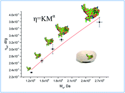
The results on the changes of molar mass distributions and average molar masses in Bombyx mori fibroin with use of size exclusion chromatography (SEC) and viscometry, respectively, are presented in terms of the determination of Mark–Houwink–Sakurada coefficients, which are lacking in the literature. SEC was applied in configuration of light scattering and differential refractive index and multi angle laser light scattering detectors (SEC-DRI-MALLS). The coefficients calculated from the correlation between intrinsic viscosities and the absolute value of molar masses obtained using the SEC-MALLS technique are equal to: K = 1.813 × 10⁻⁴ dm3 g−1 (±1.74 × 10⁻⁵), α = 0.614 (±0.068). The value of the parameter α shows that silk fibroin molecules in an LiBr solution behave like a flexible polymer molecule in a thermodynamically good solvent. The results are discussed with focus on the applicability of glass capillary viscometry and size exclusion chromatography for assessment of silk based materials condition with particular reference to the preservation artifacts and ancient textiles.


Introduction

Silk fibres have been used since 3500 B.C.E. in the textile industry, due to their incredible strength and lustre. Hence a great many historical garments, carpets, tapestries and banners collected in museums are made of silk. With an increasing call for the conditions of historic silk textiles to be assessed, and in particular of the banners from Polish museums, we turned special attention to silk degradation, covering the specific degradation processes occurring within the molecular structure which can be observed by the changes in molecular masses of polymers. On the other hand, thanks to fibroin's biocompatibility and biodegradability, silk finding numerous new applications in biotechnology1 for example, wound dressing,2 tissue engineering,3 targeted drug delivery.4,5

It is the molecular architecture of fibroin that gives the silk its characteristic properties. The primary structure is created by a sequence of amino acids, joined by a flat peptide bond, and stabilised by a mesomeric effect. The secondary structure is determined by the particular hydrophobic amino acids, which create rigid crystalline regions, arranged in the structure of beta-sheets, located in the heavy chain. This secondary structure is a result of the dominance of the hydrophobic amino acids: glycine and alanine, a part of 59-mer, which is a repeating sequence GAGAGSGAAG[SGAGAG]8Y. Ordered crystalline regions are interspersed with amorphous regions located in the area of the light chain. These regions are richer in the hydrophilic amino acids serine, threonine, arginine and lysine, which are more susceptible to degradation.

Many textile examples of cultural heritage importance, such as banners, tapestries and canvas for eastern paintings, were made of silk. Different and sometimes uncontrolled storage and exhibition circumstances have seriously affected the current condition of these objects, and can continue to act in such a way. Environmental factors such as light, temperature and humidity have many effects on the degradation of silk6,7 and the correlation between molecular structure of degraded silk and macroscopic properties can be assessed by a number of analytical methods.8 These environmental factors have substantial impact on the rate of the degradation process, manifested by the changes in glass transition temperature,9 oxidation susceptibility,7,10,11 and photoinduced –S–S– bond breakdown.12,13 The vast array of oxidation reactions were examined by Shao et al.,14 who presented results of investigations on changes in the crystallinity of fibroin as a function of the compactness of tyrosine, studied through spectroscopic methods: ATR-FTIR and FT-Raman. It was also shown that during natural ageing volatile organic compounds (VOCs) are emitted, which results in mass loss.8

The impact of humidity, UV light and temperature on the change of molecular weight distribution has already been studied, using size exclusion chromatography (SEC) with photometric detection in the ultraviolet range.15 SEC is currently regarded as the most accurate technique to determine molar mass distribution of synthetic and natural polymers, particularly when combined with absolute detection, such as multiple angle laser light scattering (MALLS). MALLS does not require synthetic standards of molar mass to be relied upon, and thus gives reliable and absolute values of both average molar masses and radii. SEC separation bases on the size of the macromolecule (the so-called hydrodynamic volume) and it can be related to molar mass for linear polymers.16–20 However, it has to be emphasized that different molar mass values and resulting mean molecular weights can be obtained for different macromolecular structure upon SEC separation from different detection systems and calibration procedures including conventional calibration (with refractive or spectrophotometric detector), universal calibration (with differential viscosimetry detection) and absolute weight-average molar mass distributions (from MALLS).21–24

The literature provides a number of studies on the characterisation of silk with conventional SEC,15,25–28 where molar masses were determined with the use of protein standards of known molar masses. Recently, SEC-MALLS analyses of silkworm and spider silk protein have also been presented.29–33 Nevertheless, silk fibroin analysis by SEC is still a challenge due to a number of problems arising from: the risk of fibroin degradation in the complicated process of its dissolution in a salt containing solvent, molecules adsorption in the stationary phase, their clogging and aggregation in a column, salt crystallisation in the solution when high salt concentration is used, and many more.

The extent of degradation of Bombyx mori silk textiles has been measured traditionally by the loss of mechanical endurance,34 pH,35 colour,15 crystalline structure by X-ray diffraction36–38 and viscosity.25,39 The latter is especially valuable because viscometry is a cheap and available method of analysis of the degradation progress in fibres in the conservation laboratory practice. However, to obtain a reliable gauge of a degradation in form of molecular mass Mark–Houwink–Sakurada (MHS) coefficients for Bombyx mori fibroin has to be known. In the literature there are no values of the coefficients available for such calculations. Tse and Dupont25 presented a comparison between the results of molar masses (MM), obtained via SEC using a conventional calibration method, and intrinsic viscosities determined with glass capillary viscometry of aged silk samples in 6.67 M lithium thiocyanate (LiSCN). Surprisingly enough, they have not computed MHS parameters from the obtained valuable results, although the correlation between MM and intrinsic viscosity was linear. It is beyond doubt that they presented a complex approach to silk analysis by SEC, but the conventional SEC provides only apparent and relative molar masses. Furthermore, the choice of LiSCN as a solvent for viscometry is not fully justified, since for silk textiles the only available standard for viscosity measurements utilizes LiBr aqueous solution.40

The MHS parameters are necessary for calculating the viscometric molecular weights of polymers from viscometric analyses. In fact, the combination of SEC-MALLS-DRI separation and detection units can be used to determine these parameters, following the approach that was elaborated in our previous paper.41 Determination of these coefficients will give both preservation scientists, and those who deal with fibroin fibre for medical applications, a simple tool for the assessment of a mean molecular weight that can be a measure of a textile's condition and the degree of polymerisation of the polypeptide chain. For the sake of microdestructivity requirement for diagnostics of historic artefacts, glass capillary viscometry should provide not only simplicity of the analysis, but most of all must guarantee possibility of non- or microdestructive analysis.

Viscometry and size exclusion chromatography in molar mass determination of silk fibroin

Dilute solution viscometry is one of most important methods of the characterisation of polymers. Its major advantage arises from its simple and cheap instrumentation. There are different but commonly known viscosity values that can be calculated from viscometric measurements: relative viscosity (ηrel, eqn (1)) specific viscosity (ηsp, eqn (2)), reduced viscosity (ηred, eqn (3)), inherent viscosity (ηinh, eqn (4)), and finally intrinsic viscosity (ηint, eqn (5), or alternatively eqn (6)), which is the theoretical viscosity of a macromolecule in a given solvent at a macromolecules concentration approaching zero.
 
image file: c6ra00871b-t1.tif(1)
 
ηsp = ηrel − 1 (2)
 
image file: c6ra00871b-t2.tif(3)
 
image file: c6ra00871b-t3.tif(4)
 
image file: c6ra00871b-t4.tif(5)
 
image file: c6ra00871b-t5.tif(6)
tx is the efflux time of a polymer dissolved in a solvent, t0 is the efflux time of a pure solvent, and c is the polymer concentration in grams per 100 cm3 (g dl−1).

According to the literature on polymer properties,19,42 the plot of ηsp/c versus c is linear with intercept equal to the intrinsic viscosity for a sufficiently low concentration. The concentration range should provide specific viscosities, ηsp, of the values between 0.1 and 0.8. In fact, the most important requirement for the choice of the concentration range is the linearity of the dependence of ηsp/c versus c or ln(ηrel)/c versus c, with no significant deviations from linear dependence at higher concentrations.

Due to its advantages and well described experiments,40 glass capillary viscometry can be used as a universal method for evaluating fibroin state of preservation. According to the Swiss Standard Association for measuring silk viscosity,40 the extent of deterioration may be assessed by calculating the viscosity number in the following way:

 
image file: c6ra00871b-t6.tif(7)
where 0.5 is the Schulz–Blaschke constant for silk fibroin in a saturated LiBr solution in water.

As mentioned above, viscometric experiments may provide the molar mass of a polymer on condition that MHS coefficients are known. Viscometric molar mass (Mv) can then be calculated from the following equation:

 
ηint = KMvα (8)
K is a constant specific for a particular system of polymer-solvent at a given temperature, and α is the MHS exponent characterising the macromolecules conformation in the solvent.

It should be noted that viscometry is not universal for the analyses of molar masses of polymers being able to provide only one parameter describing polymer which is viscosity-average molecular weight Mv mass. A lot of properties of polymers such as stiffness, strength, toughness, melting point, and viscoelasticity are dependent on other mean molar mass values.43,44

Determination of MHS coefficients and the meaning of MHS exponent α

Traditionally, MHS coefficients are determined using a series of polymer samples for which the values of the intrinsic viscosities and molar masses are known. The intrinsic viscosity is plotted against molar mass, and the MHS constant K and exponent α are obtained from the equation of the fitted curve.

The exponential parameter α characterizes conformation of a macromolecule in a given solvent and at a particular temperature. The value of α = 0 refers to spherical conformation, α < 0.5 to branched polymer structure, 0.5 < α < 0.8 to flexible chain, α > 0.8 – to more extended molecular conformation and α = 2 – rigid rod conformation.18

Experimental

Samples

Accelerated aging of silk fibroin samples. Bombyx mori silk fibroin samples were prepared from Chinese silk textile (Fei Long, Warsaw, Poland). We have chosen to use Habotai textile, which is natural coloured and unweighted degummed silk. We decided to use such silk textile as a model sample, because its macroscopic and microscopic properties are well described in the literature.7,8,11,25,26,36,45–48 Samples were cut into pieces (at least 1.00 g each) and hung in a ventilated thermostatic ageing chamber. The specimens were aged under the following conditions: dry air (oxygen atmosphere) at 120 °C for 1, 3, 5 and 7 days.

In this work, only thermal degradation process was performed to accelerate degradation of silk fibroin to obtain the samples differing in molecular weight to allow for Mark–Houwink–Sakurada coefficients calculation. This is why they differ from those used traditionally.6,45,47–50

Thermal degradation effects can be classified into two groups: one involves purely physical structural changes in a fibre, whereas the other involves chemical changes. They mainly involve melting, crystallization and glass transition. Since heat can easily penetrate the fibre, the changes are expected to occur in both crystalline and non-crystalline areas. The melting point of protein fibres drops at the temperature above which polymer undergoes thermal decomposition – chemical irreversible changes. Thermal decomposition study of silk shows weight loss recorded in the regions 220–300 °C.51

Silk fibroin structure undergoes a glass transition transformation at temperatures above 170 °C.9,52,53 Taking all that under consideration thermal aging must be performed in lower temperatures, so not to induce processes artificial for the natural degradation changes. Temperature 120 °C is much below both thermal decomposition and glass transition temperature, thus is a logic compromise between reasonable ageing temperature and relatively short time needed to obtain measurable structural changes.

Glass capillary viscometry

Sample preparation. Saturated solution of aqueous LiBr (Sigma) was prepared by gradual addition of LiBr to deionized water (Simplicity 185, Millipore) until more lithium bromide could be dissolved. Such solution was tight capped and kept at 20 °C for 24 hours to equilibrate.

Dependently on the degree of polymerisation, at least 1.00 g of silk textile was dissolved in a saturated solution of lithium bromide, as proposed by the Swiss Standard for viscosity test of silk textiles.40

Solutions were thermostated in a water bath for 3 h at 60 °C until the samples had completely dissolved. The dissolution process was followed by filtration using a Schott funnel (glass filter type G11).

Linear ranges of fibroin concentrations in LiBr were found between 30 mg cm−3 and 150 g cm−3.

Analyses. The glass capillary viscometry system consisted of an Ubbledohle type viscometer Matviskometer SCAN-C15:62, thermostated with a THERMO Haake P5 water bath. During experiments, temperature of the viscometer was maintained at a constant temperature of 20.0 ± 0.1 °C. Calibration of the viscometer and further analyses were conducted according to the procedure described in the Swiss Standard for viscosity test of silk textiles.40

The efflux time of fibroin solutions was measured at least three times for three samples for each ageing time. The analysis of the number of tests, calculated using Statistica 10, was equal 2, assuming 0.8 as the power of a test.

Size exclusion chromatography

Molar mass of fibroin was determined with use of SEC coupled with multiple angle laser light scattering detector and differential refractive index detector. The SEC system consisted of the following items: Waters 1515 isocratic pump, Waters 717 + auto sampler, a column oven, a UV-VIS detector (Waters 2487 Dual λ Absorbance, set at the wavelength 280 nm), a multiple angle laser light scattering detector Dawn Heleos (Wyatt Technology) and a differential refractive index detector Optilab T-rEX (Wyatt Technology), acting as a concentration sensitive detector. Both MALLS and RI worked at the same wavelength (658 nm). Such configuration enables determination of molar masses without conventional calibration based on protein molar mass standards. Conventional calibration with use of proteins standard can be challenging because proteins often exist as dimers, trimers and tetramers of smaller polypeptides. This phenomenon may cause abnormal elution volumes, which are not corresponding to true molar masses of proteins, and result in nonlinear calibration curve of log of molar mass versus elution volumes.26

Sample preparation

The sampling and separation conditions utilised in the SEC of fibroin were described in ref. 41. Since then we have modified some of the experimental elements, in particular composition of mobile phase. Aqueous solutions of sodium chloride are known to inhibit interaction of proteins molecules with silica based packing of a column. For this reason, we decided to change mobile phase from pure water to 0.2 M NaCl. Chromatographic separation was performed on a silica based column Yarra SEC S-4000 from Phenomenex (packed 3 μm, 300 × 4.6 mm), maintained at 30 °C (the same temperature as in the differential refractive index detector), with a downstream filter cartridge acting as guard column (Phenomenex). 0.2 M NaCl in water (HPLC grade, Carl Roth) was used as the mobile phase, with a flow rate of 0.3 mL cm−3.

Approximately 15 mg of silk textile was dissolved in 9.3 M lithium bromide (Sigma). Solutions were thermostated in a water bath for 3 h at 60 °C, until the samples had completely dissolved. It must be highlighted that no insoluble particles were observed in the lithium bromide solutions. Dissolution was followed by dialysis in a 1 cm3 dialysis capsule (Carl Roth) with a cellulose membrane tube (molecular weight cut-off 12.8 kDa, Sigma) against pure water (Millipore) for 6 hours. The dialysis solution was exchanged every 30 min to remove LiBr. The concentration of lithium bromide was monitored every 30 minutes with a XRF spectrometer. The removal of LiBr was achieved after 6 h, as evidenced by the disappearance of Br signals from the XRF spectra. The aqueous solutions of silk fibroin were prepared by mixing two volumes of 0.4 M NaCl solution with two volumes of each sample to obtain silk fibroin in 0.2 M NaCl. Although obtained solutions were transparent and free of visible particulates, solutions were centrifuged prior to injections. Silk fibroin samples were then injected in volume 50 μL. The final concentration of silk fibroin samples was about 2 mg cm−3. Each sample was analysed six times – three injections from two solutions.

The specific refractive index increment of fibroin was determined in batch-mode using a differential refractive index detector Optilab T-rEX and calculated using Astra 6.0.2 software.

Data processing

Intrinsic viscosity calculation

The determination of intrinsic viscosities was based on the extrapolation of viscosity numbers (eqn (7)) measured for four to five concentrations of silk fibroin. Extrapolation to zero concentration allowed to calculate the intrinsic viscosity. The approach based on eqn (7) was justified also by the fact that the viscosity standard recommends such a method of viscosity number calculation. What is more important, linear dependence on concentration was the best for the case of viscosity number (Fig. 1). Consequently, for all aged silk samples, intrinsic viscosities were calculated in this way.
image file: c6ra00871b-f1.tif
Fig. 1 Dependence on concentration of reduced viscosity, inherent viscosity and viscosity number.

Size exclusion chromatography measurements

The determination of absolute values of molar masses using SEC-MALLS detection is based on the Rayleigh equation:
 
image file: c6ra00871b-t7.tif(9)
k is a combined constant including the refractive index increment dn/dc:
 
image file: c6ra00871b-t8.tif(10)

Several parameters have to be known to apply the Rayleigh equation: fibroin concentration c, its specific refractive index increment dn/dc, and the second virial coefficient A2 which is optional.

Concentration values were supplied by a concentration sensitive detector, by measuring the differential refractive index. The specific refractive index value was 0.1999 ± 0.007 cm3 g−1, obtained from three batch measurements of five different concentrations of fibroin in 0.2 M sodium chloride, ranging from 0.01 mg cm−3 to 5 mg cm−3.

The second virial coefficient was omitted due to low concentrations of fibroin injected to the column (order of 10−3 mg cm−3).

Weight average molar mass from the Rayleigh equation was calculated with the use of Debye's model, which is recommended for large molecules.19,54 A plot of Rθ/k × c versus sin2(θ/2) was constructed. The plot was fitted by a linear or polynomial function to obtain the intercept at zero angle and the slope at zero angle. In this study the fit was of the 1st order for undegraded samples. For artificially aged silk fibroin, the order of polynomial was 3, which is typical of polydispersed samples.54 This curvature was present independently of fitting formalism (Debye, Zimm, Berry).

Calibration of the MALLS detector requires determination of the detector constant, which is a proportionality factor between the detector signal and the Rayleigh coefficient R(θ) for one photodiode (the 11th diode at 90°). The constant determination was performed with toluene (HPLC purity) and was found to be 1.5175 × 10⁻⁴ 1/V × cm3 (±0.0004). A further procedure includes the normalisation of other 17 photodiodes, which was done with the use of bovine serum albumin monomer (BSA) standard (66 kDa, Sigma). Interdetector and band broadening correction were performed with the use of the same BSA standard.

Calibration of differential refractive index detector was performed with use of the series of aqueous NaCl solutions, which concentrations covered range between 0.1 and 6 mg cm−3. Briefly, the idea of the calibration constant determination was very similar to dn/dc determination. The dependence between refractive indexes of the series of different concentrations solutes in a given solvent at particular temperature and laser wavelength was measured. The slope was found by fitting the experimental data using linear regression. Then calibration constant was calculated by multiplying instrumental constant (delivered by manufacturer) by slope of the dependence of DRI signal and respective concentration.

Results and discussion

Intrinsic viscosity of silk fibroin

The intrinsic viscosity of initial and aged silk samples were calculated in three ways, based on eqn (5)–(7). Intrinsic viscosity values derived from extrapolation of reduced viscosity (ηred) to zero concentration were characterised by a more rapid decrease in the ageing time of silk (Fig. 2, black line). Moreover, reduced viscosity seems to be more likely to deliver broader standard deviations.
image file: c6ra00871b-f2.tif
Fig. 2 Time progresses of intrinsic viscosities derived from reduced viscosity, inherent viscosity and viscosity number.

Another situation is observed in the case of intrinsic viscosity values derived from inherent viscosity and viscosity number. Here, the time progresses of intrinsic viscosities are close to each other (Fig. 2, red and blue lines). Both cases are less vulnerable to affect significant standard deviation.

Ultimately, intrinsic viscosity calculated with the use of viscosity number (from the Blaschke–Schulz equation) was applied to compute MHS coefficients, as described in subsection Intrinsic viscosity calculation, linear dependence on concentration was the best for the viscosity number.

Each value of ηint in Table 1 represents the η calculated from average efflux time from at least three measurements with a standard deviation between 1.5 and 4.6% (Table 2).

Table 1 Intrinsic viscosities and molar masses for thermally aged samples of silk fibroin used for determination of Mark–Houwink–Sakurada coefficients (present work)
Ageing time ηint, dl g−1 Std. dev. Mw, Da Std. dev.
0 0.379 0.0141 269[thin space (1/6-em)]700 5394
1 0.328 0.00456 187[thin space (1/6-em)]290 3750
3 0.301 0.0138 168[thin space (1/6-em)]200 3360
5 0.267 0.00536 144[thin space (1/6-em)]780 2890
7 0.233 0.01035 122[thin space (1/6-em)]100 2440


Table 2 Intrinsic viscosities and molar masses for irradiated samples of silk fibroin determined by Tse and Dupont25
Irradiance energy (kJ m−2) at 420 nm ηint, dl g−1 Std. dev. Mw, Da Std. dev.
Unaged 0.404 0.01 346[thin space (1/6-em)]000 29[thin space (1/6-em)]000
100 0.242 0.011 176[thin space (1/6-em)]000 19[thin space (1/6-em)]000
200 0.198 0.008 137[thin space (1/6-em)]000 15[thin space (1/6-em)]000
300 0.165 0.006 113[thin space (1/6-em)]000 14[thin space (1/6-em)]000


Molar masses of silk fibroin

For the purpose of MHS determination, we decided to use weight average molar masses. The relation between different mean molar mass values is as follows: Mn < Mp < Mw < Mv < Mz < Mz+1.55 Thus it seems reasonable to compare Mw to Mv, rather than to Mn (which is the smallest value) or to Mz (which value is far higher). Moreover, Mw is claimed to have greatest contribution to mechanical properties of fibres.26

Weight average molar masses was calculated from the equation:

 
image file: c6ra00871b-t9.tif(11)

The value of weight average molecular mass of the reference silk sample is close to the value determined using the SDS-PAGE electrophoresis method (350 ± 25 kDa).24 Our experiments gave a value of 270 kDa for the unaged silk textile. Unaged silk presents a bimodal MMD distribution (Fig. 4) with large dispersity, which includes both heavy chain and light fibroin molecules. Upon exposure to elevated temperature a shift of the MMD curve to lower molar masses is observed (Fig. 3, Table 1). Curves retain bimodal distribution, which is probably a result of faster degradation of the light chain in the fibroin molecule, due to its amorphous structure. However, bimodal distributions are observed only for unaged and thermoaged samples after 1 and 3 days; the bimodal shape of MMD curves indicating two macromolecular populations disappears with increasing time of ageing. Presumably merging of this two maxima into monomodal distribution is caused by high degree of degradation of the fibroin light chain.


image file: c6ra00871b-f3.tif
Fig. 3 Trend of the weight average molar masses decrease within ageing time of silk fibroin.

image file: c6ra00871b-f4.tif
Fig. 4 Molar mass distributions of silk fibroin samples.

Mark–Houwink–Sakurada coefficients

The conformation and hydrodynamic volume of fibroin molecules are different in 0.2 M NaCl and in saturated LiBr solutions. However, it must be emphasized that it is not possible to perform the SEC and viscosity experiments in the same solution. Thus we have to accept this limitation to be able to estimate molar mass of fibroin from the viscosity measurements.

The values of MHS coefficients K and α, were calculated from eqn (8). To build the experimental dependence, the [η] values were found from viscometric results and the Mw values from SEC-MALLS-DRI measurements. The parameters were found by fitting the experimental data using nonlinear regression of the power-type function expressed by eqn (8). The results are shown in Fig. 5. The optimised values amount to K = 1.813 × 10⁻⁴ dm3 g−1 ± 1.74 × 10⁻⁵, α = 0.614 ± 0.068 and R2 = 0.9641 for the Mw values in the range between 122 and 270 kDa.


image file: c6ra00871b-f5.tif
Fig. 5 Power-type correlation between weight average molar mass and intrinsic viscosity of fibroin in lithium bromide.

Because the choice of power-type function may not be obvious judging by the shape of the curve in Fig. 5, a plot constructed by fitting linear regression to the data was compared. The R2 value for linear fit of the same data was equal to 0.9424 which is lower than that found for the power-type function.

The value of α found by us refers to flexible polymer chains.18 The mutual relation of SEC and viscometry versus ageing time is depicted in Fig. 6. Both time dependence curves of Mw and ηint demonstrate similar trends. From that we conclude that MHS coefficients, computed with the use of the SEC-MALLS absolute method, can be applied to obtain reliable molar masses from viscometric measurements.


image file: c6ra00871b-f6.tif
Fig. 6 Relation of SEC and viscometry versus ageing time.

Conclusions

Traditional capillary viscometry and SEC are the most accurate analytical methods for assessing the degradation of polymers by determining molar masses. The viscometric technique could be regarded as a cheap and widely available method for molar mass determination for polymers that requires no standards of molar mass. The major problem with its utilisation for silk samples was that Mark–Houwink–Sakurada coefficients have not been available in the literature.

The method used to determine MHS coefficients developed in our previous study41 is based on the correlation between the weight average molar mass obtained from SEC-MALLS-DRI analyses for the samples aged for different periods of time with the intrinsic viscosity obtained from viscometric analyses. The values of MHS coefficients are equal to K = 1.813 × 10⁻⁴ dm3 g−1 (±1.74 × 10⁻⁵), α = 0.614 (±0.068).

Acknowledgements

The research was funded from the National Science Centre (partially 2011/01/N/ST4/03604 – chemicals, and 2014/13/N/ST4/04089 – model silk samples). European Union (“Doctus” scholarship to D.P.) is kindly acknowledged. A size exclusion chromatography system was equipped with a differential refractive index detector purchased thanks to the financial support of the European Regional Development Fund in the framework of the Polish Innovation Economy Operational Programme (contract no. POIG.02.01.00-12-023/08).

References

  1. C. Vepari and D. L. Kaplan, Prog. Polym. Sci., 2007, 32, 991–1007 CrossRef CAS PubMed.
  2. H. Teramoto, T. Kameda and Y. Tamada, Biosci., Biotechnol., Biochem., 2008, 72, 3189–3196 CrossRef CAS PubMed.
  3. Y. Wang, H.-J. Kim, G. Vunjak-Novakovic and D. L. Kaplan, Biomaterials, 2006, 27, 6064–6082 CrossRef CAS PubMed.
  4. S. Hofmann, C. T. Wong Po Foo, F. Rossetti, M. Textor, G. Vunjak-Novakovic, D. L. Kaplan, H. P. Merkle and L. Meinel, J. Controlled Release, 2006, 111, 219–227 CrossRef CAS PubMed.
  5. E. Wenk, H. P. Merkle and L. Meinel, J. Controlled Release, 2011, 150, 128–141 CrossRef CAS PubMed.
  6. F. Vilaplana, J. Nilsson, D. V. P. Sommer and S. Karlsson, Anal. Bioanal. Chem., 2015, 407, 1433–1449 CrossRef CAS PubMed.
  7. M. A. Koperska, D. Pawcenis, J. Bagniuk, M. M. Zaitz, M. Missori, T. Łojewski and J. Łojewska, Polym. Degrad. Stab., 2014, 105, 185–196 CrossRef CAS.
  8. M. A. Koperska, D. Pawcenis, J. M. Milczarek, A. Blachecki, T. Łojewski and J. Łojewska, Polym. Degrad. Stab., 2015, 120, 357–367 CrossRef CAS.
  9. N. Agarwal, D. A. Hoagland and R. J. Farris, J. Appl. Polym. Sci., 1996, 63, 401–410 CrossRef.
  10. I. V. Berghe, J. Archaeol. Sci., 2012, 39, 1349–1359 CrossRef.
  11. M. Koperska, T. Łojewski and J. Łojewska, Spectrochim. Acta, Part A, 2015, 135, 576–582 CrossRef CAS PubMed.
  12. A. Sionkowska and A. Planecka, Polym. Degrad. Stab., 2011, 96, 523–528 CrossRef CAS.
  13. S. Baltova and V. Vassileva, Polym. Degrad. Stab., 1998, 60, 53–65 CrossRef CAS.
  14. J. Shao, J. Zheng, J. Liu and C. M. Carr, J. Appl. Polym. Sci., 2005, 96, 1999–2004 CrossRef CAS.
  15. J. Kim and P. Wyeth, e-Preserv. Sci., 2009, 6, 60–67 CAS.
  16. Z. Grubisic, P. Rempp and H. Benoit, Rubber Chem. Technol., 1967, 5, 753–759 Search PubMed.
  17. H. Benoit, Z. Grubisic, P. Rempp, D. Decker and J. Zilliox, J. Chim. Phys., 1966, 63, 1507–1514 CAS.
  18. S. Mori and H. G. Barth, in Size Exclusion Chromatography, Springer Laboratory, Verlag Berlin Heidelberg New York, 1999, pp. 175–180 Search PubMed.
  19. S. Podzimek, Light Scattering, Size Exclusion Chromatography and Asymmetric Flow Field Flow Fractionation, John Wiley & Sons, Inc., Publication, 2011 Search PubMed.
  20. S. Podzimek, T. Vlcek and C. Johann, J. Appl. Polym. Sci., 2001, 82, 454–460 CrossRef CAS.
  21. A. M. Striegel, Anal. Chem., 2005, 105–113 Search PubMed.
  22. M. Gaborieau, R. G. Gilbert, A. Gray-Weale, J. M. Hernandez and P. Castignolles, Macromol. Theory Simul., 2007, 16, 13–28 CrossRef CAS.
  23. M. Gaborieau and P. Castignolles, Anal. Bioanal. Chem., 2011, 399, 1413–1423 CrossRef CAS PubMed.
  24. F. Vilaplana and R. G. Gilbert, J. Sep. Sci., 2010, 33, 3537–3554 CrossRef CAS PubMed.
  25. S. Tse and A. Dupont, in Historic Textiles, Papers, and Polymers in Museums, ed. J. M. Cardamone and M. T. Baker, ACS Symposium Series, American Chemical Society, Washington, DC, 2001; pp. 98–114 Search PubMed.
  26. N. Luxford. Reducing the Risk of Open Display: Optimising the Preventive Conservation of Historic Silks, University of Southampton, 2009 Search PubMed.
  27. Y. Joo-Hong, L. Kwang-Gill, L. Yong-Wo and K. Sun Yeou, Eur. Polym. J., 2003, 39, 1195 CrossRef.
  28. K. Hallet and D. Howell, in Scientific analysis of ancient and historic textiles: informing preservation, display and interpretation, ed. R. Janaway and P. Wyeth, Archetype Publications, London, 2005, pp. 143–150 Search PubMed.
  29. C. Jackson and J. P. O'Brien, Macromolecules, 1995, 28, 5975–5977 CrossRef CAS.
  30. W. A. Gaines, M. G. Sehorn and W. R. Marcotte, J. Biol. Chem., 2010, 285, 40745 CrossRef CAS PubMed.
  31. H. J. Cho, C. S. Ki, H. Oh, K. H. Lee and I. C. Um, Int. J. Biol. Macromol., 2012, 51, 336 CrossRef CAS PubMed.
  32. F. Hagn, C. Thamm, L. T. Scheibe and H. Kessler, Angew. Chem., Int. Ed., 2011, 50, 310 CrossRef CAS PubMed.
  33. M. Humenik and T. Scheibel, ACS Nano, 2014, 8, 1342–1349 CrossRef CAS PubMed.
  34. P. Wyeth, in Scientific analysis of ancient and historic textiles: informing preservation, display and interpretation, ed. P. Wyeth, Archetype Publications, London, 2005, pp. 137–142 Search PubMed.
  35. J. Kim, X. Zhang and P. Wyeth, e-Preserv. Sci., 2008, 5, 41–46 CAS.
  36. A. C. Hermes, R. J. Davies, S. Greiff, H. Kutzke, S. Lahlil, P. Wyeth and C. Rieke, Biomacromolecules, 2006, 7, 777–783 CrossRef CAS PubMed.
  37. P. Garside and P. Wyeth, Appl. Phys. A, 2007, 89, 871–876 CrossRef CAS.
  38. M.-Y. Li, Y. Zhao, T. Tong, X.-H. Hou, B.-S. Fang, S.-Q. Wu, X.-Y. Shen and H. Tong, Polym. Degrad. Stab., 2013, 98, 727–735 CrossRef CAS.
  39. X. Zhang and S. Yuan, Chin. J. Chem., 2010, 28, 656–662 CrossRef CAS.
  40. Text. Rundscheu, Swiss Stand. Assoc. 1964, 80, 19.
  41. D. Pawcenis, M. A. Koperska, J. M. Milczarek, T. Łojewski and J. Łojewska, Appl. Phys. A, 2013, 114, 301–308 CrossRef.
  42. S. F. Sun, in Physical Chemistry of Macromolecules, John Wiley & Sons, New Jesrey, 2004, pp. 170–175 Search PubMed.
  43. L. J. Fetters, D. J. Lohse, T. D. Richter, T. A. Witten and A. Zirkelt, Macromolecules, 1994, 27, 4639–4647 CrossRef CAS.
  44. S. R. Holding and E. Meehan, in Molecular Weight Characterisation of Synthetic Polymers, iSmithers Rapra Publishing, 1995, pp. 3–4 Search PubMed.
  45. N. Luxford, D. Thickett and P. Wyeth, in Multidisciplinary Conservation: A Holistic View for Historic Interiors, Rome, 2010, pp. 1–12 Search PubMed.
  46. P. Wyeth, in Scientific Analysis of Ancient and Historic Textiles: Informing Preservation, Display and Interpretations, ed. R. Janaway and P. Wyeth, Archetype Publications, London, 2005, pp. 137–142 Search PubMed.
  47. N. Luxford and D. Thickett, Journal of the Institute of Conservation, 2011, 34, 115–127 CrossRef.
  48. M. Y. Li, Y. Zhao, T. Tong, X. H. Hou, B. S. Fang, S. Q. Wu, X. Y. Shen and H. Tong, Polym. Degrad. Stab., 2013, 98, 727–735 CrossRef CAS.
  49. J. Nilsson, F. Vilaplana, S. Karlsson, J. Bjurman and T. Iversen, Stud. Conserv., 2010, 55, 55–65 CrossRef CAS.
  50. N. Luxford, D. Thickett and P. Wyeth, in Natural Fibres in Australasia: Proceedings of the combined (NZ and AUS) conference of The Textile Institute, ed. C. A. Wilson and R. M. Laing, The Textile Institute (NZ), Dunedin, 2009, pp. 151–157 Search PubMed.
  51. H. Kweon, I. C. Um and Y. H. Park, Polymer, 2001, 42, 6651–6656 CrossRef CAS.
  52. M. Lewin, Handbook of Fiber Chemistry, ed. M. Lewin, CRC Press, 2nd edn, 2006 Search PubMed.
  53. S. Nakamura, Y. Saegusa, Y. Yamaguchi, J. Magoshi and S. Kamiyama, J. Appl. Polym. Sci., 1986, 31, 955–956 CrossRef CAS.
  54. P. Kratochvil, Light Scattering from Polymer Solutions, ed. M. B. Huglin, Academic, New York, 1972 Search PubMed.
  55. U. Gedde, in Polymer Physics, Springer Science & Business Media, 1995, p. 9 Search PubMed.

This journal is © The Royal Society of Chemistry 2016