Toxicity assessment of precise engineered gold nanoparticles with different shapes in zebrafish embryos

Zhenjie Wang, Dan Xie, Hongzhuo Liu, Zhihong Bao* and Yongjun Wang*
School of Pharmacy, Shenyang Pharmaceutical University, Shenyang 110016, P. R. China. E-mail: wangyjspu@163.com; baozhihong@yahoo.com; Fax: +86 24 23986325; Tel: +86 24 23986325

Received 8th January 2016 , Accepted 21st March 2016

First published on 30th March 2016


Abstract

Gold nanoparticles demonstrate developmental toxicity in a shape dependent manner. In a zebrafish model, gold nanospheres exhibit more toxicity when compared with nanorods and nanopolyhedrons. After storage, nanopolyhedrons elicit obvious lethality. We believe that the results from this investigation could be used to track the toxicity of living organisms.
Due to their unique intriguing catalytic and luminescence properties, gold nanoparticles (GNPs) are widely applied in diverse roles ranging from catalysts to nanomedicine for diagnostic and therapeutic purposes.1–3 For biological applications, the biocompatibility of nanomaterials is always of paramount concern. Therefore, in vitro and in vivo toxicity assays of GNPs have routinely been carried out along with the investigations of their biomedical applications. Until now, almost all previous investigations have shown that these nanoparticles have relatively low toxicity both in cells and in animal models.4,5 Despite the need of Food and Drug Administration (FDA) to assess the effect prior to approval for human use, concerns about the development toxicity and teratogenicity of GNPs are barely founded. Current methods for evaluating the development toxicity include in vitro cell culture test and in vivo mammal segment studies; however, those methods have been shown to have limited value in predicting the effect on human embryonic and fetal development.6 Conversely, acceptance of zebrafish as a model for toxicological analysis is increasing used in worldwide for environmental assessment due to highly conserved development process, extraordinarily fast rate of development, highly sensitivity and highly genetic similarity to humans.7–9 Recently, most studies focused on the investigation of the impacts of sphere-shaped GNPs on the development of zebrafish and few of them assess the concerns of other shaped GNPs, such as gold nanorods (GNRs) and nanopolyhedrons (GNHs). In most cases, the nanotoxicity of gold nanospheres (GNSs) was almost regarded as non-toxic as compared with other metal such as silver and platinum nanoparticles in zebrafish model.10 For example, Xu et al. found about 75% of zebrafish embryos survived and few of them deformed during the 120 hours post fertilization (hpf) when incubated with 0.025–1.2 nM GNSs (12 nm), suggesting their good biocompatibility.11 They also indicated the larger GNSs (75–97 nm) were more biocompatible than the smaller ones since the majority of embryos incubated with them for 120 h developed to normal zebrafish.12 Nevertheless, previous studies also indicated that GNRs led to highly toxic to human skin cells when compared with GNSs,13 the aspect of assessing the developmental toxicity of GNRs has yet to be well established.

In this work, we investigated the developmental toxicity of GNPs in zebrafish model and to identify the dependence of their toxicity on the shape of particles. The results from this investigation can also be used to track their toxicity response to other living organisms, which assist to develop suitable methodologies for a proper nanowaste disposal. For comparative purposes, the GNRs, GNHs and GNSs were synthesized using a seed-mediated growth method in aqueous solutions, following the previously reported procedures.14,15 Among them, the GNHs were involved for the evaluation since they were usually employed as catalysts or seeds for the growth of complicated metal nanoparticles, such as nanowires. The cetyltrimethylammonium bromide (CTAB) coated nanoparticles are emphasized for this investigation since it is required as the stabilizing and structure-directing agent in the standard, wet-chemistry, seed-mediated growth method for preparing GNPs.4 Fig. 1a–c show reprehensive TEM images of nanometer sized gold material, confirming rod, sphere and polyhedron shape respectively with a narrow size distribution (see ESI Fig. S1). The GNRs have an average length, diameter, and aspect ratio of 76 ± 5 nm, 23 ± 2 nm and 3.3 ± 0.3, respectively. In the case of GNSs and freshly prepared GNHs, the average diameter are 46 ± 1 nm and 38 ± 2 nm respectively. For comparison, a TEM image of GNHs obtained after six months storage (named as old GNHs, Fig. 1d) demonstrates that its polyhedron morphology with diameter of 32–52 nm is clearly different from the freshly prepared samples, either in size or in shape. Fig. 1e exhibits the shape-dependent extinction spectra of varied GNPs. The GNRs have transverse surface plasmon resonance wavelengths (SPRWs) at 512 nm and longitudinal SPRWs at 736 nm. At the same time, the other GNPs present mostly the plasmon resonance excitation at 529 nm except old GNHs which show another excitation at 703 nm. The shoulder peak around 700 nm appeared is assigned to the few anisotropic nanorod-like aggregates observed during the storage of GNHs, which also is confirmed by TEM (Fig. 1d).


image file: c6ra00632a-f1.tif
Fig. 1 Characterization of gold nanoparticles. TEM images of the GNRs (a, 24 × 76 nm), the GNSs (b) with a diameter of 46 nm, freshly prepared GNHs (c) with a size range of 38–44 nm and the GNHs after the storage of 6 months (d) with the size range of 32–52 nm. Normalized extinction spectra of GNPs (e). GNSs and GNHs showed an absorbance maximum at ∼530 nm while GNRs at 512 nm and 736 nm.

By ICP-MS analysis, we determined the amounts of varied GNPs in the purified samples collected from the twice wash to remove free CTAB molecules. The resultant concentration of the samples from the GNSs and fresh prepared GNHs (0.74 μM and 3.3 nM respectively) were far less than those from GNRs and old GNHs (373 μM and 169 μM respectively), indicating their diverse sedimentation properties.

To test whether free CTAB arising from desorption or residual contamination was responsible for the toxicity seen in the CTAB-capped gold NPs solution, the toxicity of CTAB at the corresponding level was analyzed upon the exposure of zebrafish (all animal procedures were approved by Shenyang Pharmaceutical University Animal Care and Utilization Committee). For the determination of CTAB binding on the nanoparticles, the thermal gravimetric analysis (TGA) of the isolated NPs was carried out.16 As showed in Fig. S2 (in ESI), the weight loss of all investigating nanoparticles were observed at 200–370 °C of 2–3% due to the thermal decomposition of CTAB by a self-combustion process, yielding a highest value of 4 μM in original NPs suspensions. At these concentrations, the observed lethality due to free CTAB could be ignored since there were no significant differences in survival rates between the pure CTAB and the control group (embryo medium, Table S1 of ESI), strongly suggesting that the observed effects are the result of nanoparticles.

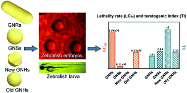
Fig. 2 describes the toxicological endpoints of the varied shaped GNPs such as zebrafish embryos or larvae survival, hatching rate and malformation at time intervals. Then teratogenic index (TI, LC50/NC50) was calculated to evaluate the teratogenicity of nanoparticles (Table 1). Exposure of embryos to fresh prepared GNHs resulted in no increase in mortality due to the rather low concentrations of particles (<0.33 nM), with no differences with embryo medium (data not shown). On the other hand, the GNRs, GNSs and old GNHs result in a dose-dependent manner in mortality. The order of potency for lethality at 80 hpf is GNSs > old GNHs > GNRs (LC50 in Table 1). Normally, the internalization of NPs was likely to be an important factor in toxicity generated by NPs.17 Early reports showed that the GNPs could diffuse into every part of zebrafish embryos from medium via passive transport mechanism, where Brownian diffusion was the rate-determining step over the whole process.11 It was then indicated that the diffusion coefficient depended on the viscosity of medium and the radius of nanoparticles. Thus, the diffusion coefficient of the GNSs was thought to be the same level of the GNHs (both freshly prepared and placed ones) although the shapes of GNHs are not perfectly spherical. However, the plots of percentages of dead zebrafish versus concentration of GNPs in Fig. 2 clearly illustrated the significant differences of toxicity between the GNSs and old GNHs (LC50 0.11 nM vs. 0.13 μM at 80 hpf) when incubated with zebrafish embryos. Moreover, the toxicity of GNPs decreased significantly when the GNRs were employed (LC50 1.54 μM at 80 hpf). One hypothesis for these differences were that GNRs may less become trapped within zebrafish embryos and retained within them for shorter periods. We then explores the metal accumulation in zebrafish embryos and larvae that appeared healthy after gold nanoparticles exposure by ICP-MS. Preliminary animal exposure data (Fig. S3 of ESI) demonstrated an average amount of loading GNRs was highest among all investigating NPs, suggesting higher numbers of GNRs incorporated into embryos and retained within them. The GNSs and old GNHs treated embryos showed similar metal content in the body whereas freshly prepared GNHs exposed embryos showed low Au accumulation inside their bodies at 80 hpf. Nevertheless, the distinct differences in lethality among the investigated varied shaped nanoparticles showed less reflection of NPs accumulation in the zebrafish. It was suggested that the majority of gold NPs toxicity is directly related to the presence of particles, and toxicity appears to occur by different mechanisms. Additional investigation for future research consideration will determine the route of entry and quantity of NPs within key sites (for example, cardiovascular system and liver) and nanomaterial distribution following exposure, which has potential to enable further understanding the developmental toxicity of varied shaped gold NPs.

Table 1 Comparative toxicities of varied shaped gold nanoparticles in zebrafish
GNPs Time (hpf) LC50a NC50b TIc (LC50/NC50)
a The concentration required for 50% lethality.b The concentration required for 50% decrease in normal animal.c Teratogenic index, the nanoparticles are considered teratogenic if the TI is >1.
GNRs 8 11.4 μM 11.8 μM 0.96
32 3.99 μM 3.73 μM 1.07
56 3.73 μM 2.28 μM 1.64
80 1.54 μM 0.96 μM 1.61
GNSs 8
32
56 0.13 nM 0.10 nM 1.31
80 0.11 nM 0.05 nM 2.24
New GNHs 8
32
56
80 0.31 nM >5
Old GNHs 8 3.67 μM 3.59 μM 1.02
32 0.53 μM 0.41 μM 1.32
56 0.31 μM 0.21 μM 1.48
80 0.13 μM 0.06 μM 2.12


image file: c6ra00632a-f2.tif
Fig. 2 Changes in normal (positive triangles), abnormal (circles), dead (squares) and hatched (inverted triangles) embryos (% total) across time and nanoparticles concentrations. Data are mean ± SEM of n independent experiments, n = 5–6.

Notably, it is the first observation that shows the discrepancy of toxicity between the fresh prepared and placed GNHs. After the storage of 6 months, the anisotropic GNHs tended to degrade due to their high surface energy vertices.18 Owing to the Gibbs–Thomson effect, the sharp tips of GNHs are even less stable than the nanorods with edges, and are therefore more rapidly degraded and decomposed into Au+, resulting in Au+ dissolved into the solution (see ESI Fig. S4). Furthermore, the color of the placed solution has changed from pink to red after six months storage. The change in GNHs morphology was also confirmed by TEM. As can be seen from the TEM images (Fig. 1d and S4b), the original sharp vertices of GNHs were no longer visible. A reduction in the overall particle size can also be observed, further supporting the idea that the observed GNHs are subjected to be degraded. Remaining toxicity of freshly prepared and placed GNHs could be attributed to the dramatic differences in the toxicity observed between particles and soluble metal ion. A number of previously reports also noted that the dissolution processes of metal NPs is crucial for an adequate assessment of the exposure and hazard of metal NPs.19

As also shown in Fig. 2, expression of the hatching rates at different development stages of zebrafish embryos exposed to the GNPs at different concentration were calculated respectively. It is demonstrated dose-dependent hatching interference by GNPs except that fresh prepared GNHs showing comparatively little effect at 80 hpf. Statistically significant hatching interference was observed at 3.73 μM GNRs, 73.6 pM GNSs and 16.9 μM old GNHs, respectively (Fig. 2, arrows). However, the hatching interference was strongly correlated to the survival rates, indicating that no obviously hatching delay for all the nanoparticles.

We also determined the GNPs mediated malformation and the results in Fig. 2 also show the dependence of normal or deformed zebrafish upon the concentrations of all four GNPs. As all tested GNPs concentration increased, the number of embryos that developed to normal zebrafish decreased, while the number of embryos that became deformed zebrafish initially increased, then decreased due to a direct transition from normal to dead. All four GNPs generated several common deformed zebrafish (Fig. 3), which included yolk sac edema/defect (Fig. 3a–c, e, f and i), lack of pigmentation (Fig. 3d), tail/spinal cord flexure and skeletal defects (Fig. 3g, h and j–m), cardiac edema (Fig. 3h) and bleeding (Fig. 3l and m, arrows). Multiple types of deformations are also observed in the same zebrafish (Fig. 3h, l and m), indicating that some of these deformations may be inter-related. The frequently observed deformed subtypes include yolk sac edema in the embryos and tail/spinal cord flexure in the developed zebrafish (>80% of all subtypes). Notably, fresh GNHs induced the highest percentage (20.7%) of deformed zebrafish for the GNPs concentrations as low as 3.3 pM at 80 hpf. Comparing LC50/NC50 ratio (TI index) at 80 hpf (Table 1) highlighted fresh GNHs as the most frequent inducer of abnormalities among the varied shaped GNPs. In contrast, GNSs, old GNHs and GNRs showed less teratogenicity as compared with fresh GNHs, with TI index of 2.24, 2.12 and 1.61 respectively.


image file: c6ra00632a-f3.tif
Fig. 3 Representative abnormalities in post- and pre-hatched control embryos and larvae.

In summary, we conclude that CTAB coated GNPs demonstrate developmental toxicity in a shape dependent manner. Indicators of toxicity mainly included high mortality while the most of embryos alive developed to normal zebrafish. In the investigated varying shaped nanoparticles, the GNRs showed less lethality but did not result in significant incidence of malformations with the investigating exposure period. Additional studies will have to be performed to confirm the role of the route of entry and nanomaterial bio-distribution on the observed toxicity. The results obtained in the present study are essential to identifying features to estimate the biological response and consequentially the material modifications that can minimize hazard.

Acknowledgements

This study was financially supported by the National Natural Science Foundation of China (Grant No. 81273450 and Grant No. 21401132) and the Key Laboratory of Functional Inorganic Material Chemistry (Heilongjiang University), Ministry of Education.

Notes and references

  1. E. C. Dreaden, A. M. Alkilany, X. Huang, C. J. Murphy and M. A. El-Sayed, Chem. Soc. Rev., 2012, 41, 2740–2779 RSC.
  2. J. Manso, M. A. Mena, P. Yanez-Sedeno and J. M. Pingarron, Anal. Biochem., 2008, 375, 345–353 CrossRef CAS PubMed.
  3. B. Perez-Lopez and A. Merkoci, Anal. Bioanal. Chem., 2011, 399, 1577–1590 CrossRef CAS PubMed.
  4. N. Khlebtsov and L. Dykman, Chem. Soc. Rev., 2011, 40, 1647–1671 RSC.
  5. A. M. Alkilany and C. J. Murphy, J. Nanopart. Res., 2010, 12, 2313–2333 CrossRef CAS PubMed.
  6. L. A. Couture, B. D. Abbott and L. S. Birnbaum, Teratology, 1990, 42, 619–627 CrossRef CAS PubMed.
  7. M. Chen, S. Chen, M. Du, S. Tang, M. Chen, W. Wang, H. Yang, Q. Chen and J. Chen, Aquat. Toxicol., 2015, 159, 208–216 CrossRef CAS PubMed.
  8. M. Ema, K. S. Hougaard, A. Kishimoto and K. Honda, Nanotoxicology, 2015, 1–22,  DOI:10.3109/17435390.2015.1073811.
  9. N. F. Beliaeva, V. N. Kashirtseva, N. V. Medvedeva, I. Khudoklinova, O. M. Ipatova and A. I. Archakov, Biomed. Khim., 2010, 56, 120–131 CrossRef CAS PubMed.
  10. P. V. Asharani, Y. Lianwu, Z. Gong and S. Valiyaveettil, Nanotoxicology, 2011, 5, 43–54 CrossRef CAS PubMed.
  11. L. M. Browning, K. J. Lee, T. Huang, P. D. Nallathamby, J. E. Lowman and X. H. Xu, Nanoscale, 2009, 1, 138–152 RSC.
  12. L. M. Browning, T. Huang and X. H. Xu, Interface Focus, 2013, 3, 20120098 CrossRef PubMed.
  13. S. Wang, W. Lu, O. Tovmachenko, U. S. Rai, H. Yu and P. C. Ray, Chem. Phys. Lett., 2008, 463, 145–149 CrossRef CAS PubMed.
  14. Z. Sun, Z. Yang, J. Zhou, M. H. Yeung, W. Ni, H. Wu and J. Wang, Angew. Chem., Int. Ed., 2009, 48, 2881–2885 CrossRef CAS PubMed.
  15. Q. F. Ruan, L. Shao, Y. W. Shu, J. F. Wang and H. K. Wu, Adv. Opt. Mater., 2014, 2, 65–73 CrossRef.
  16. M. Chakraborty, F. W. Hsiao, B. Naskar, C. H. Chang and A. K. Panda, Langmuir, 2012, 28, 7282–7290 CrossRef CAS PubMed.
  17. F. Ribeiro, J. A. Gallego-Urrea, K. Jurkschat, A. Crossley, M. Hassellov, C. Taylor, A. M. Soares and S. Loureiro, Sci. Total Environ., 2014, 466–467, 232–241 CrossRef CAS PubMed.
  18. K. E. Lee, A. V. Hesketh and T. L. Kelly, Phys. Chem. Chem. Phys., 2014, 16, 12407–12414 RSC.
  19. C. Ispas, D. Andreescu, A. Patel, D. V. Goia, S. Andreescu and K. N. Wallace, Environ. Sci. Technol., 2009, 43, 6349–6356 CrossRef CAS PubMed.

Footnote

Electronic supplementary information (ESI) available: Experimental detail, the size distribution of gold nanoparticles. See DOI: 10.1039/c6ra00632a

This journal is © The Royal Society of Chemistry 2016
Click here to see how this site uses Cookies. View our privacy policy here.