Computational-aided design of melatonin analogues with outstanding multifunctional antioxidant capacity

Annia Galano*
Departamento de Química, Universidad Autónoma Metropolitana-Iztapalapa, San Rafael Atlixco 186, Col. Vicentina. Iztapalapa, C. P. 09340, México D. F, Mexico. E-mail: agalano@prodigy.net.mx; agal@xanum.uam.mx

Received 7th January 2016 , Accepted 18th February 2016

First published on 22nd February 2016


Abstract

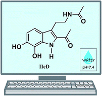
A set of 19 melatonin analogues, intended to be better antioxidants than the parent molecule, have been computationally designed. Eight of them were planned to have good primary antioxidant capacity (AOC), i.e., being good free radical scavengers. Seven of them were designed for their secondary AOC, being able to inhibit ˙OH production by acting as metal ion chelators. Based on their predicted behavior for the intended functions, four multifunctional melatonin analogues were proposed. They were found to be among the best peroxyl radical scavengers identified so far, in aqueous solution, at physiological pH. They were also found to be capable of turning off Cu(II) reduction by O2˙ and Asc, thus fully inhibiting the associated ˙OH production. Two of them, namely the IIcD and IbG analogues, were identified as the ones with the best multifunctional AOC. They both fulfill the Lipinski's and Ghose's rules for orally active drugs. However, IIcD has been chosen as the best prospect for possible application based on potential toxicity and synthetic accessibility estimations. Hopefully, these results might provide motivation for further investigations on the subject, and the synthesis of this compound, so its potential role as protector against oxidative stress – and the associated health issues – could be experimentally confirmed or refuted.


Introduction

During the last decades, oxidative stress (OS) has become the focus of numerous investigations, which had demonstrated that it represents an important risk to human health. The information gathered so far provides compelling evidence on the role of OS in the onset and development of a large number of diseases. A few examples are cancer,1 cardiovascular diseases,2,3 and neurodegenerative disorders including Parkinson's and Alzheimer's diseases, memory loss, multiple sclerosis, and depression.4–6 Accordingly, it is logical that searching for molecules that can offer protection against OS has become an active area of research.

Among such protectors, melatonin (N-acetyl-5-methoxytryptamine) has been proven to be particularly efficient.7–9 There are many reasons for that. The toxicity of melatonin is very low.10 Because of its size, high solubility in lipids and partial solubility in water, melatonin can cross physiologic barriers.11,12 Its protection against OS does not decrease after been metabolized since its metabolites also exhibit antioxidant capacity (AOC).13–15 The combined AOC of melatonin and its metabolites, which is frequently referred to as a cascade-like protection,16,17 implies that melatonin is able to deactivate several equivalents of oxidants. Such complex AOC can involve both primary AOC, via free radical scavenging processes, and secondary AOC through metal chelation.18 Moreover, it has been proposed that this family of compounds acts in a “task-division” way, with some members of the family being particularly efficient as free radical scavengers, and others mainly behaving as metal chelators.19

Inspired by the many appealing characteristics of melatonin, several attempts have been made to produce synthetic analogues with improved properties as antioxidants, but also for other purposes.13,20 This strategy relies on changing the groups in the different modifiable sites of the indole ring (Scheme 1). For example several melatonin analogous have been produced by incorporating pristine and substituted benzoyl and phenyl groups at site R1.21–23 It was found that some of them display better anxiolytic, anticonvulsant and sedative actions than melatonin. Benzoyl-melatonin, and also acetyl-melatonin, have also been found to possess enhanced anti-inflammatory effects with respect to the parent molecule.24 In addition, they have been suggested to have substantial antioxidant ability.25


image file: c6ra00549g-s1.tif
Scheme 1 Structures of melatonin and the indole ring with possible modifiable sites (R1 to R7) for producing synthetic melatonin derivatives, and some representative examples of metabolites and synthetic melatonin analogues.

Various sulfhydryl-containing melatonin derivatives have been synthesized and tested as antioxidants.26 The compounds conjugating a free sulfhydryl moiety in R7, showed improved antioxidant properties. In particular a cysteinyl-conjugated derivative was proposed as suitable for developing antioxidants species. Melatonin retinamide derivatives,27 indole-3-propionamides,28 2-phenylindole derivatives,29 and N-methylindole analogues of hydrazide/hydrazone melatonin derivatives30 have been found to inhibit lipid peroxidation. In particular, chloroindole hydrazide/hydrazone derivatives have shown significant free radical scavenging activity.31 In addition, it has been reported that the lack of a methoxy group in R3 (Scheme 1) does not significantly affect the antioxidant capacity of the melatonin derivatives.30 Several melatonin analogues with modifications in the methoxy and acylaminoethyl groups were identified as better antioxidants than melatonin itself.32 Recently, a large set of indole amino acid derivatives have been synthesized, and found to be similar to melatonin in their DPPH scavenging activity, but better than the parent molecule for inhibiting lipid peroxidation.33

Accordingly, the data gathered so far strongly indicate that melatonin analogues are very promising molecules as antioxidants. In the present work a large set of new derivatives was tested for that purpose. Their design was based on structural modifications intended to increase both the primary and secondary AOC, with respect to melatonin. In this sense they are expected to be more efficient than other melatonin derivatives that mainly present only one of these AOC, most likely free radical scavenging activity. The activity of the tested compounds was compared with those of other known antioxidants. In all the cases the group in R7 was kept identical to that of melatonin (Scheme 1). For the primary AOC, different numbers and positions of OH groups were used as substituents in sites R4 to R6. This strategy was thought based on the fact that phenolic compounds are usually very efficient free radical scavengers. In fact, some hydroxylated melatonin metabolites have been proposed as very efficient for that purpose.19 For the secondary AOC, different groups containing N and O atoms were tested in sites R1 and R2, in such a way that their location promotes metal chelation. The compounds with the highest antioxidant activity were identified and proposed as potential candidates to be implemented as protectors against OS.

Computational details

All the electronic calculations were performed with Gaussian 09 package of programs.34 The M05-2X and M05 functionals35 were used for geometry optimizations and frequency calculations for the systems without and with Cu, respectively. All the calculations were carried out with the 6-311+G(d,p) basis set and the continuum solvation model based on density (SMD),36 and water as solvent. The M05-2X functional has been chosen because it is recommended for kinetic calculations by its developers,35 and its reliability has been independently confirmed by other authors.37–40 It is among the best performing functionals for kinetic calculations in solution,41 and for calculating reaction energies involving free radicals.42 The M05 functional was chosen for the Cu involving systems because it was parameterized including both metals and non-metals, while M05-2X has double the amount of non-local exchange (2X) and was parameterized mainly for non-metals. M05 has been recommended for studies involving both metallic and non-metallic elements, and perform well not only for main-group thermochemistry but also for interactions with transition-metals.35 SMD was chosen for mimicking the solvent effects because it can be consistently used for any charged or uncharged solute in any solvent or liquid medium.36

Local minima and transition states were identified by the number of imaginary frequencies (0 or 1, respectively). In the case of the transition states, intrinsic coordinate calculations (IRC) were performed to verify that the imaginary frequency corresponds to the proper motion along the reaction coordinate. Unrestricted calculations were used for open shell systems. Thermodynamic corrections at 298.15 K were included in the calculation of relative energies.

pKa calculations

The pKa values estimated in the present work were calculated using the isodesmic method, also known as the proton exchange method, or the relative method.43 It is based on the following reaction scheme:
HA + Ref ↔ A + HRef
with HRef/Ref being an acid/base pair of a reference compound. Then the pKa can be calculated as:
 
image file: c6ra00549g-t1.tif(1)

For this method to be reliable there are two key factors to keep in mind. HRef should be structurally similar to the system of interest, and its experimental pKa should be known.

Rate constants

The rate constants (k) were calculated using the conventional transition state theory (TST)44–46 and 1 M standard state, including zero curvature tunneling corrections (ZCT).47 For the electron transfer reactions the Gibbs free energy of activation were calculated using the Marcus theory.48

The values of the total rate coefficients for the HT reactions (kHTtot) were calculated as the sum of each HT reaction path:

 
image file: c6ra00549g-t2.tif(2)

The overall rate coefficients (koverall) were calculated using the molar fractions of the acid–base species involved in each chemical route, i.e., the neutral phenol for HT and the phenolate anion for SPLET, at the pH of interest:

 
koverall = MfneutralkHTtot + MfanionkSPLET (3)

However, the ˙OOH radical is also involved in an acid–base equilibrium, with a pKa = 4.8. Therefore, in aqueous solution at pH = 7.4, the ˙OOH molar fraction is only 0.0025. Thus, to reproduce experimental rate constants of reactions involving this radical, under such conditions, this aspect must be considered. Accordingly, the rate coefficients that can be directly compared with the experiments are:

 
koverall = Mf˙OOHkoverall (4)

The percent contributions of each reaction mechanism was then estimated as:

 
image file: c6ra00549g-t3.tif(5)
 
image file: c6ra00549g-t4.tif(6)

In addition, since several of the calculated rate constants (k) are close to the diffusion-limit, the apparent rate constant (kapp) cannot be directly obtained from TST calculations. The Collins–Kimball theory is used to that purpose,49 in conjunction with the steady-state Smoluchowski50 rate constant for an irreversible bimolecular diffusion-controlled reaction, and the Stokes–Einstein51,52 approaches for the diffusion coefficient of the reactants. These computational details are in line with the quantum mechanics based test for overall free radical scavenging activity (QM-ORSA) protocol,53 which has been validated by comparison with experimental results; its uncertainties have been proven to be no larger than those arising from experiments.53

Metal chelation and Cu(II) reduction

Albeit different chemical routes may be involved in the Cu(II) chelation,54 the data estimated here correspond to the coupled-deprotonation-chelation mechanism (CDCM). This is because it has been previously demonstrated to be more likely than a direct chelation mechanism when the chelation site is susceptible to deprotonation.18,19,55 The CDCM mechanism, for a bi-dentate ligand (HnL), can be schematically represented as:
Cu(H2O)42+ + HnL ⇌ Cu(Hn−1L)(H2O)21+ + 2H2O + H+

Since it involves deprotonation, the CDCM mechanism is influenced by the pH. Under the conditions of interest in this work, i.e., physiological pH (pH = 7.4), it is possible to define a conditional Gibbs energy of reaction (ΔG′) that can be calculated, at each particular pH, according to:

 
ΔG′ = ΔG0 − 2.303RT(pH) (7)
where ΔG0 corresponds to the standard conditions (pH = 0, 1 M standard state). More detail on how to obtain eqn (7) can be found elsewhere.18,56 The ΔG0 values were calculated using ΔGgas(H+) = −4.39 kcal mol−1, and ΔGsolvation(H+) = −265.89 kcal mol−1, based on the recommendation of Camaioni and Schwerdtfeger.57 These values lead to ΔG(H+) = −270.28 kcal mol−1, in aqueous solution.

In addition, as shown in the above equilibrium, copper ions were modeled coordinated to water molecules in a near square-planar geometry, which has been previously demonstrated as the most likely one.58,59 This model is more adequate to represent “free” copper under physiological conditions than the “naked” ion, since charged species are expected to be hydrated in the aqueous phase. For consistency purposes, the hydrated Cu(I) ions were modeled also with four explicit water molecules, albeit in this case the linear two-coordinate configuration is preferred.60–62 Thus, in this model Cu(I) is coordinated to two water molecules, and the other two are solvating the system.

For calculating the reduction potential (E0) of Cu(II) a strategy similar to that proposed by Fu et al.63 has been used. It consist on calculate:

 
image file: c6ra00549g-t5.tif(8)
where F is the Faraday constant (23.06 kcal mol−1 V−1), and ΔG0 is estimated as:
 
ΔG0 = G(oxidized form)G(reduced form) − 4.44 (9)

The last term in eqn (9) is to express the data referred to the normal hydrogen electrode (NHE). The E0Cu(II)/Cu(I) value calculated this way deviates from the experimental value (E0Cu(II)/Cu(I) = 0.16 eV) by 0.52 eV. Thus this difference has been used to correct the values reported in Table 4, in such a way that the experimental E0Cu(II)/Cu(I) value is reproduced.

Synthetic accessibility and toxicity

The synthetic accessibility (SA) of the designed compounds have been estimated using the SYLVIA-XT 1.4 program (Molecular Networks, Erlangen, Germany).64 It uses several contributing criteria, including the complexity of the molecular structure and of ring systems, the number of stereo centers, and the similarity to commercially available compounds. These criteria are weighted and scaled to provide a value between 1 and 10, with the larger values corresponding to the compounds that are expected to be more difficult to synthesize. The SYLVIA program has been validated to be used for ranking virtual compound during drug discovery processes.65

On the other hand, the Toxicity Estimation Software Tool (T.E.S.T.), version 4.1, has been used to make predictions on the potential toxicity of the investigated compounds. This program estimates several descriptors using quantitative structure activity relationships (QSAR) predictions. They are intended to be used for screening untested compounds. The toxicity descriptors used in this work are:

- LD50: amount of chemical (mg kg−1) body weight that causes 50% of rats to die after oral ingestion.

- M: known as Ames mutagenicity. A chemical is positive if it induces revertant colony growth in any strain of Salmonella typhimurium.

- LCF50: concentration of the chemical (mg L−1), in water, that causes 50% of fathead minnow to die after 96 hours.

- LCD50: concentration of the chemical (mg L−1), in water, that causes 50% of Daphnia magna to die after 48 hours.

- IGC50: concentration of the chemical (mg L−1), in water, that causes 50% growth inhibition to Tetrahymena pyriformis after 48 hours.

In this work the toxicity descriptors were computed using the consensus method, which makes toxicity predictions using the simple average of the toxicities predicted from several QSAR methodologies, taking into account the applicability domain of each of them.66 The consensus method usually provides higher prediction accuracy and coverage than other methodologies.

Results and discussion

Design of melatonin analogues

Since the structural features promoting primary and secondary AOC are not necessarily the same, two separated sets of melatonin analogues were first tested for these two kinds of antioxidant protection. The first test set comprises eight melatonin analogues that were mainly designed for primary AOC (test set 1, Scheme 2). Contrary to melatonin, they present phenolic OH groups in the six member ring of the indole moiety. This particular group have been chosen based on pervious evidence that N-acetylserotonin (NAS) and 6-hydroxymelatonin (6OHM), are much better free radical scavengers than the parent molecule.19 NAS and 6OHM present an OH group in site R3 and R6, respectively.
image file: c6ra00549g-s2.tif
Scheme 2 Test set 1 of melatonin analogues, which are designed mainly for primary antioxidant activity (R1 = R2 = –H). Melatonin and N-acetylserotonin (NAS) are presented as reference compounds.

The first working test set can be divided in mono-phenolic (Ia, Ib, Ic), di-phenolic (IIa, IIb, IIc), and tri-phenolic (IIIa, IIIb) indoles. In the di-phenolic and tri-phenolic species the OH group are located in such a way that they correspond to catechol and pyrogallol-like molecules. This is because such configurations are expected to be better for scavenging free radicals than the alternative ones, i.e., with multiple OH located in non-neighboring sites.

On the other hand, a molecular framework, usually referred to as M1 (Scheme 3), has been proposed as crucial for promoting metal chelation with antioxidant implications.67 Moreover, it has been suggested that compounds with such structural feature can be considered as potentially therapeutic for the Alzheimer's disease.67 Accordingly, seven melatonin analogues have been designed to present similar chemical frameworks, in the sense that they have a N and an O atoms located in such a way that they allow the molecule to act as a bi-dentate ligand (test set 2, Scheme 4).


image file: c6ra00549g-s3.tif
Scheme 3 Structure of chelating framework M1, and some representative examples of chemical compounds presenting this structural feature.

image file: c6ra00549g-s4.tif
Scheme 4 Test set 2 of melatonin analogues, which are designed mainly for secondary antioxidant activity (R1 = R3 = R4 = R5 = R6 = –H).

After analyzing the results obtained for test sets 1 and 2, the structural features identified as most promising were combined in a third set of melatonin analogues. They are four compounds (test set 3, Scheme 5) and are intended to efficiently perform as multifunctional antioxidants, i.e. with both primary and secondary antioxidant activities. A more detailed discussion on how these species were chosen is provided in the following sections.


image file: c6ra00549g-s5.tif
Scheme 5 Test set 3 of melatonin analogues which are designed for both primary and secondary antioxidant activities (R1 = R3 = R6 = –H).

Both, the phenolic moieties for primary AOC and frameworks similar to M1 for secondary AOC, are investigated in this work for the first time in melatonin synthetic derivatives. These structural features have been chosen based on previous evidence regarding the AOC of unrelated compounds and have been intentionally combined in the pursuit of an efficient multifunctional antioxidant. The search of previous synthetic melatonin derivatives has been mainly focused on obtaining compounds with electron-enriched indole rings. They are expected to have increased primary AOC via electron transfer reactions from the antioxidant to the radical. On the contrary, the compounds presented here are designed to behave as multi-tasking antioxidants.

Acid–base equilibria in aqueous solution

Due to the presence of phenolic groups in many of the investigated melatonin analogues, they are expected to be involved in acid–base equilibria. In addition, such kind of equilibria have been previously proven to have an important role in the antioxidant activity of chemical compounds.68–71 Moreover, it has been recently shown that the acid–base equilibria play an important role on the free radical scavenging activity of hydroxylated melatonin metabolites via the sequential proton loss electron transfer mechanism (SPLET).72

Therefore the first phenolic pKa of the hydroxylated melatonin analogues studied in this work have been estimated. The chemical compounds used as HRef, the calculated first pKa values, and the corresponding molar fractions for the neutral and mono-anionic species (Mfneutral, Mfanion) at physiological pH are reported in Table 1 for each of the tested phenolic melatonin analogs.

Table 1 First phenolic pKa values for the melatonin analogues in test set 1, most likely deprotonation site (DP) for di-phenolic and tri-phenolic analogues, reference acid used to calculate the pKa (HRef), and molar fractions of the neutral and mono-anionic species (Mfneutral, Mfanion) at physiological pH (pH = 7.4)
  pKa DP HRef Mfneutral Mfanion
Ia 10.9   Phenol ∼1.000 <0.001
Ib 8.9   Phenol 0.972 0.028
Ic 10.1   Phenol 0.998 0.002
IIa 10.2 R4 Catechol 0.998 0.002
IIb 10.3 R6 Catechol 0.998 0.002
IIc 9.5 R5 Catechol 0.992 0.008
IIIa 9.4 R5 Pyrogallol 0.991 0.009
IIIb 11.2 R3 Pyrogallol ∼1.000 <0.001
IbD 8.0   Phenol 0.806 0.194
IbG 7.2   Phenol 0.378 0.622
IIcD 7.9 R5 Catechol 0.744 0.256
IIcG 7.7 R5 Catechol 0.648 0.352


It should be noted that there are several values of pKa reported for the used HRef, i.e., phenol, catechol and pyrogallol (Table 2). The ones used in eqn (2) correspond to the average of all the previously reported ones. In addition, for di-phenolic and tri-phenolic analogues there are more than one possible deprotonation site. Therefore the relative stability of the different possible mono-anions was investigated in order to identify the most likely one in each case. The proposed deprotonation sites are reported in Table 1.

Table 2 pKa values of phenol, catechol and pyrogallol
  pKa Ref.
Phenol 9.89 73
10.09 74
9.98 75
10.24 74
10.05 Average
Catechol 9.25 76
9.5 77
9.38 Average
Pyrogallol 8.94 78
8.905 79
8.20 80
8.68 Average


Melatonin analogues designed for primary antioxidant activity

To investigate the primary antioxidant activity of the melatonin analogues in test set 1 (Scheme 2) their reactions with the hydroperoxyl radical (˙OOH) have been used. This radical has been chosen for several reasons. The ˙OOH scavenging activity of melatonin and some of its metabolites has been already assessed. ˙OOH is the smallest member of the peroxyl family (˙OOR), and has been identify to play an essential role in the toxic side effects associated with aerobic respiration.81 ˙OOR can be successfully scavenged to retard OS,82 due to their moderate reactivity, which also make them appropriate for studying trends in free radical scavenging activities.8,83

Two different reaction mechanisms have been included in this part of the study, namely the hydrogen transfer (HT) and the sequential proton loss electron transfer (SPLET). The later was proposed by Litwinienko and Ingold for the reactions of the DPPH radical with substituted phenols.84 These mechanisms have been selected because, based on previous findings,71,85–88 they are expected to be the most important ones for phenolic compounds. In addition, the relative importance of HT and SPLET in the antioxidant activity of phenols, in aqueous solution, is frequently ruled by the pH. When it is lower than the pKa of the phenolic compound the dominant acid–base species is the neutral one, and the relative importance of HT is frequently higher. On the contrary, at pHs higher than the pKa, deprotonated species prevail and the SPLET route become the most important one. However, in some cases the SPLET mechanism can be the predominant one, even if the phenolate ion is present only in minor proportions.

The HT mechanism in phenolic compounds mainly takes place from the phenolic OH:

XOH + ˙R → XO˙ + HR

On the other hand, the SPLET route, comprises two elementary reaction steps:

XOH → XO + H+

XO + ˙R → XO˙ + R

The first step is just the deprotonation of the phenol yielding the phenolate ion, and as mentioned before it is ruled by the pH. The second step is the key one for kinetics, and involves an electron transfer from the phenolate ion to the free radical.

The Gibbs energies of reaction (ΔG), the Gibbs energies of activation (ΔG), the imaginary frequencies of the transition states (TS), the tunneling corrections, and rate constants for each individual HT reaction path (kHTi) are provided in Table S1, ESI. The optimized geometries of the TS are also provided as ESI. All the modeled HT pathways were found to be exergonic, i.e., thermochemically viable, with ΔG values ranging from −14.5 to −7.1 kcal mol−1, while the corresponding ΔG values are in the 7.7–12.6 kcal mol−1 range. The reorganization energies and the ΔG values for the SPLET reactions are reported in Table S2 (ESI). In this case the ΔG values range from 0.9 to 2.6 kcal mol−1, indicating that the SPLET reactions are faster than the HT ones. Therefore, the pKa values of the studied analogues would determine the relative importance of these two chemical routes, at each particular pH, regarding the peroxyl radical scavenging activity of the studied compounds.

The kinetic data and the percent contributions of the HT and SPLET routes are reported in Table 3. The data corresponding to melatonin, NAS and 6OHM was also included in the table for comparison purposes.

Table 3 Total rate coefficients of the HT reactions (kHTtot, M−1 s−1), rate constants of the SPLET reactions (kSPLET, M−1 s−1), overall rate coefficients (koverall, M−1 s−1), percent contributions of the HT (%HT) and SPLET (%SPLET) mechanisms, for the reactions of melatonin analogues in test set 1 with ˙OOH. All the data is reported at 298.15 K. Melatonin, NAS and 6OHM are included for comparison purposes
  kHTtot kSPLET koverall, pH = 7.4 koverall, pH = 7.4 %HT, pH = 7.4 %SPLET, pH = 7.4
Melatonin 1.90 × 101 8.28 × 10⁻⁴ 1.90 × 101 4.75 × 10⁻² ∼100 <0.1
NAS 1.17 × 10⁶ 7.95 × 10⁹ 5.50 × 10⁶ 1.38 × 10⁴ 21.3 78.8
6OH 3.62 × 10⁶ 8.23 × 10⁹ 8.39 × 10⁷ 2.10 × 10⁵ 4.3 95.8
Ia 9.81 × 10⁵ 7.94 × 10⁹ 3.42 × 10⁶ 8.56 × 10³ 28.6 71.4
Ib 1.43 × 10⁶ 7.39 × 10⁹ 2.07 × 10⁸ 5.18 × 10⁵ 0.7 99.3
Ic 1.58 × 10⁶ 7.91 × 10⁹ 1.68 × 10⁷ 4.21 × 10⁴ 9.4 90.6
IIa 1.47 × 10⁶ 7.79 × 10⁹ 1.33 × 10⁷ 3.33 × 10⁴ 11.0 89.0
IIb 6.69 × 10⁶ 8.01 × 10⁹ 1.80 × 10⁷ 4.49 × 10⁴ 37.2 62.8
IIc 5.12 × 10⁶ 8.09 × 10⁹ 6.46 × 10⁷ 1.61 × 10⁵ 7.9 92.1
IIIa 1.49 × 10⁶ 7.86 × 10⁹ 7.56 × 10⁷ 1.89 × 10⁵ 1.9 98.1
IIIb 2.49 × 10⁷ 8.09 × 10⁹ 2.61 × 10⁷ 6.51 × 10⁴ 95.5 4.5


The overall rate coefficients (koverall) are reported at pH = 7.4. The values of koverall, calculated using eqn (3), are also reported in Table 3 to facilitate comparisons with some data previously reported in literature, albeit koverall is the estimation intended to be directly compared with experiments. It was found that all the SPLET reactions occur within, or close to, the diffusion limited regime (kSPLET ≈ 8 × 10⁹ M−1 s−1) while the HT route is somewhat slower with rate coefficients ranging from 9.8 × 10⁵ to 2.5 × 10⁷ M−1 s−1. This in line with the above discussed ΔG values. However, HT is the main mechanism involved in the ˙OOH scavenging activity of analogue IIIb, at pH = 7.4, because of its relatively high pKa.

The most important finding, regarding primary AOC, is that all the investigated melatonin analogues are much better peroxyl radical scavengers than the parent molecule. Moreover, all the analogues except Ia, react faster with ˙OOH than NAS, which is a mono-phenolic compound with the OH group in site R3. Comparing analogue Ia with 6OHM, the only structural difference is the group at the R3 site. While they both have an OH group at site R4, R3 = –H and –OCH3 for Ia and 6OHM, respectively. Therefore, according to the calculated data it seems that the presence of a methoxy group next to the phenolic HT site slightly promotes reactivity towards peroxyl radicals.

The only of the modeled analogues that is predicted to have better ˙OOH scavenging activity than 6OHM, is Ib. However, the overall rate coefficients of analogues IIc and IIIa are very close to that of 6OHM, and the differences are probably within the method uncertainties. Analyzing the structural features of Ib, IIc and IIIa the first thing that stands out is that they have different number of OH groups (1, 2 and 3, respectively). However, all of them have an OH group at site R5. Thus, it seems that this particular feature is important for maximizing the primary antioxidant activity of melatonin analogues. Therefore, the Ib and IIc frameworks have been chosen for designing the test set 3, which is meant for multifunctional AOC.

Compared with other known antioxidants, the ˙OOH scavenging activity of the analogues in test set 1, except Ia, in aqueous solution at physiological pH, was found to be larger than those of Trolox,89 caffeine,90 capsaicin,91 α-mangostina,92 and gallic acid;70 and similar to those of ascorbic acid,93 resveratrol87 and glutathione.94 On the other hand, their primary AOC is surpassed by those of propyl gallate,95 piceatannol,87 edaravone96 and caffeic acid,69 but only moderately. Therefore, regardless of the differences among the analogues in test set 1, all of them (except Ia) can be proposed as promising primary antioxidants.

In addition, it has been previously reported that phenolic compounds can be regenerated under physiological conditions. Such regeneration takes place in such a way that these compounds can scavenge several radical equivalents in the process, two per cycle (one ˙OOH and one O2˙).56,87,95,97–101 This is expected to be the case for the compounds investigated here and to increase their antioxidant capacity.

Melatonin analogues designed for secondary antioxidant activity

Hydroxyl radical (˙OH) is one of the most reactive and dangerous of the oxidants present in biological systems. ˙OH is so reactive that it can damage almost any molecule in the vicinity of its site of formation.102 Therefore, the best strategy to prevent the oxidative damage caused by this radical is not scavenging it but preventing its production. One of the most important chemical routes yielding ˙OH is the Fenton reaction, or the metal catalyzed Haber–Weiss recombination (HWR). Therefore, because of the role of metal ions in such processes, the necessity of antioxidants able of chelating such metals has been pointed out.103,104

Copper has been chosen for testing the chelating ability of the investigated melatonin analogues in test set 2 (Scheme 4). This metal has been chosen because there is evidence supporting its role in the pathogenesis of neurodegenerative disorders.105 This has been related the formation of oxidative species,106 in particular ˙OH.105 Moreover, it has been reported that under identical experimental conditions the toxicity of Cu(II), in term of oxidative damage, is larger than that of Fe(III).103,107 In addition, the copper chelating ability of melatonin and some of its metabolites has been previously assessed,18 thus it can be used as a reference framework.

The idea here is that while Cu(I) is required for producing ˙OH, Cu(II) is the most abundant and stable oxidative state of copper. Therefore, chelating agents capable of decreasing the viability of Cu(II) reduction should be effective for preventing, or inhibiting, the ˙OH production and the consequent oxidative stress. For that strategy to be successful it is crucial that the chelation reactions yield stable complexes, i.e. they must be exergonic. Thus, this has been the first aspect explored in this work (Table 4).

Table 4 Conditional Gibbs free energies of reaction (ΔG′, kcal mol−1) and conditional equilibrium constants (K′, M), at pH = 7.4, for the Cu(II) chelation by melatonin analogues in test set 2. Reduction potential vs. NHE (E0, eV), Gibbs free energies (ΔG, kcal mol−1) and rate constants (k, M−1 s−1) for the Cu(II) reduction reactions by O2˙ and ascorbate. All the data was calculated at 298.15 K
  ΔG K E0 ΔG (O2˙) ΔG (Asc) k (O2˙) k (Asc)
Cu(II)     0.16 −13.01 −2.81 6.68 × 10⁸ 1.83 × 10⁷
A −2.34 5.2 × 101 0.01 −3.87 6.33 7.13 × 10⁶ 7.59 × 10⁴
B 1.09            
C −1.33 9.5 × 100 0.04 −4.73 5.47 1.07 × 10⁷ 1.44 × 10⁵
D −5.35 8.3 × 10³ −0.20 1.02 11.21 3.45 × 10⁵ 7.89 × 10²
E 0.61            
F −0.61 2.8 × 100 0.15 −7.23 2.96 6.89 × 10⁶ 1.63 × 10⁵
G −4.89 3.9 × 10³ −0.16 −0.08 10.11 2.87 × 10⁵ 1.08 × 10³


It was found that all the reactions involving Cu(II) chelation by the designed melatonin analogues in test set 2, except B and E, are exergonic at room temperature (Table 4). Therefore, analogues A, C, D, F and G are predicted to be able of acting as Cu(II) ligands. Their optimized geometries are provided as ESI. The most stable chelates were found to be those involving analogues D and G, with conditional equilibrium constants equal to 8.3 × 10³ and 3.9 × 10³ M, respectively, at physiological pH.

Additional data, regarding Cu(II) reduction, were computed for all the chelates yield by reactions with negative ΔG values. It was found that chelation by melatonin analogues A, C, D and G significantly lower the reduction potential of Cu(II), with respect to free copper, with D and G showing the maximum effect (Table 4). Accordingly, A, C, D and G are expected to decrease the feasibility of the Cu(II) reduction, being D and G the most efficient for that purpose.

The Gibbs free energies and rate constants of the reactions of Cu(II) with two reductants, i.e., O2˙ and ascorbate (Asc), have also been calculated (Table 4). The reaction with O2˙ corresponds to the first step of the HWR, and the reaction with Asc was modeled to include the possible presence of other, less strong, reductants in the biological systems. In addition, copper–ascorbate mixtures are frequently used in experiments to generate oxidative conditions. O2˙ was calculated including 4 explicit water molecules, which is a model previously validated for this species.108 In the calculation of the Gibbs energies the correction to E0Cu(II)/Cu(I) was included, as well as equivalent corrections for the pairs 3O2/O2˙ and Asc/Asc. These empirical corrections are expected to facilitate possible further comparisons with experiments.

The ideal behavior for a secondary antioxidant acting as a Cu(II) chelating agent should be to fully inhibit the reactions with reductants, O2˙ and Asc in this case. It was found that all the stable chelates with melatonin analogues turn the Cu(II) + Asc reaction into a non-viable process, i.e., with positive ΔG values. On the other hand, only the D analogue makes the Cu(II) reduction by O2˙ an endergonic process, albeit for analogue G the reaction becomes isoergonic (Table 4). Regarding the kinetics, these two species, D and G, are also the ones slowing the most the Cu(II) reduction. The rate constants for “free” Cu(II) were estimated to be 6.7 × 10⁸ and 1.8 × 10⁷ M−1 s−1, for the reactions with O2˙ and Asc respectively. According to the values in Table 4, chelation by melatonin analogs D and G decrease these values by about 2000 and 20[thin space (1/6-em)]000 times. The reorganization energies and the ΔG values used in the calculations of the rate constants are reported in Table S3, ESI.

In addition, the melatonin analogues that chelates Cu(II) through endergonic reactions are expected to act as ˙OH-inactivating ligand (OIL) in biological systems.109 Some of them (D and G) can directly sequester metal ions from reductants. The others (A, C and F), which only downgrade the Cu(II) reduction by O2˙, should still be able of deactivating OH radicals immediately after formed via Fenton-like reactions. This is because the high reactivity of this radical towards organic frameworks would facilitate the reaction with the ligands in the Cu chelates, which would be just in the ˙OH formation site.

Analyzing altogether the data regarding the secondary AOC of the investigated melatonin analogs by chelating metal ions, it becomes evident that D and G are the most promising species for inhibiting ˙OH production, and thus the consequent oxidative stress. Therefore, their structural features have been those included in the design of melatonin analogues with multifunctional AOC.

Melatonin analogues designed for multifunctional antioxidant activity

Based on the results discussed in the two previous sections, the structural features of primary antioxidants Ib and IIc and of secondary antioxidants D and G have been included in the set of melatonin analogs designed to be multifunctional antioxidants (test set 3, Scheme 5). The IIIa structure has not been included to avoid having many H donor groups in the structure (please see the next section for more details).

The estimated first pKa values of the analogues in test set 3 are reported in Table 5, together with the most likely deprotonation site for the two di-phenols, and the molar fractions of the neutral and mono-anionic species at physiological pH. They are all predicted to be dominantly in their neutral form at this pH, but with significant amounts of the mono-anions, except IbG, which shows the opposite trend.

Table 5 First phenolic pKa values, most likely deprotonation site (DP) for melatonin analogues in test set 3, reference acid used to calculate the pKa (HRef), and molar fractions of the neutral and mono-anionic species (Mfneutral, Mfanion) at physiological pH (pH = 7.4)
  pKa DP HRef Mfneutral Mfanion
IbD 8.0   Phenol 0.806 0.194
IbG 7.2   Phenol 0.378 0.622
IIcD 7.9 R5 Catechol 0.761 0.239
IIcG 7.7 R5 Catechol 0.670 0.330


The information on the primary and secondary antioxidant activities of analogues in test set 3 is reported in Tables 6 and 7, respectively. The ΔG, ΔG, imaginary frequencies of the TS, tunneling corrections and kHTi, are provided in Table S3, ESI. The optimized geometries of the TS are also provided as ESI, as well as the reorganization energies and the ΔG values for the SPLET reactions (Table S4).

Table 6 Total rate coefficients of the HT reactions (kHTtot, M−1 s−1), rate constants of the SPLET reactions (kSPLET, M−1 s−1), overall rate coefficients (koverall, M−1 s−1), percent contributions of the HT (%HT) and SPLET (%SPLET) mechanisms, for the reactions of melatonin analogues in test set 3 with ˙OOH. All the data is reported at 298.15 K
  kHTtot kSPLET koverall, pH = 7.4 koverall, pH = 7.4 %HT, pH = 7.4 %SPLET, pH = 7.4
IbD 2.47 × 10⁵ 5.23 × 10⁹ 1.02 × 10⁹ 2.54 × 10⁶ <0.1 ∼100
IbG 4.97 × 10⁵ 4.37 × 10⁹ 2.72 × 10⁹ 6.79 × 10⁶ <0.1 ∼100
IIcD 7.48 × 10⁷ 7.79 × 10⁹ 1.92 × 10⁹ 4.79 × 10⁶ 3.0 97.0
IIcG 9.61 × 10⁶ 7.84 × 10⁹ 2.59 × 10⁹ 6.48 × 10⁶ 0.2 99.8


Table 7 Conditional Gibbs free energies of reaction (ΔG′, kcal mol−1) and conditional equilibrium constants (K′, M), at pH = 7.4, for the most likely Cu(II) chelation routes by melatonin analogues in test set 3. Reduction potential vs. NHE (E0, eV), Gibbs free energies (ΔG, kcal mol−1) and rate constants (k, M−1 s−1) for the Cu(II) reduction reactions by O2˙ and ascorbate. All the data was calculated at 298.15 K
  ΔG K E0 ΔG (O2˙) ΔG (Asc) k (O2˙) k (Asc)
IbD −5.38 8.76 × 10³ −0.22 1.49 11.68 2.29 × 10⁵ 4.49 × 10²
IbG −7.52 3.23 × 10⁵ −0.31 3.54 13.74 4.58 × 10⁴ 4.17 × 101
IIcD −6.39 4.85 × 10⁴ −0.30 3.17 13.36 4.91 × 10⁴ 5.46 × 101
IIcG −6.47 5.55 × 10⁴ −0.25 2.15 12.34 5.08 × 10⁴ 9.53 × 101


The overall rate coefficients for the reactions of these analogues with ˙OOH were found to be within, or near to, the diffusion limit. This indicates that they all should be excellent peroxyl radical scavengers, contrary to what has been reported for the parent molecule. Moreover, including the structural features of the secondary antioxidants D and G in the phenolic melatonin analogues does not interfere with the primary antioxidant activity. On the contrary, the analogs in test set 3 are all predicted to be better peroxyl radical scavengers than those in test set 1. For the multifunctional melatonin analogues the primary AOC is dominated by the SPLET route, with very small contributions from HT. The largest one appears to be for IIcD, and is only about 3%.

Compared to other known antioxidants, the melatonin analogues in test set 3 are predicted to be among the best peroxyl radical scavengers reported so far. They seem to be able of reacting with these radicals as fast as possible, considering the limit imposed by diffusion, at least in aqueous solution, at physiological pH. Such fast reactions are rare to find, which supports the idea that the designed melatonin analogues should be exceptionally good primary antioxidants.

Regarding their secondary AOC, the analogues in test set 3 can form different chelates with Cu(II), due to the presence of phenolic groups in the six membered ring of the indole moiety. Some of them are equivalent to the ones formed by molecules in test set 2, i.e., involving the indolic N atom, and the O atom in the R2 group (c1). Others involve the OH in R5 and the indolic N (c2), and for IIcD and IIcG there are also possible chelates involving the OH groups in R4 and R5 (c3). The Gibbs energies of all these chelation reactions, and the corresponding equilibrium constants are reported in Table S5, ESI. It was found that, in all the cases, the c1 chelates are the lowest in energy. Thus, the following analyses are just for them, as they should be the most abundant ones.

The data on the secondary AOC of these compounds is reported in Table 7, while the reorganization energy and the ΔG values used in the calculations of the corresponding rate constants are provided in Table S4, ESI. It was found that the reactions yielding the c1 chelates are systematically more exergonic for the molecules in the test set 3 than those involving any of the chelating agents in test set 2. This indicates that the presence of OH groups in the six membered ring of the indole moiety reinforces the chelating ability of melatonin analogues, with the exception of IbD which has a ΔG′ value at pH = 7.4 almost identical to that of the D analogue.

The same applies for all the other calculated properties. Chelates with IbD, IbG, IIcD and IIcG as ligands, all inhibit the Cu(II) reduction to a larger extent than the chelates involving the melatonin analogues in test set 2. They all turn endergonic the reactions with both O2˙ and Asc and dramatically slow them down. The most efficient as OH inhibitors, via metal chelation are expected to be IbG and IIcD. Considering that they also are excellent for scavenging free radicals, these two melatonin analogues are proposed as the most promising multifunctional antioxidants, especially IIcD due to its better secondary AOC.

Other relevant features

In addition to their reactivity towards oxidants, there are other aspects to consider regarding the potential use of the investigated melatonin analogues as potential drugs with therapeutic applications against oxidative stress and related health issues. To that purpose several molecular descriptors have been calculated. They were chosen to check if the designed compounds fulfill the Lipinski's rule of five110 and the Ghose's rule.111 These two sets of rules establish some criteria that any orally active drug must have. According to Lipinski's rule they should have no more than 5 hydrogen bond donors (HBD), no more than 10 (5 × 2) hydrogen bond acceptors (HBA), a molecular weight (MW) under 500 (5 × 100) g mol−1, and an octanol/water partition coefficient (log[thin space (1/6-em)]P) lower than 5. Molecules violating more than one of these rules may have problems with bioavailability. On the other hand the Ghose's rule establishes that orally active drugs must have a log[thin space (1/6-em)]P ranging from −0.4 to 5.6, molar refractivity (MR) from 40 to 130, MW from 160 to 480, and a number of heavy atoms (XAt) from 20 to 70. Otherwise the intended effects should be impeded by poor permeation or absorption. Different methods have been used to estimate log[thin space (1/6-em)]P (Table 8), and the values are all in the range 0.36–2.64, i.e., within the limits established in both Lipinski's and Ghose's rules. Discrepancies among the log[thin space (1/6-em)]P values calculated using different methods are attributed to the fact that the strategies involved in the calculations are also different. For example milog[thin space (1/6-em)]P uses molecular fragments while ALOGPS is an atomic-contribution method. More details on this topic can be found elsewhere.112
Table 8 Molecular descriptors for the whole set of melatonin analogues designed in the present work. log[thin space (1/6-em)]P, number of hydrogen bond acceptors (HBA), number of hydrogen bond donors (HBD), molecular weight (MW), molar refractivity (MR), number of heavy atoms (XAt), topological polar surface area (TPSA) and rotatable bonds (RB). Melatonin is included for comparison purposes
  log[thin space (1/6-em)]Pa log[thin space (1/6-em)]Pb log[thin space (1/6-em)]Pc HBA HBD MW MRd XAt TPSAe RB
a Calculated using Crippen's method.115b Calculated using ALOGPS 2.1.116c Calculated using milog[thin space (1/6-em)]P 2.2 from Molinspiration online property calculation toolkit.d Calculated using the Ghose & Crippen method.117e Reported in Å2, calculated using the Molinspiration Property Calculation Service (http://www.molinspiration.com).
Melatonin 1.75 1.42 1.45 4 2 232 65.6 17 54.12 4
Ia 1.65 1.42 1.45 4 3 218 60.9 16 65.12 4
Ib 1.65 0.98 0.91 4 3 218 60.9 16 65.12 4
Ic 1.65 1.02 0.91 4 3 218 60.9 16 65.12 4
IIa 1.26 0.98 1.15 5 4 234 62.7 17 85.34 4
IIb 1.26 0.95 1.15 5 4 234 62.7 17 85.34 4
IIc 1.26 0.92 0.42 5 4 234 62.7 17 85.34 4
IIIa 0.87 0.87 0.65 6 5 250 64.5 18 105.57 4
IIIb 0.87 0.93 0.65 6 5 250 64.5 18 105.57 4
A 1.56 0.58 0.36 4 2 230 65.6 17 61.96 5
B 1.39 0.55 0.36 4 2 244 69.6 18 61.96 6
C 1.48 1.53 1.44 5 2 260 69.9 19 71.19 6
D 1.63 1.98 1.65 4 2 244 70.2 18 61.96 5
E 0.92 1.78 1.48 5 2 259 74.0 19 65.20 6
F 1.90 1.78 1.55 5 2 273 74.8 20 65.20 6
G 1.36 1.15 1.02 4 2 282 85.4 21 61.96 7
IbD 1.24 1.56 1.00 5 3 260 72.0 19 82.19 5
IbG 0.97 2.64 2.36 5 3 298 87.1 22 82.19 7
IIcD 0.85 1.36 1.28 6 4 276 73.8 20 102.42 5
IIcG 0.58 2.37 2.07 6 4 314 88.9 23 102.42 7


The values of all these molecular descriptors for the whole set of melatonin analogues designed in this work are provided in Table 8. As the values in this table show all the investigated compounds fulfill all the criteria in the Lipinski's rule. Only two exceptions were found (IIIa and IIIb), which belong to test set 1 and have 5 HBD. That is why their structural features were not included in the test set for multifunctional antioxidant activity. Regarding the Ghose's rule, the most frequent problem is related to the XAt value that is slightly below 20 for most of the studied compounds. However, it is not expected to be a major inconvenience since melatonin itself has XAt = 17, and its beneficial effects as an oral drug has been extensively documented. The melatonin analogues that fulfill all the criteria in both Lipinski's and Ghose's rules are analogues F, G, IbG, IIcD and IIcG.

It should be noted, however, that these rules are empirical, and based on statistical analyses using the existing data on the bioavailability of oral drugs. Thus, they are meant to be guidelines, not rigorous laws. Moreover, viable drugs must also have other important features including manufacturability and safety.113 Therefore, these aspects are also addressed here. In addition, it is currently considered that physicochemical properties other than those included in Lipinski's and Ghose's rules should also be explored for newly developed drugs.114 Some examples are topological polar surface area (TPSA) and the number of rotatable bonds (RB). Accordingly, they have also been estimated and are included in Table 8.

The estimated synthetic accessibility, of each designed compound, is reported in Table 9 where melatonin and other – currently in use – drugs have been included for comparison purposes. The SA values can be grouped into 3 categories, using the default thresholds of the SYLVIA program: easy (SA ≤ 3), medium (3 < SA < 6), and difficult (SA ≥ 6). According to this classification all the compounds designed in this work correspond to medium SA, with values ranging from 3.5 to 4.3. Even the largest value is close enough to that of ciprofloxacin, which suggests that their manufacturability should not be problematic. In the particular case of the two compounds identified here as the most promising multifunctional antioxidants, the SA value for IIcD (3.9) is lower than that of IbG (4.2).

Table 9 Synthetic accessibility (SA), oral rat 50 percent lethal dose (LD50), Ames mutagenicity (M), 96 hour fathead minnow 50 percent lethal concentration (LCF50), 48 hour Daphnia magna 50 percent lethal concentration (LCD50), and Tetrahymena pyriformis 50 percent growth inhibition concentration (IGC50). Melatonin, and some other known drugs, are included for comparison purposes
  SA LD50 M LCF50 LCD50 IGC50
Melatonin 2.46 1298.11 0.05 (−) 23.51 2.25 66.62
Ia 3.50 1558.61 0.08 (−) 28.67 2.56 78.32
Ib 3.54 1526.48 −0.01 (−) 21.41 3.72 78.60
Ic 3.56 1248.74 −0.08 (−) 25.59 4.66 78.12
IIa 3.61 1061.32 0.18 (−) 20.55 4.44 58.25
IIb 3.63 1066.44 0.26 (−) 9.19 2.98 59.47
IIc 3.61 1273.36 0.12 (−) 13.71 6.64 58.17
IIIa 3.70 1646.33 0.36 (−) 5.73 6.65 52.64
IIIb 3.71 1163.93 0.41 (−) 8.09 3.17 34.20
A 3.65 1442.93 0.17 (−) 5.86 4.58 47.98
B 3.67 1732.22 0.01 (−) 3.26 N/A 34.81
C 3.86 997.81 0.03 (−) 4.04 11.85 48.55
D 3.74 1352.30 0.07 (−) 3.89 5.39 28.51
E 3.92 573.98 0.30 (−) 14.30 N/A N/A
F 4.03 709.19 0.46 (−) 6.99 25.19 75.83
G 4.10 1135.35 0.51 (+) 0.62 0.72 2.40
IbD 3.82 1660.01 0.09 (−) 3.68 6.25 21.02
IbG 4.22 526.23 0.47 (−) 0.17 0.19 2.03
IIcD 3.90 1203.01 0.30 (−) 3.11 9.80 20.96
IIcG 4.30 265.38 0.40 (−) 0.25 0.33 2.46
Aspirin 2.20 757.21 0.43 (−) 80.28 472.51 472.51
Floxetin 3.98 1002.16 0.13 (−) 0.41 6.10 × 10⁻² 2.33
Ciprofloxacin 4.26 36.25 0.70 (+) 0.11 0.16 N/A


Regarding the toxicity descriptors, the lower the value of M, and the larger the values of LD50, LCF50, LCD50 and IGC50, the lower the toxicity of the tested chemical. G is the only of the designed compounds that was identified as potentially mutagenic. Between compounds IIcD and IbG, the later have a larger value of M, but still its mutagenicity is predicted as negative and with a value similar to that of aspirin. Regarding the LD50, LCF50, LCD50 and IGC50 descriptors, the range taken from melatonin and other currently in use drugs are 1298.11–36.25, 80.28–0.11, 472.51–0.06, and 472.51–2.33, respectively. The values corresponding to most of the designed compounds are within these ranges. Comparing the IIcD and IbG compounds, the first one was systematically found to have the lowest predicted toxicity, regardless of the descriptor used.

Therefore, considering the values of the molecular descriptors and the primary and secondary AOC of all the investigated compounds, altogether, melatonin analogue IIcD is identified as the most promising agent against oxidative stress, and its deleterious effects. It is expected to efficiently act as a multifunctional antioxidant, contrary to what has been previously described for naturally occurring melatonin derivatives, i.e., most of them are only good as primary or secondary antioxidant, but not as both simultaneously. IIcD is also predicted to be synthetically accessible and with low toxicity. Hopefully, it might be synthesized in the near future, thus its activity could be experimentally corroborated.

Conclusions

A set of 19 melatonin derivatives, intended to be better antioxidants than the parent molecule, have been computationally designed. Eight of them were planned to be good primary AOC, i.e., good free radical scavengers. Seven of them were designed for their secondary AOC, to be able of inhibiting ˙OH production by acting as metal ion chelators. Based on their predicted behavior for the intended functions, four multifunctional melatonin analogues were proposed. They were found to be among the best peroxyl radical scavengers identified so far (in aqueous solution, at physiological pH) with rate constants within, or near to, the diffusion limited regime. They are predicted to be better antioxidants than Trolox, resveratrol, ascorbic acid, and other known antioxidants. Three of the multifunctional analogues were also found to be capable of turning off the Cu(II) reduction by O2˙ and Asc, thus fully inhibiting the associated ˙OH production. Two of them, namely the IbG and IIcD were identified as the most promising multifunctional antioxidants. In addition, they fulfill both the Lipinski's and Ghose's rules for orally active drugs. However, based on potential toxicity and synthetic accessibility estimations, IIcD has been chosen as the best prospect for possible application. Hopefully, this investigation might provide motivation for further investigations in the subject, and the synthesis of this compound, so its potential role as protector against oxidative stress, and the associated health issues, could be experimentally tested.

Acknowledgements

The Laboratorio de Visualización y Cómputo Paralelo at Universidad Autónoma Metropolitana-Iztapalapa is acknowledge for computing time. This work was partially supported by project SEP-CONACyT 167491.

Notes and References

  1. B. Tekiner-Gulbas, A. D. Westwell and S. Suzen, Curr. Med. Chem., 2013, 20, 4451–4459 CrossRef CAS PubMed.
  2. M. Matsuda and I. Shimomura, Rev. Endocr. Metab. Disord., 2014, 15, 1–10 CrossRef CAS PubMed.
  3. E. Eren, H. Y. Ellidag, Y. Cekin, R. U. Ayoglu, A. O. Sekercioglu and N. Yilmaz, Redox Rep., 2014, 19, 34–39 CrossRef CAS PubMed.
  4. B. Halliwell, Drugs Aging, 2001, 18, 685–716 Search PubMed.
  5. M. Pohanka, Curr. Med. Chem., 2014, 21, 356–364 CrossRef CAS PubMed.
  6. C. Pimentel, L. Batista-Nascimento, C. Rodrigues-Pousada and R. A. Menezes, Oxid. Med. Cell. Longevity, 2012, 2012, 132146 Search PubMed.
  7. M. R. Ramis, S. Esteban, A. Miralles, D. X. Tan and R. J. Reiter, Curr. Med. Chem., 2015, 22, 2690–2711 CrossRef CAS PubMed.
  8. A. Galano, D. X. Tan and R. J. Reiter, J. Pineal Res., 2011, 51, 1–16 CrossRef CAS PubMed.
  9. R. Hardeland, Endocrine, 2005, 27, 119–130 CrossRef CAS PubMed.
  10. G. Jahnke, M. Marr, C. Myers, R. Wilson, G. Travlos and C. Price, Toxicol. Sci., 1999, 50, 271–279 CrossRef CAS PubMed.
  11. L. Ceraulo, M. Ferrugia, L. Tesoriere, S. Segreto, M. A. Livrea and V. Turco Liveri, J. Pineal Res., 1999, 26, 108–112 CrossRef CAS PubMed.
  12. D. Bonnefont-Rousselot and F. Collin, Toxicology, 2010, 278, 55–67 CrossRef CAS PubMed.
  13. H. Gurer-Orhan and S. Suzen, Curr. Med. Chem., 2015, 22, 490–499 CrossRef CAS PubMed.
  14. A. Galano, D. X. Tan and R. J. Reiter, J. Pineal Res., 2013, 54, 245–257 CrossRef CAS PubMed.
  15. R. J. Reiter, D. X. Tan, M. J. Jou, A. Korkmaz, L. C. Manchester and S. D. Paredes, Neuroendocrinol. Lett., 2008, 29, 391–398 Search PubMed.
  16. D. X. Tan, L. C. Manchester, R. J. Reiter, W. B. Qi, M. Karbownik and J. R. Calvo, Biol. Signals Recept., 2000, 9, 137–159 CrossRef CAS PubMed.
  17. R. J. Reiter, D. X. Tan and A. Galano, Physiology, 2014, 29, 325–333 CrossRef CAS PubMed.
  18. A. Galano, M. E. Medina, D. X. Tan and R. J. Reiter, J. Pineal Res., 2015, 58, 107–116 CrossRef CAS PubMed.
  19. R. Álvarez-Diduk, A. Galano, D. X. Tan and R. J. Reiter, J. Phys. Chem. B, 2015, 119, 8535–8543 CrossRef PubMed.
  20. S. Suzen, Curr. Drug Delivery, 2013, 10, 71–75 CrossRef CAS.
  21. A. Lira-Rocha, O. Espejo-González and E. B. Naranjo-Rodríguez, Eur. J. Med. Chem., 2002, 37, 945–951 CrossRef CAS PubMed.
  22. R. Bustamante-García, E. B. Naranjo-Rodríguez, A. S. Lira-Rocha, A. Ortiz-Osornio and O. Picazo, Behav. Brain Res., 2013, 236, 148–156 CrossRef PubMed.
  23. R. Bustamante-García, A. S. Lira-Rocha, O. Espejo-González, A. E. Gómez-Martínez and O. Picazo, Prog. Neuro-Psychopharmacol. Biol. Psychiatry, 2014, 51, 133–139 CrossRef PubMed.
  24. C. Phiphatwatcharaded, A. Topark-Ngarm, P. Puthongking and P. Mahakunakorn, Drug Dev. Res., 2014, 75, 235–245 CrossRef CAS PubMed.
  25. J. R. Johns and J. A. Platts, Org. Biomol. Chem., 2014, 12, 7820–7827 Search PubMed.
  26. P. L. Tsia and M. K. Hu, J. Pharm. Pharmacol., 2003, 55, 1655–1660 CrossRef CAS PubMed.
  27. Z. Ates-Alagoz, T. Coban and E. Buyukbingol, Arch. Pharm., 2006, 339, 193–200 CrossRef CAS PubMed.
  28. Z. Ateş-Alagöz, T. Coban and S. Suzen, Med. Chem. Res., 2005, 14, 169–179 CrossRef.
  29. S. Suzen, P. Bozkaya, T. Coban and D. Nebioǧlu, J. Enzyme Inhib. Med. Chem., 2006, 21, 405–411 CrossRef CAS PubMed.
  30. H. Shirinzadeh, B. Eren, H. Gurer-Orhan, S. Suzen and S. Özden, Molecules, 2010, 15, 2187–2202 CrossRef CAS PubMed.
  31. A. D. Yilmaz, T. Coban and S. Suzen, J. Enzyme Inhib. Med. Chem., 2012, 27, 428–436 CrossRef CAS PubMed.
  32. G. Gürkök, T. Coban and S. Suzen, J. Enzyme Inhib. Med. Chem., 2009, 24, 506–515 CrossRef PubMed.
  33. S. Suzen, S. S. Cihaner and T. Coban, Chem. Biol. Drug Des., 2012, 79, 76–83 Search PubMed.
  34. M. J. Frisch, G. W. Trucks, H. B. Schlegel, G. E. Scuseria, M. A. Robb, J. R. Cheeseman, G. Scalmani, V. Barone, B. Mennucci, G. A. Petersson, H. Nakatsuji, M. Caricato, X. Li, H. P. Hratchian, A. F. Izmaylov, J. Bloino, G. Zheng, J. L. Sonnenberg, M. Hada, M. Ehara, K. Toyota, R. Fukuda, J. Hasegawa, M. Ishida, T. Nakajima, Y. Honda, O. Kitao, H. Nakai, T. Vreven, J. A. Montgomery Jr, J. E. Peralta, F. Ogliaro, M. J. Bearpark, J. Heyd, E. N. Brothers, K. N. Kudin, V. N. Staroverov, R. Kobayashi, J. Normand, K. Raghavachari, A. P. Rendell, J. C. Burant, S. S. Iyengar, J. Tomasi, M. Cossi, N. Rega, N. J. Millam, M. Klene, J. E. Knox, J. B. Cross, V. Bakken, C. Adamo, J. Jaramillo, R. Gomperts, R. E. Stratmann, O. Yazyev, A. J. Austin, R. Cammi, C. Pomelli, J. W. Ochterski, R. L. Martin, K. Morokuma, V. G. Zakrzewski, G. A. Voth, P. Salvador, J. J. Dannenberg, S. Dapprich, A. D. Daniels, Ö. Farkas, J. B. Foresman, J. V. Ortiz, J. Cioslowski and D. J. Fox, Journal, 2009 Search PubMed.
  35. Y. Zhao, N. E. Schultz and D. G. Truhlar, J. Chem. Theory Comput., 2006, 2, 364–382 CrossRef PubMed.
  36. A. V. Marenich, C. J. Cramer and D. G. Truhlar, J. Phys. Chem. B, 2009, 113, 6378–6396 CrossRef CAS PubMed.
  37. M. Altarawneh and B. Z. Dlugogorski, Environ. Sci. Technol., 2013, 47, 5118–5127 CrossRef CAS PubMed.
  38. M. Dargiewicz, M. Biczysko, R. Improta and V. Barone, Phys. Chem. Chem. Phys., 2012, 14, 8981–8989 RSC.
  39. J. Murillo, D. Henao, E. Vélez, C. Castaño, J. Quijano, J. Gaviria and E. Zapata, Int. J. Chem. Kinet., 2012, 44, 407–413 CrossRef CAS.
  40. K. P. Prasanthkumar and J. R. Alvarez-Idaboy, RSC Adv., 2014, 4, 14157–14164 RSC.
  41. A. Galano and J. R. Alvarez-Idaboy, J. Comput. Chem., 2014, 35, 2019–2026 CrossRef CAS PubMed.
  42. Y. Zhao and D. G. Truhlar, J. Phys. Chem. A, 2008, 112, 1095–1099 CrossRef CAS PubMed.
  43. J. Ho and M. L. Coote, Theor. Chem. Acc., 2010, 125, 3–21 Search PubMed.
  44. H. Eyring, J. Chem. Phys., 1935, 3, 63–71 CrossRef.
  45. M. G. Evans and M. Polanyi, Trans. Faraday Soc., 1935, 31, 875–894 RSC.
  46. D. G. Truhlar, B. C. Garrett and S. J. Klippenstein, J. Phys. Chem., 1996, 100, 12771–12800 CrossRef CAS.
  47. A. Kuppermann and D. G. Truhlar, J. Am. Chem. Soc., 1971, 93, 1840–1851 CrossRef CAS.
  48. R. A. Marcus, Pure Appl. Chem., 1997, 69, 13–29 CrossRef CAS.
  49. F. C. Collins and G. E. Kimball, J. Colloid Sci., 1949, 4, 425–437 CrossRef CAS.
  50. M. Smoluchowski, Z. Phys. Chem., 1917, 92, 129–168 Search PubMed.
  51. A. Einstein, Ann. Phys., 1905, 17, 549–560 CrossRef CAS.
  52. G. G. Stokes, Mathematical and Physical Papers, Cambridge University Press, Cambridge, 1903 Search PubMed.
  53. A. Galano and J. R. Alvarez-Idaboy, J. Comput. Chem., 2013, 34, 2430–2445 CrossRef CAS PubMed.
  54. M. Francisco-Marquez, M. Aguilar-Fernández and A. Galano, Comput. Theor. Chem., 2016, 1077, 18–24 CrossRef CAS.
  55. Y. Villuendas-Rey, J. R. Alvarez-Idaboy and A. Galano, J. Chem. Inf. Model., 2015, 55, 2552–2561 CrossRef CAS PubMed.
  56. R. Álvarez-Diduk and A. Galano, J. Phys. Chem. B, 2015, 119, 3479–3491 CrossRef PubMed.
  57. D. M. Camaioni and C. A. Schwerdtfeger, J. Phys. Chem. A, 2005, 109, 10795–10797 CrossRef CAS PubMed.
  58. V. S. Bryantsev, M. S. Diallo and W. A. Goddard Iii, J. Phys. Chem. A, 2009, 113, 9559–9567 CrossRef CAS PubMed.
  59. J. Ortega-Castro, M. Adrover, J. Frau, J. Donoso and F. Muñoz, Chem. Phys. Lett., 2009, 475, 277–284 CrossRef CAS.
  60. T. Fujii, F. Moynier, M. Abe, K. Nemoto and F. Albarède, Geochim. Cosmochim. Acta, 2013, 110, 29–44 CrossRef CAS.
  61. J. L. Fulton, M. M. Hoffmann and J. G. Darab, Chem. Phys. Lett., 2000, 330, 300–308 CrossRef CAS.
  62. J. L. Fulton, M. M. Hoffmann, J. G. Darab, B. J. Palmer and E. A. Stern, J. Phys. Chem. A, 2000, 104, 11651–11653 CrossRef CAS.
  63. Y. Fu, L. Liu, H. Z. Yu, Y. M. Wang and Q. X. Guo, J. Am. Chem. Soc., 2005, 127, 7227–7234 CrossRef CAS PubMed.
  64. K. Boda, T. Seidel and J. Gasteiger, J. Comput.–Aided Mol. Des., 2007, 21, 311–325 CrossRef CAS PubMed.
  65. P. Bonnet, Eur. J. Med. Chem., 2012, 54, 679–689 CrossRef CAS PubMed.
  66. H. Zhu, A. Tropsha, D. Fourches, A. Varnek, E. Papa, P. Gramatical, T. Öberg, P. Dao, A. Cherkasov and I. V. Tetko, J. Chem. Inf. Model., 2008, 48, 766–784 CrossRef CAS PubMed.
  67. C. Rodriguez-Rodriguez, N. Sanchez de Groot, A. Rimola, A. Alvarez-Larena, V. Lloveras, J. Vidal-Gancedo, S. Ventura, J. Vendrell, M. Sodupe and P. Gonzalez-Duarte, J. Am. Chem. Soc., 2009, 131, 1436–1451 CrossRef CAS PubMed.
  68. R. M. Han, D. D. Li, C. H. Chen, R. Liang, Y. X. Tian, J. P. Zhang and L. H. Skibsted, J. Agric. Food Chem., 2011, 59, 10367–10372 CrossRef CAS PubMed.
  69. J. R. León-Carmona, J. R. Alvarez-Idaboy and A. Galano, Phys. Chem. Chem. Phys., 2012, 14, 12534–12543 RSC.
  70. T. Marino, A. Galano and N. Russo, J. Phys. Chem. B, 2014, 118, 10380–10389 CrossRef CAS PubMed.
  71. A. Pérez-González, A. Galano and J. R. Alvarez-Idaboy, New J. Chem., 2014, 38, 2639–2652 RSC.
  72. R. Álvarez-Diduk, A. Galano, D. X. Tan and R. J. Reiter, Theor. Chem. Acc., 2016, 135, 1–5 Search PubMed.
  73. A. D[a with combining cedilla]browski, P. Podkościelny, Z. Hubicki and M. Barczak, Chemosphere, 2005, 58, 1049–1070 CrossRef PubMed.
  74. J. R. Torres-Lapasió, M. C. García-Alvarez-Coque, E. Bosch and M. Rosés, J. Chromatogr., 2005, 1089, 170–186 CrossRef.
  75. M. D. Liptak, K. C. Gross, P. G. Seybold, S. Feldgus and G. C. Shields, J. Am. Chem. Soc., 2002, 124, 6421–6427 CrossRef CAS PubMed.
  76. N. Schweigert, R. W. Hunziker, B. I. Escher and R. I. L. Eggen, Environ. Toxicol. Chem., 2001, 20, 239–247 CAS.
  77. G. Ivanova, E. Bratovanova and D. Petkov, J. Pept. Sci., 2002, 8, 8–12 CrossRef CAS PubMed.
  78. H. S. Kim, T. D. Chung and H. Kim, J. Electroanal. Chem., 2001, 498, 209–215 CrossRef CAS.
  79. I. F. Sedeh, L. O. Ohman and S. Sjöberg, Acta Chem. Scand., 1992, 46, 933–940 CrossRef CAS.
  80. T. Stalin, P. Vasantha Rani, B. Shanthi, A. Sekar and N. Rajendiran, Indian J. Chem., 2006, 45, 1113–1120 Search PubMed.
  81. A. D. N. J. De Grey, DNA Cell Biol., 2002, 21, 251–257 CrossRef CAS PubMed.
  82. P. Terpinc and H. Abramovič, Food Chem., 2010, 121, 366–371 CrossRef CAS.
  83. R. C. Rose and A. M. Bode, FASEB J., 1993, 7, 1135–1142 CrossRef CAS PubMed.
  84. G. Litwinienko and K. U. Ingold, Acc. Chem. Res., 2007, 40, 222–230 CrossRef CAS PubMed.
  85. L. Cao, H. Yu, S. Shao, S. Wang and Y. Guo, Anal. Methods, 2014, 6, 7149–7153 RSC.
  86. C. Iuga, J. R. Alvarez-Idaboy and N. Russo, J. Org. Chem., 2012, 77, 3868–3877 CrossRef CAS PubMed.
  87. M. Cordova-Gomez, A. Galano and J. R. Alvarez-Idaboy, RSC Adv., 2013, 3, 20209–20218 RSC.
  88. J. M. Dimitrić Marković, D. Milenković, D. Amić, M. Mojović, I. Pašti and Z. S. Marković, RSC Adv., 2014, 4, 32228–32236 RSC.
  89. M. E. Alberto, N. Russo, A. Grand and A. Galano, Phys. Chem. Chem. Phys., 2013, 15, 4642–4650 RSC.
  90. J. R. León-Carmona and A. Galano, J. Phys. Chem. B, 2011, 115, 4538–4546 CrossRef PubMed.
  91. A. Galano and A. Martínez, J. Phys. Chem. B, 2012, 116, 1200–1208 CrossRef CAS PubMed.
  92. A. Martínez, A. Galano and R. Vargas, J. Phys. Chem. B, 2011, 115, 12591–12598 CrossRef PubMed.
  93. A. Galano and J. R. Alvarez-Idaboy, J. Comput. Chem., 2013, 34, 2430–2445 CrossRef CAS PubMed.
  94. A. Galano and J. R. Alvarez-Idaboy, RSC Adv., 2011, 1, 1763–1771 RSC.
  95. M. E. Medina, C. Iuga and J. R. Alvarez-Idaboy, Phys. Chem. Chem. Phys., 2013, 15, 13137–13146 RSC.
  96. A. Pérez-González and A. Galano, J. Phys. Chem. B, 2011, 115, 1306–1314 CrossRef PubMed.
  97. A. Galano and A. Pérez-González, Theor. Chem. Acc., 2012, 131, 1–13 Search PubMed.
  98. A. Galano, M. Francisco Marquez and A. Pérez-González, Chem. Res. Toxicol., 2014, 27, 904–918 Search PubMed.
  99. M. E. Medina, A. Galano and J. R. Alvarez-Idaboy, Phys. Chem. Chem. Phys., 2014, 16, 1197–1207 RSC.
  100. M. E. Medina, C. Iuga and J. R. Álvarez-Idaboy, RSC Adv., 2014, 4, 52920–52932 RSC.
  101. C. Caicedo, C. Iuga, R. Castañeda-Arriaga and J. R. Alvarez-Idaboy, RSC Adv., 2014, 4, 38918–38930 RSC.
  102. G. R. Buettner, Arch. Biochem. Biophys., 1993, 300, 535–543 CrossRef CAS PubMed.
  103. M. E. Letelier, S. Sánchez-Jofré, L. Peredo-Silva, J. Cortés-Troncoso and P. Aracena-Parks, Chem.–Biol. Interact., 2010, 188, 220–227 CrossRef CAS PubMed.
  104. R. R. Crichton and R. J. Ward, Met. Ions Biol. Syst., 2004, 41, 185–219 CAS.
  105. G. J. Brewer, Exp. Biol. Med., 2007, 232, 323–335 CAS.
  106. L. M. Gaetke and C. K. Chow, Toxicology, 2003, 189, 147–163 CrossRef CAS PubMed.
  107. O. I. Aruoma, B. Halliwell, E. Gajewski and M. Dizdaroglu, Biochem. J., 1991, 273, 601–604 CrossRef CAS PubMed.
  108. I. Janik and G. N. R. Tripathi, J. Chem. Phys., 2013, 139, 014302 CrossRef PubMed.
  109. G. Berthon, Agents Actions, 1993, 39, 210–217 CrossRef CAS PubMed.
  110. C. A. Lipinski, F. Lombardo, B. W. Dominy and P. J. Feeney, Adv. Drug Delivery Rev., 2001, 46, 3–26 CrossRef CAS PubMed.
  111. A. K. Ghose, V. N. Viswanadhan and J. J. Wendoloski, J. Comb. Chem., 1999, 1, 55–68 CrossRef CAS PubMed.
  112. R. Mannhold and H. Van De Waterbeemd, J. Comput.–Aided Mol. Des., 2001, 15, 337–354 CrossRef CAS PubMed.
  113. P. D. Leeson and A. M. Davis, J. Med. Chem., 2004, 47, 6338–6348 CrossRef CAS PubMed.
  114. F. López-Vallejo, M. A. Giulianotti, R. A. Houghten and J. L. Medina-Franco, Drug Discovery Today, 2012, 17, 718–726 CrossRef PubMed.
  115. A. K. Ghose, A. Pritchett and G. M. Crippen, J. Comput. Chem., 1988, 9, 80–90 CrossRef CAS.
  116. I. V. Tetko and G. I. Poda, J. Med. Chem., 2004, 47, 5601–5604 CrossRef CAS PubMed.
  117. A. K. Ghose and G. M. Crippen, J. Chem. Inf. Comput. Sci., 1987, 27, 21–35 CrossRef CAS PubMed.

Footnote

Electronic supplementary information (ESI) available: Gibbs energies of reaction, Gibbs energies of activation, imaginary frequencies, tunneling corrections, and rate constants for each individual HT reaction path. Reorganization energies and Gibbs energies of activation for the SPLET reactions. Optimized geometries of the transition states, and of the thermically viable copper chelates. See DOI: 10.1039/c6ra00549g

This journal is © The Royal Society of Chemistry 2016
Click here to see how this site uses Cookies. View our privacy policy here.