Microwave-assisted synthesis of void-induced graphene-wrapped nickel oxide hybrids for supercapacitor applications

Rajesh Kumar*a, Rajesh Kumar Singh*b, Raluca Savua, Pawan Kumar Dubeyc, Pradip Kumard and Stanislav A. Moshkaleva
aCentre for Semiconductor Components, State University of Campinas (UNICAMP), 13083-870 Campinas, Sao Paulo, Brazil. E-mail: rajeshbhu1@gmail.com
bDepartment of Physics, Indian Institute of Technology (Banaras Hindu University), Varanasi 221005, India. E-mail: rksbhu@gmail.com
cNanotechnology Application Centre, University of Allahabad, Allahabad 211002, India
dCentre for Materials Architecturing, Korea Institute of Science and Technology, Hwarangno 14-gil 5, Seongbuk-gu, Seoul 136-791, Republic of Korea

Received 6th January 2016 , Accepted 3rd March 2016

First published on 4th March 2016


Abstract

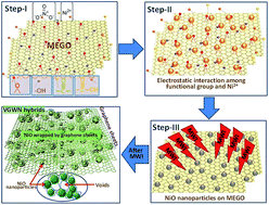
Here we demonstrate a simple strategy for the large-scale synthesis of void-induced and graphene-wrapped nickel oxide (VGWN) hybrids using domestic microwave irradiation. When a homogeneously mixed aqueous suspension of partially microwave exfoliated graphene oxide (MEGO) sheets and nickel nitrate nanoparticles is irradiates with a microwave, the in situ formation of voids with wrapping of NiO nanoparticles with graphene sheets is easily realized. The novel VGWN hybrid materials were used for investigation of electrochemical capacitive behaviours. Remarkably, such hybrid structure provides the right combination of electrode properties for high-performance supercapacitors and cyclic stability. The wrapping of graphene sheets on the NiO nanoparticles can offer highly conductive pathways by bridging individual NiO nanoparticles together, thus facilitating the charge/discharge rate and cycling performance of supercapacitors. The hybrid materials displayed enhanced capacitive performance (549 F g−1 at 10 mV s−1). Additionally, over 88% of the initial capacitance was retained after repeating the cyclic voltammetry test for 2500 cycles. The enhanced electrochemical performance can be ascribed to the synergic effects of the two components' voids and wrapping, suggesting VGWN hybrids as novel electrode materials promising potential applications as high-performance energy storage devices.


Introduction

For a new generation, systems as promising candidates for energy storage supercapacitors with high performance such as high power, longer stable cycle life, fast charge–discharge rate and low maintenance cost are required.1–4 Supercapacitors have two energy storage mechanisms, namely electric double layer capacitance (EDLC) and pseudocapacitance. Pseudo-capacitive materials, such as transition metal oxides, are being explored for use in supercapacitors with a large specific capacitance and high energy density.5 However, pseudocapacitors often suffer from the low charge–discharge rate capability and poor stability, since the metal oxide materials are usually insulating or semiconducting, which hinders the fast electron transport required for high charge/discharge rates. Recently, hybrids nanomaterials made by chemically modified with graphene and transition metal oxides have attracted wide attention in the field of supercapacitors due to their synergetic effect, arising from the combination of the redox reaction of metal oxides with the high surface area/conductivity of graphene, to improve the electrochemical performance.6 The high theoretical surface area of graphene (2630 m2 g−1),7 high young modulus (∼1.0 TPa)8 as well as good thermal properties (∼5000 W m−1 K−1)9 and high electrical conductivity10 makes it an attractive material for applications in supercapacitors and batteries.11

It has been reported that the electrochemical performance of supercapacitor hybrids are highly dependent on the quality and conductivity performance of graphene.12,13 The strong π–π stacking and van der Waals force between the planar basal planes of graphene sheets result in self-agglomeration of graphene, creating particulate graphite platelets which lose the unique ultrahigh surface area of graphene sheets. This adversely affects the potential applications of graphene based macro scale structures in the fields of supercapacitors and batteries. It was found that the loading of metal oxide nanoparticles can prevent or decrease the possibility of agglomeration and restacking of graphene, and consequently provide a higher electrochemically active surface area, resulting in higher electrochemical performance. To prevent self-aggregation of graphene, currently a number of strategies have been developed for fabricating porous macro scale structures. These includes the addition of “spacers”, template-assisted growth,14 crumpling of graphene sheets,15 graphene wrapped on Co3O4,16 reduced graphene oxide wrapped FeS nanocomposite,17 graphene wrapped Si sphere,18 graphene wrapped carbon nanotubes19 and graphene wrapped Fe2O3.20,21 The wrapping of graphene on metal oxide or on spacers as voids, also enhance the properties of synthesized hybrids for supercapacitor applications such as specific capacitance, stability, long time durability etc. Therefore, a method to prepare the hybrid materials in which metal oxide particles are wrapped, encapsulated, intercalated sandwich matrix still remains a challenge. In these models metal oxide tightly bonds and are confined within graphene matrix which limits its mobility during the electrochemical measurements.

Herein we report the scalable microwave assisted synthesis of voids-induced graphene-wrapped NiO (VGWN) nanoparticles with improved performance as electrode materials for supercapacitor applications. We believe that an effective strategy for the production of uniformly dispersed and wrapped NiO nanoparticle is to reduce graphene oxide by employing microwave irradiation. Moreover, we also demonstrate that the VGWN hybrids are excellent materials due to presence of wrapped metal oxide and voids which makes it naturally favourable for the use as supercapacitor electrodes. Also, the unique structure of graphene and NiO with a large specific surface area and voids present allows the rapid access of electrolyte ions to the NiO surface. As an electrode material for electrochemical supercapacitor, it can deliver 515 F g−1 specific capacitance. Our synthesis method presents a promising general route for the large-scale production of wrapped graphene–metal oxide nanoparticles with voids as energy storage materials.

Experimental section

Synthesis of partially microwave exfoliated graphene oxide (MEGO)

In the present investigation, graphite oxide was synthesized by chemical oxidation of graphite powder, the modified Staudenmaier's method.22 The graphite oxide powder was partially exfoliated employing microwave irradiation for short time at relatively low temperature.23 After irradiation, its structure becomes porous with some functional groups attached on the surfaces. The microwave exfoliated graphene oxide (MEGO) were characterized and used in the next step for wrapping onto the surface of metal oxide nanoparticles.

Synthesis of VGWN hybrids

For synthesis of voids-assisted graphene wrapped NiO hybrids, a series of systematic experiments was carried out for optimization. In a typical synthesis experiment, partially MEGO (15 mg) was dispersed in de-ionized (DI) water (50 mL) using ultrasonicator for 10 min. After that, 0.20 mg nickel nitrate (Ni(NO3)2) was dissolved in 20 mL DI water and added to the above MEGO dispersed solution. The final solution was subject to further ultrasonication for 10 min, giving the nickel ions present in the solution full access to the MEGO surface. Then, 3 mL of diluted ammonia (NH3·H2O) was added to the above mixture solution under stirring (30 min). Subsequently, 0.15 mL of hydrogen peroxide (H2O2) was added slowly into the solution and stirred for 30 min to obtain a homogeneous solution. The black precipitated products were filtered out and washed with DI water several times and then dried under a vacuum atmosphere at room temperature for overnight for complete evaporation of moisture. Finally, this material was exposed to microwave irradiation for 3 minutes for the formation of VGWN hybrids. The as-synthesized final porous materials were characterized and used for the supercapacitor electrode application.

Materials characterization

The morphology and microstructures of the samples was examined by a FEI Nova Nano SEM 450 field emission scanning electron microscopy (FE-SEM) and Transmission Electron Microscope (TEM FEI Tecnai F20 microscope). The structural characterization for crystalline nature of the synthesized materials was analyzed using an X-ray diffraction (XRD, D/MAX-2500/PC; Rigaku Co., Tokyo, Japan). For surface area and pore size distribution, nitrogen adsorption–desorption isotherms were obtained at 77 K using a Micromeritics Tristar 3000 gas adsorption analyzer. The Raman spectrum was recorded on a LabRAM HR UV/vis/NIR (Horiba Jobin Yvon, France) using a CW Ar-ion laser (514.5 nm) as an excitation source focused through a confocal microscope (BXFM, Olympus, Japan) equipped with an objective lens (50 μm, numerical aperture 0.50) at room temperature. The X-ray photoelectron spectroscopy (XPS) spectrum was recorded on a MultiLab 2000 photoelectron spectrometer (Thermo-VG Scientific, USA) with Al Kα (1486.6 eV) as the X-ray source. All XPS spectra were corrected using the C 1s line at 284.6 eV. The changes in the surface chemical bonding and surface composition were characterized by Fourier transform infrared spectroscopy (FTIR, Jasco FT-IR 4100) and the test samples were pressed into tablets with KBr.

Preparation of electrode

The glassy carbon electrodes (GCEs) (5 mm diameter) were first polished with alumina slurry and then sonicated in absolute ethanol and distilled water for about 5 min, respectively. Then, 5.0 mg of the VGWN hybrid material was dispersed in 1.0 mL of dimethylformamide (DMF) with the aid of ultrasonic agitation to give a 5.0 mg mL−1 black suspension. Then, 10 μL of the black suspension was dropped on a cleaned GCE electrode with the help of a micro-syringe. After complete dehydration, 5 μL of Nafion (5 wt%) was casted and used as a net to hold the samples on the electrode surface stably. Thus, a uniform film of VGWN hybrids coats on the surface of GCE. The solvent was allowed to evaporate before the electrochemical test. All electrochemical measurements were done in a three electrode system. The electrochemical measurements, such as cyclic voltammetry (CV) and long-term cycling stability were carried out by employing an electrochemical workstation (SI 1287, Solartron Analytical). The as prepared VGWN hybrids electrode behave as working electrode, Pt (platinum) wire as a counter electrode, and Ag/AgCl electrode as a reference electrode. The electrochemical properties and specific capacitances of the VGWN hybrids electrode materials were investigated in 5 M KOH electrolyte.

Results and discussion

The microstructural morphology of the MEGO and VGWN hybrids nanostructures were characterized by SEM and TEM. Fig. 1(a) shows the SEM image which indicates that the prepared MEGO has crumpled, curly and wavy morphology. The observed wrinkles and folds over the entire surface of MEGO are characteristic nature of graphene sheets.24 The edges of the MEGO can be clearly seen in Fig. 1(a), which shows the open porous structure, with several voids on the edges. During the formation of VGWN hybrids, these open voids play crucial role for the wrapping of NiO nanoparticles. The SEM analysis in Fig. 1(b) demonstrates the morphology of the as synthesized VGWN hybrids, depicting the well-distributed and dispersed NiO nanoparticle inside the voids and no detectable particle agglomeration is seen as shown in Fig. 1(b). The average size of the NiO nanoparticles inside the graphene sheets is approximately 30–60 nm. The NiO nanoparticles in VGWN hybrids could be easily bridged by graphene sheets owing to their relatively larger lateral sizes, yielding a wrapped interconnected VGWN hybrid network. The strong electrostatic interaction between oppositely charged NiO and graphene oxide together with the flexibility of graphene oxide can be responsible for this. It is expected that the NiO nanoparticles are homogeneously distributed and tightly attached in voids and are covered by graphene layers. These conductive graphene surfaces provides conducting channels for electrons and generates synergism effects in VGWN hybrids.25 Inset of Fig. 1(c) shows the voids available inside the graphene sheets near the NiO nanoparticles. Meanwhile, NiO nanoparticles wrapped by graphene sheets and the voids inside graphene surfaces are clearly seen through the transparent graphene sheets. The graphene wrapping is more visible in Fig. 1(d). This also shows the wrapped graphene sheets structure on the NiO nanoparticles. Additionally, the voids with wrapped graphene network could serve as promising electrodes and/or supporting matrixes for advanced electrodes in energy storage and conversion systems owing to the enhanced host capability, structural stability, and electrical conductivity. It is seen from Fig. 1(c) and (d) that the VGWN hybrids consists of thin, crumpled closely connected transparent network structure and NiO nanoparticles are nearly spherical in shape. It is clear that the voids are seen inside graphene which exhibits a porous structure as seen in Fig. 1(d) (magnified view can be seen in ESI Fig. S1). These voids containing spaces inside graphene are expected to increase the active surface area and therefore may increase the specific capacitance as well as cycle stability of the VGWN hybrids. The unique VGWN hybrids, combined with nanostructured wall and/or pores seamlessly, can serve as freeway networks for quick electron transfer and easy electrolyte ion diffusion in the electrodes. We have also tried to observe the morphological changes in VGWN hybrids for longer microwave irradiation. For long time microwave irradiation (4 minutes), shows that the VGWN hybrids nanostructure starts to damage their internal morphologies and structural defect appears on the surfaces (ESI Fig. S2). Also, NiO nanoparticles started to agglomerate due to high temperature for long time during the microwave irradiation. The wrapped graphene sheets are broken in nearby locations on the surfaces and voids are open on the top surfaces. In another word we can say that the microwave irradiation for 3 minutes is well suited for the good networked VGWN hybrids formation.
image file: c6ra00426a-f1.tif
Fig. 1 SEM micrographs of MEGO and VGWN hybrids. (a) MEGO (b) low magnification and (c and d) high-magnification images of VGWN hybrids.

Further the nitrogen (N2) adsorption–desorption isotherms was used to measure the surface areas and the corresponding pore size distribution for porous structure of VGWN hybrids. The surface area of VGWN hybrids was evaluated by the Brunauer–Emmett–Teller (BET) model, while the pore size distribution was estimated with Barrett–Joyner–Halenda (BJH) theory. It can be seen in Fig. 2 that measured N2 adsorption–desorption curve display type IV adsorption isotherms with clear hysteresis loop indicating that the VGWN hybrids contain mesopores (according to IUPAC classification) in their structures. As a result, larger BET areas and higher pore volumes are obtained. The BET surface area of as synthesized VGWN hybrids in this work is 398 m2 g−1, which is high due to the insertion of voids and wrapped morphology structure. However, this value is much smaller than the theoretical value (2630 m2 g−1 for a single graphene sheet) due to the overlapping of graphene sheets and multi layers structure.7 It can be seen that most pores are distributed in a wide range from 5 to 15 nm (inset of Fig. 2), suggesting that as synthesized VGWN hybrids exhibit porous structures mainly composed of mesopores due to voids.26


image file: c6ra00426a-f2.tif
Fig. 2 Nitrogen adsorption–desorption isotherms of VGWN hybrids (inset: the BJH pore size distribution plot).

The wrapping of graphene onto NiO nanoparticles was also confirmed by TEM images in Fig. 3. Both the NiO nanoparticles and wrinkled structured graphene sheets can be clearly seen. However, the NiO nanoparticles were not agglomerated and are spherical within the graphene sheets. Surface of graphene-wrapped NiO spheres appears crinkled and rough, which can provide more space for the electrolyte to transport more freely.25 Inset of Fig. 3(c) shows the lattice spacing of 0.21 nm which corresponds to the d spacing between adjacent (200) crystallographic planes of graphene wrapped NiO crystal.27


image file: c6ra00426a-f3.tif
Fig. 3 TEM images of MEGO and VGWN hybrids. (a) MEGO, (b) low magnification and (c and d) high-magnification images of VGWN hybrids.

Fig. 4(a) shows the EDX analysis of MEGO, and VGWN hybrids. The EDX data of MEGO shows the presence of elemental content as C and O, indicating that MEGO has a lower O content which is due to the functional group attached on the graphene basal plane and edges. Based on the obtained results, the atomic weight percentages of C and O were 83.7% and 16.3%, respectively. These functional groups play a major role for the binding of Ni nanoparticles. The EDS analysis of the VGWN hybrid shows the presence of C, Ni, and O elements with 76.7, 11.1 and 12.2 wt%, respectively. The existence of O and Ni with an approximate ratio of 1[thin space (1/6-em)]:[thin space (1/6-em)]1 implies its stoichiometry as NiO.


image file: c6ra00426a-f4.tif
Fig. 4 (a) Energy dispersive X-ray spectroscopy (b) X-ray diffraction pattern (c) Raman spectra and (d) FT-IR spectra of MEGO and VGWN hybrids.

Fig. 4(b) shows the diffraction pattern of the as-synthesized VGWN hybrids, which completely matches to the crystalline phase of nickel oxide. Nickel oxide shows four major diffraction peaks at 2θ = 37.3°, 43.3°, 62.8° and 75.1° which can be assignable to (111), (200), (220) and (311) crystal planes of face-centred cubic NiO. All the peaks are in good agreement with the standard profile of NiO (JCPDS card no. 78-0643).28 The one broad characteristic peak (002) at 2θ = 25.1° can be seen, which represent the presence of graphene. The corresponding interlayer spacing is more as compared to MEGO because the inset of Fig. 3(b) shows that the (002) peak in pristine MEGO appears at 2θ = 26.4°. The small angle shift (∼1.3°) due to the formation of VGWN hybrids, suggest that the interlayer spacing of graphene layers are increased due to stress and voids creations during the wrapping of NiO nanoparticles by the graphene sheets.

Raman spectrum is strongly sensitive to the electronic structure and has been widely used to characterize carbon based materials.29 The Raman spectra of MEGO and VGWN hybrids are shown in Fig. 4(c). As shown in Fig. 4(c), two characteristic bands were observed in Raman spectra i.e., the G band arising from the in-plane bond stretching of all sp2 C bonded pairs and the D band associating with the defect sites or lattice distortion.30 Two prominent peaks in MEGO (1391 cm−1 and 1604 cm−1) and VGWN (1374 cm−1 and 1588 cm−1) which corresponds to the D and G bands are observed. It is generally accepted that the intensity ratio of D to G band (ID/IG) reflects the graphitization degree of carbonaceous materials and the defect density. The intensity ratios (ID/IG) for MEGO and VGWN are 0.84 and 0.91, respectively. An increased in ID/IG intensity ratio indicates that a decrease in the size of the in-plane sp2 domains and a spatially ordered crystal structure of the MEGO,31,32 indicating the introduction of defect due to NiO nanoparticles during the wrapping of graphene sheets and creations of voids inside the graphene layers. This disordered structure may be beneficial to enhance specific capacitance and facilitate ion diffusion through graphene sheets in electrochemical measurements. There are also two very small peaks located at 370 cm−1 and 530 cm−1 which corresponding to the shaking peaks of NiO.33,34

The FTIR spectra of the products are shown in Fig. 4(d). The broad absorption peak centred at 3426 cm−1 and 3418 cm−1 in MEGO and VGWN hybrids is associated with the asymmetric and symmetric stretching vibrations of the –OH group of moisture and the surface hydroxyls.35 The intensity of –OH band in VGWN hybrids located at 3418 cm−1 is reduced, which is likely due to detachment of –OH and removal of moisture from the graphene surfaces. Furthermore, a new absorption peak located at 1560 cm−1 corresponding to the aromatic skeletal of C[double bond, length as m-dash]C stretching vibration is observed, which further instruct the reduction of MEGO.36 Another peak at 1076 cm−1 corresponds to the vibration adsorption peak of C–O–C.37 The strongest broad peaks in the range of 400–1000 cm−1 are derived from the stretching vibration of NiO.38

The surface chemical states, composition change and interaction of VGWN hybrids were investigated by X-ray photoelectron spectroscopy (XPS). XPS spectrum of VGWN hybrids as (a) XPS survey region (b) high-resolution spectra of Ni 2p, (c) C 1s, and (d) O 1s regions are shown in Fig. 5. In the XPS survey spectrum of VGWN (Fig. 5(a)), it shows that the hybrids mainly consists of carbon C 1s (284.5 eV), oxygen O 1s (531.8 eV) and nickel Ni 2p (855.6 and 873.2 eV). No other elemental signals are detected in the general XPS spectrum. Fig. 5(b), shows the high resolution of Ni 2p spectrum, which is associated with two main peaks Ni 2p3/2 and Ni 2p1/2, in which the Ni 2p3/2 centred at 855.6 eV and Ni 2p1/2 at 873.2 eV, respectively. The energy separation between Ni 2p3/2 and Ni 2p1/2 peaks is observed to be 17.6 eV and is in good agreement with the formation of NiO nanoparticles.39,40 This formation of NiO is also consistent with Raman and XRD results. The intense peak at 284.5 eV can be attributed to sp2 C[double bond, length as m-dash]C network in graphene, while the other peaks are associated with groups arising from oxygen containing groups bonded with carbon.41 The others minor deconvoluted peaks at 286.1, 286.9, 288.8 eV corresponds to single bonded components of hydroxyl in C–O (286.1 eV), carbonyl carbon in C[double bond, length as m-dash]O (286.9 eV) and the carboxylate carbon in O–C[double bond, length as m-dash]O (288.8 eV), respectively.42,43 The intensity of the peak related to the C[double bond, length as m-dash]C group is much greater than those associated with oxygen containing groups, which indicates that most of the oxygen-containing groups are removed during the attachment of metal oxides on the graphene surface. For O 1s, (Fig. 5(d)), the spectra deconvoluted into three peaks. The peak at 529.8 eV is due to the oxygen bonded with nickel in NiO, while 531.8 eV can be ascribed to the oxygen that binds to Ni 2p, and the other at higher binding energy of 532.7 can be assigned to the oxygen of functional groups (COO–).44,45


image file: c6ra00426a-f5.tif
Fig. 5 XPS spectra of VGWN hybrids. (a) Complete survey (b) Ni 2p spectra (c) C 1s spectra and (d) O 1s spectra.

Electrochemical measurements

To investigate the electrochemical potential application, the samples were fabricated in the form of electrodes and the relevant electrochemical behaviour was characterized by electrochemical workstation in three electrodes system. The CV curves of VGWN hybrids electrodes was measured at different scan rates in the potential window ranging from 0 to 0.8 V (vs. Ag/AgCl). Fig. 6(a) presents the CV curves of the VGWN hybrids electrode at scan rate of 10, 25, 50, 75 and 100 mV s−1, respectively. The voltammetry cycles characteristics show good capacitive response even at a high scan rate. The rectangular-like shape and the appearance of small humps in the CV curves indicate that the capacitive response mainly comes from the electric double-layer capacitance (EDLC) behaviour. Reduction peaks move toward lower potentials when the scan rate is increased which may be due to the electric polarization and irreversible reactions at a higher scan rate.46 This void contains NiO nanoparticles completely wrapped by conducting graphene sheets, effectively facilitate the transportation of electrolyte ions on its surfaces during the cycling process and, thus, improves the supercapacitance. Therefore, both the presence of voids in graphene and wrapping of NiO by graphene contribute to the observed high specific capacitance for VGWN hybrids. Fig. 6(b) show the specific capacitances of the VGWN hybrid electrodes as 549, 469, 421, 407 and 408 F g−1 at scan rates of 10, 25, 50, 75 and 100 mV s−1, respectively. The electrochemical measurements for partially MEGO prepared under similar conditions were carried out for the comparison purpose. The MEGO have lower specific capacitance as 362, 298, 264, 249 and 248 at scan rates of 10, 25, 50, 75 and 100 mV s−1, respectively. The improvement in specific capacitance of VGWN hybrids as comparison to MEGO may be due to the wrapping of NiO nanoparticles and voids formation inside graphene layers during wrapping. These two reasons are mainly responsible for the enhanced specific capacitance of VGWN hybrids.
image file: c6ra00426a-f6.tif
Fig. 6 Electrochemical performance of VGWN hybrids. (a) Cyclic voltammetry test (b) specific capacitance and (c) long-term cycling stability of the electrode at a constant current density of 8 A g−1 over 2500 cycles; the inset shows charge/discharge curve of the VGWN hybrids in the potential range from 0 to 0.8 V at 8 A g−1.

Fig. 6(c) shows the long cycling life and multiple cycle performance of VGWN hybrids, which are important requirements for a supercapacitor electrode. The cycle stability of the electrode was carried out by repeating the charge/discharge cycle measurement for 2500 cycles at constant current density of 8 A g−1. The specific capacitance of the VGWN hybrid electrode decreases up to 12% of initial value for the first 115 cycles, which was due to an activation process and the degradation of microstructure of electrode material in the supercapacitor electrode. However, the value remained stable as 88%, exhibiting the better long-term electrochemical stability of as synthesized hybrids. The achieved specific capacitance, long term stability and retention properties of as synthesized VGWN hybrids shows better performance than the works reported by others on NiO–graphene hybrids based supercapacitors.47–60 (ESI Table S1. Comparison of electrochemical performance of NiO–graphene hybrids based supercapacitors.)

The capacitance was maintained without obvious aging or performance degradation, which demonstrate the excellent electrochemical performance of the VGWN hybrid electrode material for application in practical energy storage devices. The reason is that the NiO nanoparticles wrapped and inserted into the VGWN hybrids prevent not only the loss of surface contact, but also the insertion of NiO nanoparticles attributed to their volume expansions during cycling at higher current density. The void volume inside graphene can allow easy penetration of the electrolyte, further shortening the ion transfer pathway to their surfaces. The repeated cycling would help electrolyte ions move into the spaces among NiO nanoparticles and graphene and utilize the larger space of hybrids. During charge/discharge process, the confined NiO nanoparticles in voids are stables, and these particles can not move inside the graphene layers. Therefore, the hybrids can greatly reduce the diffusion length, resulting in an improvement of electrochemical properties of the hybrid materials. These results further confirms that the wrapped and voids like structure for metal oxide are important for enhancing the specific capacitance of VGWN hybrids. Also, the interfacial contact and increased contact area between NiO and MEGO can significantly improve the accessibility of VGWN hybrids to the electrolyte ions and shorten the ion diffusion and migration pathways. It is concluded that the synergistic effect between wrapped conducting graphene sheets with voids and NiO nanoparticles is responsible for the excellent electrochemical performance.

Mechanism for the formation of VGWN hybrids

The procedure for the preparation of the VGWN hybrids is illustrated in Fig. 7. The MEGO suspension was first dispersed in nickel nitrate solution with NH3H2O. Under ultrasonication the Ni2+ ions were adsorbed on the MEGO surface due to the functional groups on the MEGO surface. These functional groups attached on the graphene surfaces are responsible for the bonding of the NiO nanoparticle with graphene surface. Step-I shows that MEGO treated with NH3·H2O provides alkaline media for the creation of amine group on the graphene surfaces. Also the MEGO has a negatively charged surface due to the presence of carboxylic, hydroxyl and epoxy groups which bind nickel oxide through electrostatic interactions.61–64 The decomposition of nickel nitrate salt provides, positively charged Ni2+ ions, which gets attracted towards these functional group sites due to the electrostatic force between Ni2+ ions and polar negatively charged functional groups on the surface.64–66 The Ni2+ ions react with functional groups containing MEGO sheets, leading to the local creation of bridge, as Ni–O–C bonding.67 This leads to the formation of Ni(OH)2 with hydroxyl group or NiO with epoxy group on the surfaces of MEGO and acts as a nucleation centre for the further growth of NiO nanoparticles.68 In Step-II, previously MEGO modified with Ni(OH)2 was stirring with H2O2, which would lead to the oxidation of Ni2+ into NiO nanoparticles. The other possible reaction is that Ni2+ ions are bonded on the surface of MEGO and they also form Ni–O bonding with help of functional group (epoxy group). These functionalities provide oxygen bridges between MEGO and NiO nanoparticles and also contribute to the good conductivity of the VGWN hybrid, which directly correlates to excellent electrochemical performance.67 Including Step I and II, the reaction pathway can be written as69
 
Ni2+ + 2OH → Ni(OH)2 (1)
 
2Ni(OH)2 + H2O2 → 2NiO + 2H2O (2)

image file: c6ra00426a-f7.tif
Fig. 7 Schematic illustration of four step process for the formation of VGWN hybrids.

In Step III, MEGO with NiO nanoparticles are irradiated with microwave and during this process, defects and disorders were induced on the surface of MEGO containing NiO nanoparticles.70 The MEGO structure gets changed by the incorporation of pentagon–heptagon defects and strained domains induced structural defect, giving rise to accommodation of edge dislocations.71 The local positive and negative defects on graphene sheets show the curvature in form of voids. These defects can be created through the thermal energy transformation from microwave irradiation to graphene sheets within short time.72 These defects sites appear on the graphene surface creates active sites for wrapping. The remaining functional groups on the surface of MEGO serve as nucleation and anchoring sites for NiO nanoparticles during the microwave irradiation. Thus, NiO nanoparticles were covered by outer graphene layer of MEGO and are dispersed inside the graphene sheets in the MEGO to fill the inner voids as shown in Fig. 7.73,74 The partially reduced MEGO shows this crucial phenomena for the unique voids induced graphene wrapped NiO nanostructure. Consequently, due to voids formation during the wrapping of NiO into MEGO, the electrochemical performance and structural integrity of the VGWN remains superior.75–77

Conclusions

In summary, a simple approach is used to fabricate the VGWN hybrids via the microwave exfoliation method. The unique structure of graphene and NiO exhibits enhanced electrode performance for supercapacitors with high specific capacitance and cycling performance. The introduction of graphene into the hybrids can offer highly conductive pathways by bridging adjacent NiO together, facilitating the kinetics for both charge transfer and ion transport throughout the electrodes. Therefore, the hybrids can greatly reduce the diffusion length, resulting in an improvement of electrochemical properties of the hybrid materials. The hollow voids interior could provide larger specific surface area, better electrolyte penetration and less inner stress. Additionally, the graphene surfaces afford good contact with NiO towards all the sides. Furthermore, the VGWN hybrids as a supercapacitor electrode offered high specific capacitance of 549 F g−1 at scan rate of 10 mV s−1. Furthermore, the VGWN hybrids as electrode materials have a good cycling stability, and the capacitance was maintained at 88% after 115 cycles at a current density of 8 A g−1. The unique nanostructure of NiO wrapped up with highly flexible and conductive graphene nanosheets represents an effective strategy that may be applied for various metal oxide electrodes to mitigate the mechanical degradation and capacity fading, critical for developing advanced electrochemical energy storage systems with long cycle life and high rate performance.

Acknowledgements

RK, RS and SAM would like to acknowledge CNPq and FAPESP (Brazil) for financial support. PKD acknowledge the financial support from UGC, New Delhi for providing DSK postdoctoral fellowship.

Notes and references

  1. R. Celiešiūtė, R. Trusovas, G. Niaura, V. Švedas, G. Račiukaitis, Ž. Ruželė and R. Pauliukaite, Electrochim. Acta, 2014, 132, 265–276 CrossRef.
  2. P. G. Bruce, S. A. Freunberger, L. J. Hardwick and J.-M. Tarascon, Nat. Mater., 2012, 11, 19–29 CrossRef CAS PubMed.
  3. D. Bresser, S. Passerini and B. Scrosati, Chem. Commun., 2013, 49, 10545–10562 RSC.
  4. L. L. Zhang and X. S. Zhao, Chem. Soc. Rev., 2009, 38, 2520–2531 RSC.
  5. Y. Wang and I. Zhitomirsky, Langmuir, 2009, 25, 9684–9689 CrossRef CAS PubMed.
  6. K. Gil-Pyo, N. Inho, P. Soomin, P. Junsu and Y. Jongheop, Nanotechnology, 2013, 24, 475402 CrossRef PubMed.
  7. M. D. Stoller, S. Park, Y. Zhu, J. An and R. S. Ruoff, Nano Lett., 2008, 8, 3498–3502 CrossRef CAS PubMed.
  8. C. Lee, X. Wei, J. W. Kysar and J. Hone, Science, 2008, 321, 385–388 CrossRef CAS PubMed.
  9. A. A. Balandin, S. Ghosh, W. Bao, I. Calizo, D. Teweldebrhan, F. Miao and C. N. Lau, Nano Lett., 2008, 8, 902–907 CrossRef CAS PubMed.
  10. Y. Zhu, S. Murali, W. Cai, X. Li, J. W. Suk, J. R. Potts and R. S. Ruoff, Adv. Mater., 2010, 22, 3906–3924 CrossRef CAS PubMed.
  11. C. Liu, Z. Yu, D. Neff, A. Zhamu and B. Z. Jang, Nano Lett., 2010, 10, 4863–4868 CrossRef CAS PubMed.
  12. H. Wang, H. S. Casalongue, Y. Liang and H. Dai, J. Am. Chem. Soc., 2010, 132, 7472–7477 CrossRef CAS PubMed.
  13. D. Bitao, Z. Han, L. Jin, Z. Lusi, G. Guoxin and D. Shujiang, Nanotechnology, 2014, 25, 435403 CrossRef PubMed.
  14. X. Cao, Y. Shi, W. Shi, G. Lu, X. Huang, Q. Yan, Q. Zhang and H. Zhang, Small, 2011, 7, 3163–3168 CrossRef CAS PubMed.
  15. J. Luo, H. D. Jang, T. Sun, L. Xiao, Z. He, A. P. Katsoulidis, M. G. Kanatzidis, J. M. Gibson and J. Huang, ACS Nano, 2011, 5, 8943–8949 CrossRef CAS PubMed.
  16. H. Sun, X. Sun, T. Hu, M. Yu, F. Lu and J. Lian, J. Phys. Chem. C, 2014, 118, 2263–2272 CAS.
  17. L. Fei, Q. Lin, B. Yuan, G. Chen, P. Xie, Y. Li, Y. Xu, S. Deng, S. Smirnov and H. Luo, ACS Appl. Mater. Interfaces, 2013, 5, 5330–5335 CAS.
  18. P. Wu, H. Wang, Y. Tang, Y. Zhou and T. Lu, ACS Appl. Mater. Interfaces, 2014, 6, 3546–3552 CAS.
  19. P. Nayak, P. N. Santhosh and S. Ramaprabhu, J. Phys. Chem. C, 2014, 118, 5172–5179 CAS.
  20. G. Zhou, D.-W. Wang, F. Li, L. Zhang, N. Li, Z.-S. Wu, L. Wen, G. Q. Lu and H.-M. Cheng, Chem. Mater., 2010, 22, 5306–5313 CrossRef CAS.
  21. J. Zhao, B. Yang, Z. Zheng, J. Yang, Z. Yang, P. Zhang, W. Ren and X. Yan, ACS Appl. Mater. Interfaces, 2014, 6, 9890–9896 CAS.
  22. L. Staudenmaier, Ber. Dtsch. Chem. Ges., 1898, 31, 1481–1487 CrossRef CAS.
  23. I. Janowska, K. Chizari, O. Ersen, S. Zafeiratos, D. Soubane, V. Costa, V. Speisser, C. Boeglin, M. Houllé, D. Bégin, D. Plee, M.-J. Ledoux and C. Pham-Huu, Nano Res., 2010, 3, 126–137 CrossRef CAS.
  24. W. Gao, N. Singh, L. Song, Z. Liu, A. L. M. Reddy, L. Ci, R. Vajtai, Q. Zhang, B. Wei and P. M. Ajayan, Nat. Nanotechnol., 2011, 6, 496–500 CrossRef CAS PubMed.
  25. Y.-L. Zhang, L. Guo, H. Xia, Q.-D. Chen, J. Feng and H.-B. Sun, Adv. Opt. Mater., 2014, 2, 10–28 CrossRef.
  26. X. Ma, L. Gan, M. Liu, P. K. Tripathi, Y. Zhao, Z. Xu, D. Zhu and L. Chen, J. Mater. Chem. A, 2014, 2, 8407–8415 CAS.
  27. L. Liu, Y. Li, S. Yuan, M. Ge, M. Ren, C. Sun and Z. Zhou, J. Phys. Chem. C, 2010, 114, 251–255 CAS.
  28. K. K. Purushothaman, I. Manohara Babu, B. Sethuraman and G. Muralidharan, ACS Appl. Mater. Interfaces, 2013, 5, 10767–10773 CAS.
  29. W. Yunwei, T. Xili, G. Xiaoning, W. Yingyong, J. Guoqiang and G. Xiangyun, Nanotechnology, 2013, 24, 475602 CrossRef PubMed.
  30. C. Hang and F. Tobin, Nanotechnology, 2015, 26, 135702 CrossRef PubMed.
  31. Y. Wang, S. Gai, N. Niu, F. He and P. Yang, J. Mater. Chem. A, 2013, 1, 9083–9091 CAS.
  32. Y. Zhang, F. Xu, Y. Sun, Y. Shi, Z. Wen and Z. Li, J. Mater. Chem., 2011, 21, 16949–16954 RSC.
  33. Y. Jiang, D. Chen, J. Song, Z. Jiao, Q. Ma, H. Zhang, L. Cheng, B. Zhao and Y. Chu, Electrochim. Acta, 2013, 91, 173–178 CrossRef CAS.
  34. X. Rong, F. Qiu, C. Zhang, L. Fu, Y. Wang and D. Yang, Powder Technol., 2015, 275, 322–328 CrossRef CAS.
  35. R. Peng-Gang, Y. Ding-Xiang, J. Xu, C. Tao and L. Zhong-Ming, Nanotechnology, 2011, 22, 055705 CrossRef PubMed.
  36. Z.-S. Wu, W. Ren, L. Gao, J. Zhao, Z. Chen, B. Liu, D. Tang, B. Yu, C. Jiang and H.-M. Cheng, ACS Nano, 2009, 3, 411–417 CrossRef CAS PubMed.
  37. J. Ye, H. Zhang, Y. Chen, Z. Cheng, L. Hu and Q. Ran, J. Power Sources, 2012, 212, 105–110 CrossRef CAS.
  38. X. H. Xia, J. P. Tu, J. Zhang, X. L. Wang, W. K. Zhang and H. Huang, Sol. Energy Mater. Sol. Cells, 2008, 92, 628–633 CrossRef CAS.
  39. Y. Hattori, T. Konishi and K. Kaneko, Chem. Phys. Lett., 2002, 355, 37–42 CrossRef CAS.
  40. G.-f. Cai, J.-p. Tu, J. Zhang, Y.-j. Mai, Y. Lu, C.-d. Gu and X.-l. Wang, Nanoscale, 2012, 4, 5724–5730 RSC.
  41. C. Caiying, C. Chaoqiu, H. Peipei, D. Feifei, Z. Shichao, L. Ping, F. Jinchuan, S. Weiguo and Q. Yong, Nanotechnology, 2014, 25, 504001 CrossRef PubMed.
  42. V. Datsyuk, M. Kalyva, K. Papagelis, J. Parthenios, D. Tasis, A. Siokou, I. Kallitsis and C. Galiotis, Carbon, 2008, 46, 833–840 CrossRef CAS.
  43. S. Park, J. An, I. Jung, R. D. Piner, S. J. An, X. Li, A. Velamakanni and R. S. Ruoff, Nano Lett., 2009, 9, 1593–1597 CrossRef CAS PubMed.
  44. C. H. Kim and B.-H. Kim, J. Power Sources, 2015, 274, 512–520 CrossRef CAS.
  45. E. T. Kinzer and C. M. Chambers, Surf. Sci., 1965, 3, 261–279 CrossRef CAS.
  46. D. Han, P. Xu, X. Jing, J. Wang, P. Yang, Q. Shen, J. Liu, D. Song, Z. Gao and M. Zhang, J. Power Sources, 2013, 235, 45–53 CrossRef CAS.
  47. L. Wang, H. Tian, D. Wang, X. Qin and G. Shao, Electrochim. Acta, 2015, 151, 407–414 CrossRef CAS.
  48. B. Zhao, H. Zhuang, T. Fang, Z. Jiao, R. Liu, X. Ling, B. Lu and Y. Jiang, J. Alloys Compd., 2014, 597, 291–298 CrossRef CAS.
  49. L.-B. Zhang, S.-R. Yang, J.-Q. Wang, Y. Xu and X.-Z. Kong, Chin. Chem. Lett., 2015, 26, 522–528 CrossRef CAS.
  50. N. B. Trung, T. V. Tam, D. K. Dang, K. F. Babu, E. J. Kim, J. Kim and W. M. Choi, Chem. Eng. J., 2015, 264, 603–609 CrossRef CAS.
  51. H. Yan, J. Bai, B. Wang, L. Yu, L. Zhao, J. Wang, Q. Liu, J. Liu and Z. Li, Electrochim. Acta, 2015, 154, 9–16 CrossRef CAS.
  52. Y. F. Liu, G. H. Yuan, Z. H. Jiang, Z. P. Yao and M. Yue, J. Alloys Compd., 2015, 618, 37–43 CrossRef CAS.
  53. L. Wang, X. Li, T. Guo, X. Yan and B. K. Tay, Int. J. Hydrogen Energy, 2014, 39, 7876–7884 CrossRef CAS.
  54. L. Niu, J. Wang, W. Hong, J. Sun, Z. Fan, X. Ye, H. Wang and S. Yang, Electrochim. Acta, 2014, 123, 560–568 CrossRef CAS.
  55. X. Su, H. Chai, D. Jia, S. Bao, W. Zhou and M. Zhou, New J. Chem., 2013, 37, 439–443 RSC.
  56. X. Zhu, H. Dai, J. Hu, L. Ding and L. Jiang, J. Power Sources, 2012, 203, 243–249 CrossRef CAS.
  57. P. Cao, L. Wang, Y. Xu, Y. Fu and X. Ma, Electrochim. Acta, 2015, 157, 359–368 CrossRef CAS.
  58. Y.-G. Zhu, G.-S. Cao, C.-Y. Sun, J. Xie, S.-Y. Liu, T.-J. Zhu, X. B. Zhao and H. Y. Yang, RSC Adv., 2013, 3, 19409–19415 RSC.
  59. X. Xia, J. Tu, Y. Mai, R. Chen, X. Wang, C. Gu and X. Zhao, Chem.–Eur. J., 2011, 17, 10898–10905 CrossRef CAS PubMed.
  60. C. Wu, S. Deng, H. Wang, Y. Sun, J. Liu and H. Yan, ACS Appl. Mater. Interfaces, 2014, 6, 1106–1112 CAS.
  61. D. Li, M. B. Muller, S. Gilje, R. B. Kaner and G. G. Wallace, Nat. Nanotechnol., 2008, 3, 101–105 CrossRef CAS PubMed.
  62. T. Szabó, O. Berkesi, P. Forgó, K. Josepovits, Y. Sanakis, D. Petridis and I. Dékány, Chem. Mater., 2006, 18, 2740–2749 CrossRef.
  63. A. Lerf, H. He, M. Forster and J. Klinowski, J. Phys. Chem. B, 1998, 102, 4477–4482 CrossRef CAS.
  64. B. Fatima, F. Jabeen, Z. Padashbarmchi and M. Najam-ul-Haq, RSC Adv., 2015, 5, 23658–23665 RSC.
  65. B. Zhao, J. Song, P. Liu, W. Xu, T. Fang, Z. Jiao, H. Zhang and Y. Jiang, J. Mater. Chem., 2011, 21, 18792–18798 RSC.
  66. R. Kumar, R. K. Singh, P. K. Dubey, D. P. Singh, R. M. Yadav and R. S. Tiwari, Adv. Mater. Interfaces, 2015, 2, 1500191 Search PubMed.
  67. G. Zhou, D.-W. Wang, L.-C. Yin, N. Li, F. Li and H.-M. Cheng, ACS Nano, 2012, 6, 3214–3223 CrossRef CAS PubMed.
  68. V. Senthilkumar, F. B. Kadumudi, N. T. Ho, J.-W. Kim, S. Park, J.-S. Bae, W. M. Choi, S. Cho and Y. S. Kim, J. Power Sources, 2016, 303, 363–371 CrossRef CAS.
  69. S.-J. Li, N. Xia, X.-L. Lv, M.-M. Zhao, B.-Q. Yuan and H. Pang, Sens. Actuators, B, 2014, 190, 809–817 CrossRef CAS.
  70. L. Kashinath, K. Namratha and K. Byrappa, Appl. Surf. Sci., 2015, 357, 1849–1856 CrossRef CAS.
  71. L. Nilsson, M. Andersen, J. Bjerre, R. Balog, B. Hammer, L. Hornekær and I. Stensgaard, Surf. Sci., 2012, 606, 464–469 CrossRef CAS.
  72. R. Kumar, P. K. Dubey, R. K. Singh, A. R. Vaz and S. A. Moshkalev, RSC Adv., 2016, 6, 17669–17677 RSC.
  73. B.-M. Goh, Y. Wang, M. V. Reddy, Y. L. Ding, L. Lu, C. Bunker and K. P. Loh, ACS Appl. Mater. Interfaces, 2014, 6, 9835–9841 CAS.
  74. L. Yue, W. Zhang, J. Yang and L. Zhang, Electrochim. Acta, 2014, 125, 206–217 CrossRef CAS.
  75. Y. Yang, C. Han, B. Jiang, J. Iocozzia, C. He, D. Shi, T. Jiang and Z. Lin, Mater. Sci. Eng., R, 2016, 102, 1–72 CrossRef.
  76. X. Li, Y. Hu, J. Liu, A. Lushington, R. Li and X. Sun, Nanoscale, 2013, 5, 12607–12615 RSC.
  77. H. Kim, Z. Wen, K. Yu, O. Mao and J. Chen, J. Mater. Chem., 2012, 22, 15514–15518 RSC.

Footnote

Electronic supplementary information (ESI) available. See DOI: 10.1039/c6ra00426a

This journal is © The Royal Society of Chemistry 2016
Click here to see how this site uses Cookies. View our privacy policy here.