DOI:
10.1039/C6RA00071A
(Paper)
RSC Adv., 2016,
6, 24418-24429
Synthesis of chitosan/PEO/silica nanofiber coating for controlled release of cefepime from implants
Received
2nd January 2016
, Accepted 8th February 2016
First published on 16th February 2016
Abstract
Nanofibers, which have good properties such as high surface to volume ratio, high porosity, very small pores, and the ability to load drugs, can be considered for a variety of medical applications. Silica/chitosan/poly(ethylene oxide) (PEO)/cefepime nanofibers are suitable as an antibacterial coating for orthopedic implants. This bioactive coating reduced adhesion of bacteria to the surface of the implants and prevented the formation of biofilms. Electrospinning is known to be the best way to produce the nanofibers because of the low cost, simplicity of the process and production of polymeric nanofibers from biodegradable materials. The morphology of electrospun nanofibers was studied by the use of a scanning electron microscope (SEM). The average diameters of the prepared nanofibers was determined by Image J software. Nanofibers cross linked by glutaraldehyde are stable in different pH of 5.5, 7.4 and 8.4 of SBF buffer for 24 h at 37 °C. These nanofibers are effective on E. coli, S. aureus and S. epidermidis bacteria. Cefepime release from the nanofibers was investigated by UV-vis spectroscopy at λmax = 258.4 nm and continued for 16 days. The chemical structures of the nanofibers were evaluated by FT-IR. The growth and viability percentage of fibroblast cells with nanofibers are at desirable levels after 6 days. The aim of this work is to improve the known methods of forming antibacterial coatings on orthopedic implants to prevent the development of biofilms.
1. Introduction
Nanofibers have unique characteristics such as flexibility, high surface area to volume ratio and high porosity with very small pore size. Therefore nanofibers are promising materials for many biomedical applications for instance in the production of artificial blood vessels, biochemical agent detection, drug delivery, tissue engineering,1 wound healing2 and filtration.3 Electrospinning is a simple and efficient procedure for the production of uniform and continuous nanofibers of synthetic and natural polymers and some inorganic materials.4,5
An electrospinning apparatus includes a syringe with a metal needle mounted on a syringe pump, a high voltage power supply that is connected to the needle and a metal collector plate. The polymer, together with any additives such as antibiotics, is dissolved in a solvent at a suitable concentration and loaded into the syringe. During the electrospinning process, the polymer solution is slowly pushed to the needle tip by the syringe pump. The electrical field provided by the high power supplier induces charges within the polymer solution at the tip and causes a jet of the polymer solution to fly towards the collection plate and form nanofibrous membranes. After the jet is formed, the solvent begins to evaporate immediately (Fig. 1).6
 |
| | Fig. 1 Schematic image of a typical electrospinning device. | |
Silica is an attractive material to apply in metallic substrates for example implants and has found wide applications in the fields of drug delivery, sensors, separations, optical and magnetic devices, because it is known to have excellent bioactivity and exhibit chemical bonding to the surrounding tissues, particularly bone.7
Silica nanofibers can be used in different medicinal applications and bone replacement, but have several problems, they have weak mechanical characteristics including high brittleness and low strength, fast reaction with surrounding tissues, which limit their long-term sustainability. In addition, silica has a poorly controlled release specification, which limits the drug delivery effect to a short term of time.8 In this work, we combined the silica precursor with a natural polymer chitosan, in order to increase mechanical properties and control of the drug releasing profile. Because chitosan is flexible and easily formable as compared with inorganic materials. Nanofibers of the silica/chitosan hybrid with 10, 20, 30 and 50 vol% of silica were synthesized by using electrospinning. Chitosan is a deacetylated derivative of chitin from the shells of crustaceans such as shrimps, crabs and lobsters, and has been found to inhibit the growth of microbes in a large body of work. Chitosan has been applied widely in biomedical applications, because of its cell compatibility, biodegradability, hemostatic activity, anti-infection activity and non-toxic properties.9–11 These characteristics of chitosan make it an appropriate material for hybridization with silica as a coating on metallic implants. Chitosan promotes the formation of ordered bone tissue as it allows for the growth, replication and cell shape retention of osteoblast cells. The positively charged chains of chitosan attract to proteins, for give better cell attachment and promote cell adhesion. Chitosan is soluble in water, methanol, ethanol and acetone mixtures, in the presence of a small amount of acid. In a pH below 2–6, chitosan has free amino groups that make it a positively charged polyelectrolyte.12 One of the most important factors in electrospinning is viscosity of the polymeric solution. The viscosity of a chitosan solution is too high, because of the strong hydrogen bonding between NH2 and OH groups of the chitosan chains. Decrease in viscosity with the addition of PEO can be attributed to the change in intra and intermolecular interactions of the chitosan chains. PEO molecules bind onto the chitosan backbone and increased the solubility of the chitosan and decrease the solution viscosity. Disruption of the self-association of chitosan chains decreased by forming new hydrogen bonds between OH groups of PEO and water molecules.13
Cefepime is a fourth generation cephalosporin antibiotic with a wide antimicrobial spectrum and good activity against both of Gram-negative and Gram-positive bacteria.14 Cefepime is a bactericidal factor that acts by inhibition of bacterial cell wall synthesis and exhibits rapid penetration into Gram-negative bacterial cells. Penicillin binding proteins are molecular targets of cefepime within bacterial cells.15
Infections associated with implants may occur during the surgical process, in the course of (disturbed) postoperative wound healing or from hematogenous infections. Most implant infections are caused by bacteria of the Gram-positive family of Staphylococci. The most commonly known microorganisms in implant infections are Staphylococcus aureus, Staphylococcus epidermidis, Pseudomonas aeruginosa, Enterococcus and Enterobacteria such as E. coli.16 Bacterial infections at the site of implanted medical devices have created different problems in the biomedical arena. Resistant of biofilms to the immune response and systemic antibiotic therapies and their spread is the primary cause of infection of implants. The formation of a pathogenic biofilm ensues from the initial adhesion of bacteria to an implant surface. Thus, often prevention of adhesion of different bacteria is regarded as the most critical way to inhibit implant infections. We hybridized silica with chitosan and cefepime antibiotic to produce a new antibacterial coating for orthopaedic implants, to prevent biofilm formation and spread of bacterial infection in the tissue around implants.
2.1. Materials and instruments
Medium molecular weight chitosan, PEO with molecular weight of 900![[thin space (1/6-em)]](https://www.rsc.org/images/entities/char_2009.gif) 000, tetra-ethyl-ortho-silicate (TEOS 99%), αMEM, glutaraldehyde 25% (v/v) in water were purchased from Sigma-Aldrich, hydrochloric acid (HCl, 37%), DMSO, acetic acid, Tween 80 and Mueller Hinton agar cultivation environment were prepared from Merck and antibiotic cefepime was from Tehran Drug Co., two different types of bacteria including Escherichia coli (E. coli), Staphylococcus epidermidis and Staphylococcus aureus (S. aureus) were used. The following reagents from Sigma-Aldrich were used to prepare simulated body fluid (SBF) solution: NaCl, KCl, Na2HPO4·(7H2O), Na2HPO4·(2H2O), Na2HPO4·(12H2O), KH2PO4, KH2PO4.
000, tetra-ethyl-ortho-silicate (TEOS 99%), αMEM, glutaraldehyde 25% (v/v) in water were purchased from Sigma-Aldrich, hydrochloric acid (HCl, 37%), DMSO, acetic acid, Tween 80 and Mueller Hinton agar cultivation environment were prepared from Merck and antibiotic cefepime was from Tehran Drug Co., two different types of bacteria including Escherichia coli (E. coli), Staphylococcus epidermidis and Staphylococcus aureus (S. aureus) were used. The following reagents from Sigma-Aldrich were used to prepare simulated body fluid (SBF) solution: NaCl, KCl, Na2HPO4·(7H2O), Na2HPO4·(2H2O), Na2HPO4·(12H2O), KH2PO4, KH2PO4.
The electrospinning process was performed with a Fanavaran Nano-meghyas Co. (model ES100) electrospinning device. A scanning electron microscope (Camscan SEM, model MV2300) was employed to study the surface morphology of the prepared nanofibers. An inverted microscope was used for investigating fibroblast cells. An FT-IR model Magna-IR-550 was used to record IR spectra of the prepared nanofibers. A UV-vis spectrometer was used to study the drug delivery profile of cefepime from the nanofiber scaffold. Sample stirring and heating was carried out with a heating magnetic stirrer (model IKA-RCT-B).
2.2. Preparation of chitosan/PEO/TEOS/cefepime solution
The chitosan/PEO mixture was prepared by slowly adding 0.27 g of chitosan powder with medium molecular weight to 0.04 g PEO in the appropriate volume of 50% acetic acid. Afterward, 0.25 mL Tween 80 was added to this solution as an emulsifier. To improve the spin ability of the polymer solution, a small amount of DMSO was introduced into the stock solution as a co-solvent. The prepared solution was stirred using a magnetic stirrer at 200 rpm for 12 h at room temperature, to yield a homogeneous solution. For preparation of the silica solution, TEOS was mixed with water, ethanol and HCl in a molar ratio of 1:3:8:0.04, respectively. First, TEOS and ethanol were mixed. The HCl/water solution was added drop by drop to the TEOS/ethanol solution under stirring. The solution was heated at 60 °C for 1 h and then cooled to room temperature. Then, this solution was added to the chitosan/PEO solution with 0, 10, 20, 30 and 50 volume percent of TEOS and mixed. Finally, cefepime added to the chitosan/PEO/TEOS solution to obtain solutions containing 0.3–3 wt% and stirred for 3 h.
2.3. Electrospinning process
Electrospinning was performed under environmental conditions using three basic components: a spinneret (syringe filled with polymer solution), a high voltage supply connected to the needle and a grounded target placed at a defined distance from the spinneret. A grounded aluminium plate with a fixed aluminium foil was used as the target. The following electrospinning parameters were applied for the experiments: a high voltage of 21 kV, a tip-to-target distance of 11 cm and a feeding rate of the polymer solution of 0.5 mL h−1.
2.4. Sample characterization
The surface morphology of the samples was analysed by SEM. An AIS-0122 SEM-FEG was used to obtain images of gold sputter coated nanofibers at 5 kV. The coating was performed with a 5200E sputter coater to reduce charging of the electrospun sample. Image J software was used to determine the average nanofiber diameter and standard deviation by measuring the diameter of at least 50 fibers. Infrared (IR) spectra of the composite fibers were recorded with a Shimadzu 8400S FTIR spectrometer in transmittance mode at room temperature. All samples were scanned from 4000 to 400 cm−1 with a resolution of 4 cm−1.
2.5. Cross linking of nanofibers
In order to improve the hydrophilic properties of the nanofibers, the prepared nanofibers were cross linked by placing them under a vapour of glutaraldehyde. For this purpose, electrospun nanofibers were placed on top of 25% aqueous glutaraldehyde in a desiccator for 24 h at room temperature to be exposed to glutaraldehyde vapour. After 24 h, the nanofibers were removed from the desiccator and were dried at room temperature for 24 h to remove the unreacted glutaraldehyde.17
2.6. Stability of nanofibers
SBF buffers at pH 5.5, 7.4, 8.5 and 37 °C temperature (which are stirred during the experiments to be homogeneous) was used to simulate blood pH in natural conditions and after implantation, when infections and biofilms of bacteria form on the surface of the implant. In order to examine the stability and drug releasing properties of the prepared nanofibers, the stability of the cross linked and non cross linked nanofibers were studied by immersing segments of chitosan/PEO/TEOS/cefepime nanofiber in this solution for 24 h. After this time, nanofibers were dried at room temperature for another 24 h. Then, the morphology of the nanofibers were studied by SEM.
2.7. Drawing a calibration curve for the environment
A UV-vis spectrometer is used for to draw a calibration curve. First a stock concentration of the drug is prepared in water. Then standard solutions with sequential concentrations of the drug are prepared. The drug absorption of each concentration is measured at the maximum wavelength (λmax) of cefepime which is 258.4 nm.18
2.8. Cefepime release from nanofibers
The profile of cefepime release from the nanofiber scaffold was studied by UV-vis spectrometery. First, 5 × 5 cm segments of chitosan/PEO/TEOS/cefepime nanofiber were immersed in 5 mL of SBF buffer at 37 °C. At different release times, 200 μL of the buffer solution was removed and analysed by a UV-vis spectrometer at 200–800 nm wavelengths, to determine the concentration of cefepime and then replaced back into the release solution to maintain the volume.
2.9. Antibacterial activity
In order to evaluate the antimicrobial properties of the electrospun nanofiber with/without cefepime against microbial pathogens and to compare this effect, inhibition zone studies were performed against Gram-positive bacteria S. aureus (ATCC 6538) and S. epidermidis and the Gram-negative bacterium E. coli (ETEC ATCC 35401). To perform the test, first nanofibers were sterilized by using ethanol 75% treatment. Several colonies of each strain obtained from a fresh culture in a blood agar plate, were suspended in 5 mL of Mueller-Hinton broth to achieve turbidity equal to the 0.5 Mac-Farland (1–2 × 10⁸ CFU mL−1) standards. The suspensions were inoculated with sterile swabs onto 150 mm diameter Mueller-Hinton agar plates. After the agar surfaces were allowed to dry, nanofibers were applied on each plate. Plates were incubated at 37 °C for 24 h and then inhibition zones were measured.
2.10. In vitro cellular study
Fibroblast cell culture and staining the nanofiber scaffold for cell culture were performed. To investigate cellular level and in vitro bioactivity of nanofibers, SBF was prepared. Before starting the cell culture experiments, the nanofibers were sterilized by UV for 45 min for each side. Cells were expanded in minimal essential medium (α-MEM) supplemented with 10% fetal calf serum (FCS), 0.22 g NaHCO3, penicillin (100 U mL−1) and streptomycin antibiotic (100 U mL−1) in a humidified atmosphere (37 °C, 7%CO2). The chitosan/PEO/TEOS/cefepime nanofibers were placed in plates and soaked in cell culture medium for 24 h. Cells were allowed to adhere for 30 min in the incubator before filling up the wells with additional medium. The test samples were seeded at a density of 103 × 5 cells and were incubated in 300 μL of complete medium at 37 °C in a humidified atmosphere (5% CO2). Every second day, samples were transferred to a new well plate and replenished with fresh medium. Cell counting was done using trypan blue dye. To check the growth and proliferation of fibroblasts cells, each of the wells of the plate on the second, fourth and sixth days was investigated by using an inverted microscope.
3. Results and discussion
3.1. Effect of TEOS concentration and chitosan![[thin space (1/6-em)]](https://www.rsc.org/images/entities/h3_char_2009.gif) :TEOS ratio on nanofiber morphology
:TEOS ratio on nanofiber morphology
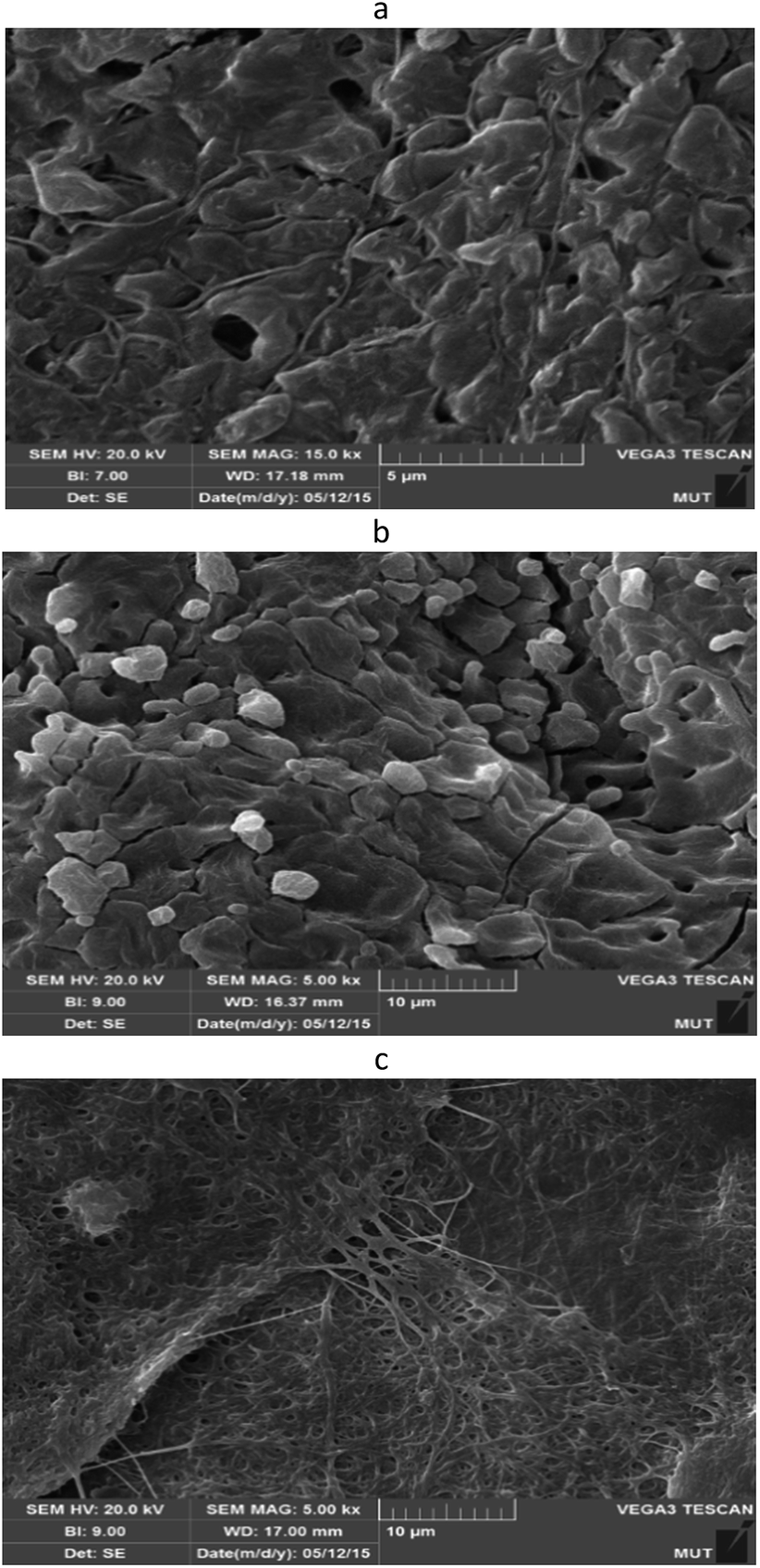
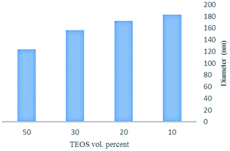
For study the effect of varying TEOS content on the morphology of the chitosan/PEO/TEOS nanofibers, these solutions were electrospun with 10–50 vol% TEOS. Fig. 2 shows the effect of chitosan/PEO/TEOS volume ratio on nanofiber diameter resulting from electrospinning of each solution. Uniform nanofibers with a smooth surface were obtained within a composition window of chitosan/PEO/TEOS ranging from 90:10 to 50:50 vol% ratios. Spinning conditions such as voltage, temperature, TCD and relative humidity were kept fixed, so that only the polymer, solvent and viscosity properties could influence the nanofiber morphology. Fig. 3 shows SEM images of samples generated after electrospinning from solutions containing the silica precursor (TEOS solution containing HCl, ethanol and water) in different concentrations with chitosan/PEO. All nanofibers were electrospun regularly and without knots. The larger nanofiber diameter is a result of the increased viscosity of the solution, the diameter of the nanofibers in 50:50 solution chitosan/TEOS is lower than others but this solution has low viscosity and it is more difficult for electrospinning. For this reason, we selected 20:80 and 30:70 volume ratio of TEOS/chitosan to perform the next step means addition of drug. Using software Image J, the diameters of 50 nanofibers were measured and their average diameter was calculated. Table 1 lists nanofiber diameters of different volume ratios of TEOS:chitosan. The average diameters of the as spun nanofibers are 171.745 nm for a chitosan/TEOS ratio of 80:20 vol%. The chitosan/TEOS 70:30 vol% ratio has a smaller average diameter of 156.242 nm.
 |
| | Fig. 2 Effect of chitosan/TEOS volume ratio on nanofibers diameter. | |
 |
| | Fig. 3 SEM images of the electrospun nanofibers from solutions of: (a) 10% TEOS/chitosan, (b) 20% TEOS/chitosan, (c) 30% TEOS/chitosan and (d) 50% TEOS/chitosan. | |
Table 1 Nanofiber diameters of different volume ratios of TEOS:chitosan
| TEOS:chitosan vol% |
Nanofiber diameters (nm) |
| 10:90 |
182.984 |
| 20:80 |
171.745 |
| 30:70 |
156.242 |
| 50:50 |
123.592 |
3.2. Electrospun chitosan/PEO/TEOS/cefepime nanofibers
Fig. 4 and 5 show the SEM images of electrospun nanofibers with different percentages of antibiotic. It has been shown that electrospinning with a solution at concentration lower than 1 wt% results in nanofibers with no beads. In 2 and 3 wt% antibiotic concentration many beads are created on the structure of nanofibers, because particles of antibiotic did not dissolve in the solution of chitosan/TEOS completely.
 |
| | Fig. 4 SEM images of the electrospun nanofibers: chitosan/20% TEOS/cefepime solutions with different amounts of cefepime: (a) 0.5 wt%, (b) 1 wt%, (c) 2 wt% and (d) 3 wt%. | |
 |
| | Fig. 5 SEM images of the electrospun nanofibers: chitosan/PEO/ 30% TEO/cefepime solutions with different amounts of cefepime: (a) 0.3 wt%, (b) 0.5 wt%, (c) 1 wt% and (d) 2 wt%. | |
On increasing the cefepime concentration to 1 wt%, good quality of nanofibers are obtained which can be attributed to the increased antibacterial activity of the nanofibers. On further increase of antibiotic concentration, the bead defects return (Fig. 5d) and the nanofiber diameter increased and did not show uniformity on the structure of nanofibers. Uniform nanofibers with a smooth surface were obtained with a composition of chitosan/PEO/30% TEOS/1% cefepime (Fig. 5c). We selected nanofibers with chitosan/PEO/30% TEOS/1% cefepime as appropriate nanofibers. This as-spun nanofiber presented also smooth surfaces and possessed a smaller average diameter of 112 nm.
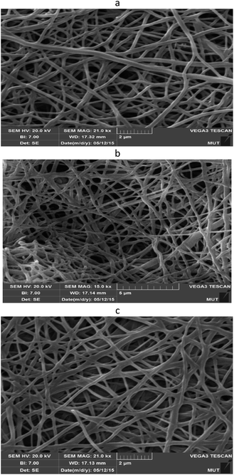
3.3. Nanofibers cross linked by glutaraldehyde
Fig. 6 show SEM images of electrospun chitosan/PEO/TEOS/cefepime nanofibers before and after cross linking the nanofibers. The nanofibers in the mats kept their morphology after cross linking with glutaraldehyde. This process is caused by activation of the carboxylic group of glutaraldehyde and the amine groups of polysaccharide chains of chitosan and production of C![[double bond, length as m-dash]](https://www.rsc.org/images/entities/char_e001.gif) N bonds. Therefore nanofiber networks are formed and their properties such as swelling, solubility and stability increased and biodegradability of nanofibers decreased.20
N bonds. Therefore nanofiber networks are formed and their properties such as swelling, solubility and stability increased and biodegradability of nanofibers decreased.20
 |
| | Fig. 6 SEM images of electrospun chitosan/PEO/TEOS/cefepime nanofibers (a): before and (b): after making cross linked nanofibers. | |
3.4. Stability of nanofibers
Fig. 7 and 8 show the SEM images of chitosan/PEO/TEOS/cefepime nanofibers before and after placing them in SBF buffer. Non cross linked nanofibers were unstable and lose their structures in the buffer solutions, whereas the cross linked nanofibers were stable and their morphology didn’t differ even after 24 h. Cross linked nanofibers have the ability to keep their structures at three pH values, after loading onto implants and implantation in the body. This high stability originates from the bonding of chitosan chains together during cross linking process which prevents water from entering into the nanofiber scaffold.
 |
| | Fig. 7 Stability study of non-cross linked nanofibers of chitosan/PEO/TEOS/cefepime after placing them in buffer at pH: (a) 5.5, (b) 7.4 and (c) 8.5. | |
 |
| | Fig. 8 Stability study of cross linked nanofibers of chitosan/PEO/TEOS/cefepime after placing them in buffer at pH: (a) 5.5, (b) 7.4 and (c) 8.5. | |
3.5. Antibiotic release from nanofibers
The in vitro release of cefepime antibiotic from the nanofibers was studied by immersing samples in SBF buffer 37 °C at pH 5.5, 7.4. Over a period of time, the release of the drug was measured. First, it is necessary to drawn the calibration curve. The calibration curve of cefepime was determined by taking the absorbance of cefepime concentrations between 0 and 2 mg mL−1 as parameters and the calibration curve was fitted to the Lambert–Beer law. A linear relationship between the cefepime concentration (y) and the optical absorbance (x) was obtained (y = 0.3539x + 0.1082 and R2 = 0.9703). All experiments were repeated three or more times and the experimental data are expressed as means ± standard error deviation. Fig. 9 shows the relationship between absorption and antibiotic concentration. To evaluate the amount of drug loaded by nanofibers, a UV-vis spectrometer was used for analysis.
 |
| | Fig. 9 Calibration curve of cefepime antibiotic. | |
Fig. 10 and 11 show the release profiles of cefepime into buffer at 37 °C. The initial burst release was observed in both of samples. The released cefepime reached a plateau after 22 hours.
 |
| | Fig. 10 Release profile of cefepime from the nanofiber scaffold with time at pH = 5.5. | |
 |
| | Fig. 11 Release profile of cefepime from the nanofiber scaffold with time at pH = 7.4. | |
However, the morphology of the chitosan structure could affect the release of cefepime. The fastest release was observed from the sample prepared in earlier times. The fast release from the fibrous structure was due to higher permeability, shorter diffusion distance and fast swelling activity.
Most of the loaded cefepime in the nanofibers was released in the first hours. After a few hours, the release was much slower and reached a plateau after 23 h. This release profile can be attributed to the porous structure of the prepared nanofiber which causes penetration of active substance into the nanofibers pores, thus the releasing process occurs slowly. The drug wasn’t released suddenly and the delivery happened moderately during the 16 days. The slower release percentage of cefepime could be attributed to the interaction between chitosan and cefepime (covalent bonding). A large amount of cefepime interacted with chitosan, thus a higher percentage of antibiotic was retained during the release. The result of moderate delivery of active substance is more antibacterial activity for a longer time.
3.6. FT-IR spectroscopy
The FT-IR spectrum of chitosan/PEO electrospun nanofibers is demonstrated in Fig. 12. The broad band between 3600–3200 cm−1 is assigned to the stretching mode of the O–H and N–H bonds in the chitosan and the O–H bond in the PEO backbone. The bands located between 2890–2960 cm−1 are ascribed to the symmetrical stretching mode of the C–H bond. These results imply that hydrogen bonding occurs between chitosan and PEO molecules.
 |
| | Fig. 12 FT-IR spectrum of chitosan/PEO nanofiber. | |
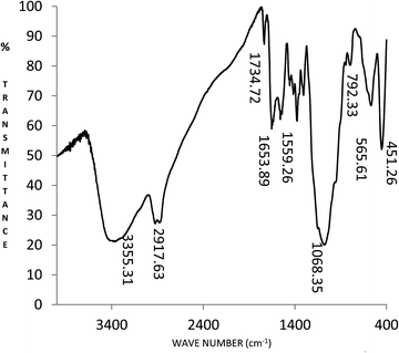
FT-IR spectrum of chitosan/PEO/TEOS nanofibers is shown in the Fig. 13. The broad band at 3355 cm−1 is assigned to the stretching mode of the O–H and N–H bonds in the chitosan and the O–H bond in the PEO backbone. The medium bands at 2862 cm−1 and 2917 cm−1 are attributed to the C–H stretch and C–O–C of the PEO, respectively. Characteristic bands of silica are observed: those at 1068 and 950 cm−1 and 792 cm−1 are attributed to Si–O–Si and Si–OH stretching vibrations, respectively, which is consistent with the reported results. It has been recently considered that covalent bonds between silane and chitosan can only proceed via the hydroxyl groups of chitosan due to the formation of strong Si–O bonds rather than weaker Si–N bonds. Also terminal –OH groups are easily available for bonding, hydrogen bonding between chitosan/PEO and silica is possible which might result in the formation of composite nanofiber.21,22
 |
| | Fig. 13 FT-IR spectrum of chitosan/PEO/TEOS nanofiber. | |
The FT-IR spectra of as-spun nanofibers indicate which bonds are formed or lost during the electrospinning processes and elucidate whether chitosan/PEO/TEOS/cefepime nanofibers are a physical mixture or a covalently bound network. Fig. 14 shows a wide peak at 3333.63 cm−1 assigned to the stretching of the chitosan hydroxyl groups. The medium band at 2921.58 cm−1 and the strong triplet bands at 1147 cm−1, 1104 cm−1 and 1064 cm−1 with a maximum at 1104 cm−1 are attributed to the C–H stretch and C–O–C respectively of the PEO. The 1622 cm−1 peak shows stretching of the COOH of carboxylic acid and β-lactam group of cefepime. Silica shows characteristic peaks at 1074, 559 and 443 cm−1 corresponding to the asymmetrical stretching, symmetric stretching and bending vibrations in Si–O–Si bonds, respectively. The probability of formation of covalent bonds between the chitosan and cefepime is very high.23–25
 |
| | Fig. 14 FT-IR spectrum of chitosan/PEO/TEOS/cefepime nanofiber. | |
3.7. Antibacterial performance
The antibacterial activity of chitosan/PEO/TEOS and chitosan/PEO/TEOS/cefepime nanofibers was tested by the agar plate method. The antimicrobial effects of the composite nanofibers on the growth of the bacteria E. coli, S. aureus and S. epidermidis and the inhibition zones of them are tested. Many antimicrobial properties of chitosan are created in an acidic environment. At a higher pH of 6.5 chitosan solubility decreases and poly cationic properties are lost. The result of the reaction between positive and negative charges of the cell surface of Gram-negative bacteria and the surface charge of chitosan is to cause damage to the growth of the bacterial cell wall. The results show these nanofibers have a bacteriostatic antibacterial effect and affected only the growth of the Gram-positive bacteria (S. aureus) and show no effect on the Gram-negative bacteria.26,27 Fig. 15 shows that the chitosan/PEO/TEOS/cefepime nanofiber is effective on Gram-positive and negative bacteria. Scaffolding nanofibers with and without antibiotic were effective on Gram-positive and negative bacteria and have an inhibitory and bactericide effect on them. Cefepime disrupts the synthesis of the peptidoglycan layer of bacterial cell walls, which causes them to break down and eventually the bacteria die.15
 |
| | Fig. 15 The antibacterial activity of chitosan/PEO/TEOS and chitosan/PEO/TEOS/cefepime nanofibers in the presence of (a): E. coli and (b): S. aureus and (c): S. epidermidis. | |
It was found that nanofibers with TEOS and cefepime show the most antibacterial activity of 25 and 40 mm inhibition zone against S. aureus and E. coli, respectively. Furthermore, as can be seen from Fig. 15c, chitosan/PEO/TEOS/cefepime nanofibers are effective on S. epidermidis, but chitosan/PEO/TEOS nanofibers (blue flashes) show no effect, yellow flashes show antibacterial activity of cefepime in chitosan/PEO/TEOS/cefepime nanofibers. In contrast, the nanofibers without antibiotic showed the least antimicrobial activity among the nanofibers (Table 2). It was observed that nanofibers with cefepime are the most effective against all tested microbes.
Table 2 Antibacterial activity of chitosan/PEO/TEOS and chitosan/PEO/TEOS/cefepime nanofibers on both Gram-negative and positive bacteria
| Sample nanofiber |
Inhibition zone of E. coli (mm) |
Inhibition zone of S. aureus (mm) |
| Chitosan/PEO/TEOS |
5 |
12 |
| Chitosan/PEO/TEOS/cefepime |
40 |
25 |
The antimicrobial mechanism includes the initial deposition of a chitosan coating on the anionic cell wall with subsequent alteration of biochemical functions and damage to internal organelles by internalized chitosan oligomers.19
3.8. Cell cultured study
Fig. 16 shows a microscopic study of mouse fibroblast cells which were wide with a round nucleus and big. The cells adhere to the bottom of the plate on the second day, and the cytoplasm of the fibroblast cells begin to spread and were seen as broad and multifaceted forms.
 |
| | Fig. 16 Fibroblasts cultured in DMEM containing 10% serum respectively for (A) 2, (B) 4, (C) 6 and (D) 8 days. | |
Also the cells were viewed with eosin–hematoxylin colouring using an inverted microscope. The cells appear yellow because we used an inverted microscope with a yellow filter (Fig. 17).
 |
| | Fig. 17 Fibroblast cells in DMEM under an inverted microscope with a magnification of (a) 10×, (b) 40× and (c) 100×. | |
Fig. 18 shows SEM images of mouse fibroblast cells cultured on the nanofibers, the cells lay among the nanofibers and have grown wide and have good form. Chitosan has been shown to possess many properties desirable in implant coatings such as cell attachment, growth and encouraging ordered bone tissue formation.19
 |
| | Fig. 18 SEM image of fibroblast cells on chitosan/PEO/TEOS/cefepime nanofibers at various magnifications. | |
The appearance of the fibroblast cells remained almost the same in terms of width, length and size. These cells have suitable adhesion to the nanofibers. This indicates that fibroblast cells in the presence of nanofibers have a good interaction and growth is possible. The percent of live and dead cells at the end of sixth day is indicative, at the end of the second, fourth and sixth days, after coloring the cells with trypan blue, more than 90% of the cells are alive. Dead cells which had been penetrated by trypan blue were seen as blue, while living cells were observed transparent and non-stained. Fig. 19 shows fibroblast cells cultured on nanofibers colored with trypan blue and yellow arrows indicate dead cells which are purple or blue while living cells remain colorless and clear.
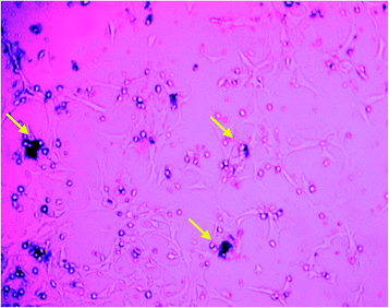 |
| | Fig. 19 Fibroblast cells cultured on nanofibers with trypan blue coloring. | |
To determine the live and dead fibroblasts, cells were counted using a neobar lam under an inverted microscope. The yellow arrow shows a dead cell and uncolored cells are live cells (Fig. 20).
 |
| | Fig. 20 Fibroblast cell count using a neobar lam under an inverted microscope. | |
After cell counting below the neobar lam on the second, fourth and sixth days, the live cells percentage was calculated. After 6 days of culture, no significant difference was observed among control and chitosan/PEO/TEOS/cefepime nanofiber samples. The proliferation of cells on all samples increased over the time of study. An important aspect to mention is the significantly (P < 0.05) lower percentage of cells on the chitosan/PEO/TEOS/cefepime sample compared to the control throughout the investigation (Table 3 and Fig. 21). Thus nanofibers can be prepared as biocompatible scaffold and used in the clinical and medical applications.
Table 3 Percentage of live cells on the chitosan/PEO/TEOS/cefepime sample compared to the control
| Sample |
Days |
| Sixth |
Forth |
Second |
| Control |
96.1 |
94.7 |
92.5 |
| Chitosan/PEO/TEOS/cefepime nanofiber |
93.3 |
92 |
91 |
 |
| | Fig. 21 Diagram of percentage of live cells after 2, 4 and 6 days. | |
4. Conclusion
We have prepared chitosan/PEO/TEOS/cefepime nanofibers by the electrospinning method. Chitosan acted as a natural antibacterial polymer and PEO is a synthetic polymer that decreases the viscosity of the chitosan. Chitosan/30% TEOS/1% cefepime is an appropriate nanofiber. This as-spun nanofiber presented smooth surfaces and possessed a small average diameter of 112 nm. This composite nanofiber can be used for applications such as antibacterial coating for orthopaedic implants. Chitosan/PEO/TEOS/cefepime nanofibers are effective on Gram-positive S. epidermidis and S. aureus and Gram-negative bacteria E. coli. Where chitosan serves as a flexible scaffold to support cell attachment, growth of fibroblast cells and improved the mechanical weakness of silica. The results of drug release showed cefepime delivery happened moderately during the 16 days. Thus nanofibers of chitosan/PEO/TEOS/cefepime can be used as a promising approach for medical implant coverage and to prevent bacterial infections during the time after implantation.
References
- Z. Rozek, W. Kaczorowski, D. Lukas, P. Louda and S. Mitura, Journal of Achievements in Materials and Manufacturing Engineering, 2008, 27, 33–38 Search PubMed.
- J. P. Chen, G. Y. Chang and J. K. Chen, Colloids Surf., A, 2008, 313, 183–188 CrossRef.
- K. M. Yun, C. J. Hogan Jr., Y. Matsubayashi, M. Kawabe, F. Iskandar and K. Okuyama, Chem. Eng. Sci., 2007, 62, 4751–4759 CrossRef CAS.
- Y. Gao, Y. Truong, Y. Zhu and L. Kyratzis, J. Appl. Polym. Sci., 2014, 131, 40797–44810 Search PubMed.
- Z. Huang, Y. Zhang, M. Kotaki and S. Ramakrishna, Compos. Sci. Technol., 2003, 63, 2223–2253 CrossRef CAS.
- S. S. Choi and S. G. Lee, J. Mater. Sci. Lett., 2003, 22, 891–893 CrossRef CAS.
- S. H. Jun, E. J. Lee, S. W. Yook, H. E. Kim, H. W. Koh and J. H. Jang, J. Mater. Sci.: Mater. Med., 2010, 21, 207–214 CrossRef PubMed.
- H. Guo, H. Qian, S. Sun, D. Sun, H. Yin and X. Cai, et al., Chem. Cent. J., 2011, 5, 1 CrossRef CAS PubMed.
- S. H. Jun, E. J. Lee, S. W. Yook, H. E. Kim, H. W. Kim and Y. H. Koh, Acta Biomater., 2010, 6, 302–307 CrossRef CAS PubMed.
- N. Banik, A. Hussain, A. Ramteke, H. K. Sharma and T. K. Maji, RSC Adv., 2012, 2, 10519–10528 RSC.
- D. Archana, B. K. Singh, J. Dutta and P. K. Dutta, Int. J. Biol. Macromol., 2015, 73, 49–57 CrossRef CAS PubMed.
- H. Homayoni, S. A. H. Ravandi and M. Valizadeh, Carbohydr. Polym., 2009, 77, 656–661 CrossRef CAS.
- N. Bhattarai, D. Edmondson, O. Veiseh, F. Matsen and M. Zhang, Biomaterials, 2005, 26, 6176–6184 CrossRef CAS PubMed.
- E. Moreno, I. Davila, E. Laffond, E. Macias, M. Isidoro and A. Ruiz, J. Invest. Allergol. Clin. Immunol., 2007, 17, 52–54 CAS.
- W. Long, Fast Analysis of Cefepime and Related Impurities on Poroshell 120 EC-C18, Agilent Technologies, 2011, pp. 5990–7492EN Search PubMed.
- K. Bruellhoff, J. Fiedler, M. Möller, J. Groll and R. E. Brenner, Int. J. Artif. Organs, 2010, 33, 646–653 CAS.
- M. A. Abdelgawada, S. M. Hudsona and O. J. Rojasb, Carbohydr. Polym., 2014, 100, 166–178 CrossRef PubMed.
- A. L. Behera, S. K. Sahoo and S. V. Patil, Int. J. PharmTech Res., 2010, 2(799), 798–803 CAS.
- H. J. Martin, K. H. Schulz, J. D. Bumgardner and J. A. Schneider, Thin Solid Films, 2008, 516, 6277–6286 CrossRef CAS.
- B. Sarmento, Chitosan-Based Systems for Biopharmaceuticals, John Wiley & Sons, Ltd., 2012, ch. 7, pp. 107–124 Search PubMed.
- G. Toskasa, C. Cherifa, R. D. Hunda, E. Laourinea, B. Mahltigb and A. Fahmic, Carbohydr. Polym., 2013, 94, 713–722 CrossRef PubMed.
- T. Pirzada, S. A. Arvidson, C. D. Saquing, S. S. Shah and S. A. Khan, Langmuir, 2012, 28, 5834–5844 CrossRef CAS PubMed.
- P. Yang, S. Gai and J. Lin, Chem. Soc. Rev., 2012, 41, 3679–3698 RSC.
- C. Shao, H. Y. Kim, J. Gong, B. Ding, D. R. Lee and S. J. Park, Mater. Lett., 2003, 57, 1579–1584 CrossRef CAS.
- T. Pirzada, S. A. Arvidson, C. D. Saquing, S. S. Shah and S. A. Khan, Langmuir, 2014, 30, 15504–15513 CrossRef CAS PubMed.
- Z. M. Zhong, R. G. Xing, S. Liu, L. Wang, S. B. Cai and P. C. Li, Carbohydr. Res., 2008, 33, 566–570 CrossRef PubMed.
- Y. B. Wu, S. H. Yu, F. L. Mi, C. W. Wu, S. S. Shyu, C. K. Peng and A. C. Chao, Carbohydr. Polym., 2004, 57, 435–440 CrossRef CAS.
|
| This journal is © The Royal Society of Chemistry 2016 |
Click here to see how this site uses Cookies. View our privacy policy here.