DOI:
10.1039/C5RA27955K
(Paper)
RSC Adv., 2016,
6, 27922-27932
A pH-driven molecular shuttle based on rotaxane-bridged periodic mesoporous organosilicas with responsive release of guests†
Received
29th December 2015
, Accepted 9th March 2016
First published on 11th March 2016
Abstract
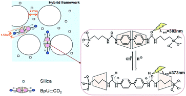
As a strategy for achieving integration of two-state rotaxane based molecular switches and ordered solid-state frameworks, a pH-driven molecular shuttle was immobilized into the framework of the periodic mesoporous organosilicas (PMOs) that possessed enough free space to accommodate the mechanical motion of β-cyclodextrins (β-CDs). In this molecular shuttle, β-CDs threaded a symmetrical molecular thread composed of a biphenyl unit, two ureido and propyl groups, and were end-trapped mechanically by two siloxane stoppers. The β-CDs, as the shuttles, could be reversibly translocated along the thread by the pH stimuli in the rigid framework, accompanied with the change of fluorescence emission of the biphenyl units. Particularly, the PMOs could be employed as a pH-controllable smart-release platform via the reciprocating movement properties of the molecular shuttle, and accelerated cargo release was achieved after acidification. Furthermore, the PMO materials show very low cytotoxicity and fine biocompatibility, which ensure their potential in biomedical applications.
1. Introduction
A rotaxane is described as a molecular system in which a macrocycle threads a linear molecule with two bulky stoppers. This macrocycle could shuttle between two or more stations connected by linkers under external stimuli,1–4 and this unique property makes rotaxanes promising candidates for molecular machines and devices.5–8 Among the suitable macrocycles, cyclodextrins (CDs), which have a good capability to associate various inorganic/organic/biological molecules, are attractive wheel components in constructing mechanically interlocked assemblies.9,10 Moreover, CDs containing both primary and secondary hydroxyl groups, are water-soluble, nontoxic, commercially available, and readily functionalized.
Within the past decade, mesoporous silica nanoparticles have increased in popularity for drug delivery purposes due to their ability to encapsulate a payload of therapeutic compounds, transporting them to specific locations in the body, and making a sustained release of the drug. Furthermore, the mesoporous silica nanoparticles could be covered and mechanized with nanovalves and other artificial molecular or supramolecular machines, converting them into smart drug delivery vehicles, which could release their cargo in response to target physical (e.g. light or temperature),11–15 chemical (e.g. pH or redox changes),16–21 and biochemical (e.g. enzymes or DNA) stimuli.22–26 Among these stimuli, pH-responsive activation represents a very convenient and feasible method because the measurement of the pH in solution is simple and swift. In addition, tumors exhibit lower pH environments compared with normal cells, especially in intracellular endosomes and lysosomes,27 which is another advantage for pH-responsive release systems. However, most of end-caps of nanovalves are shed by opening the sealants in response to stimuli, leading to a one-off release for surface functionalization system. Hence, developments concerning the reversible and repeated response release system are still challenging. Compared with mesoporous pure silica, periodic mesoporous organosilicas (PMOs) are a new class of organic–inorganic hybrid materials in which the organic groups are located within the channel walls as bridges between Si centers.28–30 PMOs have remarkable features such as a large surface area, tunable pore size, modifiable surface properties, and have organic groups inside the channel walls that provide new opportunities for controlling the physicochemical and mechanical properties of the materials.31–34 In a short time, an amazing variety of functional groups, morphologies and applications have been developed.35,36 However, very few studies have reported on rotaxanes functionalized PMOs in which rotaxanes were inserted into the frameworks of mesostructure by the co-condensation method. It belongs to the solid-state shuttles in which the β-CD or Hp-β-CD needs enough free space to accomplish back and forth of the mechanical movement.37–39 Therefore, the PMOs materials could be very appropriate and convenient platform for the solid-state shuttling.
Here, we prepared a rotaxane siloxane precursor (BpU⊂CD2, Fig. 1) composed of β-CD and a derivative of biphenyl (BpU) with triethoxysilyl groups at both ends. It is difficult to assemble these rotaxane complexes into the frameworks of ordered mesoporous structures due to the large molecules of β-CD and complicated interactions between host and guest molecules of rotaxane. In this work, a novel oligomeric cationic surfactant sym-Ph(1-3-14)3 (Fig. S1†) was employed in the preparation of β-CD-rotaxane PMOs in order to enhance the ordering mesoporous structure of the hybrid materials. We successfully fabricate the mesoporous materials with β-CD rotaxane units inside pore walls using sym-Ph(1-3-14)3 as the template. The incorporation of β-CD into PMOs possesses significant characteristics. Firstly, cyclodextrin rotaxanes could improve the hydrophilicity and biocompatibility of pore walls of the mesoporous silica, and easily disperse in aqueous solution. Secondly, the β-CD component of the rotaxane acts as a molecular shuttle, responding to the pH change, which could be used as a pH switch. In the BpU⊂CD2 hybrid PMOs (BpU⊂CD2-PMOs), fluorescent biphenyl groups involved in β-CDs in the rotaxane act as the sensors that provide self-feedback on the position of the β-CDs in the rotaxane (biphenyl or propyl groups) through the fluorescent properties of the hybrid materials. Ureido units in BpU⊂CD2-PMOs, containing two amino groups, are as pH triggers connected with biphenyl and propyl groups in the sides of the ureido units. As the pH in the acidic range, a protonation takes place for the amino groups, and carbonyl groups in the ureido units are transformed into the enol groups due to the isomerization, making the benzidine groups more hydrophilic, leading to the detachment of the β-CDs from the biphenyl to the propyl units and vice versa. The pH-driven molecular shuttles of the smart PMOs could be applied for the controllable release of drug. Upon addition of acid solution, the movement of β-CD could disrupt the interactions between the cargoes and pore walls, allowing the loaded Rhodamine 6G (Rh6G) to release. Even more important, a reversible and repeated response release is realized by control of molecular shuttle in the solid-state system. In addition, hydroxypropyl-β-cyclodextrin (Hp-β-CD) based rotaxane PMOs (BpU⊂HpCD-PMOs) were also prepared by the same synthetic method and they exhibited similar pH-responded fluorescence properties as the BpU⊂CD2-PMOs.
 |
| Fig. 1 Synthesis of β-CD based [3] rotaxane siloxane precursor (BpU⊂CD2) and the proposed mechanism of the pH-induced shuttling movement processes of molecular shuttle in BpU⊂CD2-PMOs. | |
2. Experimental
2.1 Chemicals and reagents
4,4′-Diaminobiphenyl, 3-(triethoxysilyl) propyl isocyanate, β-cyclodextrin, (2-hydroxypropyl)-β-cyclodextrin and N,N,N′,N′-tetramethyl-1,3-propanediamine were purchased from Shanghai Jingchun Reagent Company. Acetone, pyridine and ethanol were obtained from Tianjin Fuyu Reagent Company. Tetraethoxy orthosilane (TEOS) and aqueous ammonia were bought from Tianjin Guangcheng Reagent Company. The oligomeric cationic surfactant sym-Ph(1-3-14)3 was prepared according to the ref. 40 except that the N,N,N′,N′-tetramethylethylenediamine was replaced by N,N,N′,N′-tetramethyl-1,3-propanediamine.
2.2 Synthesis
Synthesis of 4,4-bis(3-triethoxysilylpropylureido) biphenyl without CDs for the control sample (BpU). 4,4-Diaminobiphenyl (1.84 g, 10.0 mmol) and 3-(triethoxysilyl) propyl isocyanate (5.90 g, 24.0 mmol) were dissolved in anhydrous ethanol (150 mL). The mixture was refluxed in the N2 atmosphere at 80 °C for 24 h. After cooling to room temperature, the grayish solid was filtered and washed with hexane three times, and then dried in vacuum for 3 days. (Yield, 82%) 1H NMR (300 MHz, DMSO-d6, δ): 8.43 (2H, PhNHCO), 7.47 (8H, ArH), 6.16 (2H, NH in NHCH2), 3.75 (12H, ethoxy CH2), 3.06 (4H, CH2 in CH2NH), 1.47 (4H, CH2–CH2–Si), 1.15 (18H, ethoxy CH3), and 0.56 (4H, Si–CH2) (Fig. S2†). 13C NMR (75 MHz, DMSO-d6, δ) 7.21, 18.08, 23.29, 41.70, 57.63, 117.87, 126.03, 132.58, 139.35, 155.10 (Fig. S3†). HR-MS (ESI) calcd for [BpU + H]+, 679.96, found 679.34.
Synthesis of benzidine-β-CD inclusion complex (benzidine⊂CD2). β-CD (11.35 g, 10.0 mmol) was dissolved in distilled water (200 mL) and heated to 70 °C, then benzidine (1.38 g, 7.50 mmol) dissolved in acetone (120 mL) was slowly dropped into β-CD solution with sufficient stirring, and then stirred at 70 °C for 24 h. After the excess benzidine was removed by filtration, the obtaining yellow precipitate was washed with distilled water and acetone, in order to clean the residual host and guest molecules respectively, and then it was dried in vacuum oven at 50 °C for 48 h (yield, 80%). The structure and molar ratio of inclusion complex were confirmed by 1H NMR spectra. 1H NMR (300 MHz, D2O, δ): 7.20 (d, 2H, ArH), 6.82 (d, 2H, ArH), 4.96 (7H, β-CD), 3.81–3.72 (21H, β-CD), 3.62–3.46 (21H, β-CD) (see Fig. S4†). The results revealed that benzidine forms 1
:
2 complexes with β-CD. 13C NMR (75 MHz, DMSO-d6, δ) 59.86, 71.97, 72.33, 72.99, 81.46, 101.87, 114.29, 125.92, 128.60, 146.7 (Fig. S5†).
Synthesis of [3] rotaxane siloxane precursor (BpU⊂CD2). Benzidine⊂CD2 inclusion complex (9.82 g, 4.00 mmol) and 3-(triethoxysilyl) propyl isocyanate (2.37 g, 9.60 mmol) were dissolved in anhydrous pyridine (250 mL), respectively, and kept stirring in a N2 atmosphere at 70 °C for 24 h. A yellow solid was obtained with the addition of hexane (100 mL), the solid was filtered and washed with hexane, and then dried in a vacuum oven at 70 °C for 48 h. (Yield, 78%) 1H NMR (300 MHz, DMSO-d6, δ): 8.39 (2H, PhNHCO), 7.37 (8H, ArH), 6.13 (2H, NHCH2), 3.78 (12H, ethoxy CH2), 3.06 (4H, CH2NH), 1.16 (18H, ethoxy CH3), 0.55 (4H, Si–CH2), 5.72 (14H, β-CD), 5.67 (14H, β-CD), 4.84 (14H, β-CD), 4.44 (14H, β-CD), 3.78–3.55 (56H, β-CD), 3.38–3.27 (28H, β-CD) (Fig. S6†). 13C NMR (75 MHz, DMSO-d6, δ) 7.20, 18.15, 23.28, 41.70, 57.64, 59.85, 71.98, 72.35, 72.98, 81.49, 101.88, 117.88, 126.24, 132.54, 139.33, 155.08 (Fig. S7†). HR-MS (ESI) calcd for [BpU⊂CD2 + 2H]2+, 1475.46, found 1475.05.
Synthesis of benzidine and Hp-β-CD inclusion complex (benzidine⊂HpCD). The preparation of benzidine⊂HpCD was similar to the synthesis of benzidine⊂CD2, except that β-CD was replaced by (2-hydroxypropyl)-β-cyclodextrin (Hp-β-CD). (Yield, 75%) 1H NMR (300 MHz, DMSO-d6, δ): 7.20 (4H, ArH), 6.56 (4H, ArH), 6.10–5.55 (7H, Hp-β-CD), 4.99–4.53 (21H, Hp-β-CD), 3.75–3.22 (63H, Hp-β-CD) and 1.05 (21H, Hp-β-CD) (see Fig. S8†). The results reveal that benzidine forms 1
:
1 complexes with Hp-β-CD. 13C NMR (75 MHz, DMSO-d6, δ) 19.70, 59.86, 65.27, 72.20, 73.02, 77.40, 81.61, 100.84, 114.59, 125.98, 128.93, 146.19 (Fig. S9†).
Synthesis of [2] rotaxane siloxane precursor (BpU⊂HpCD). The preparation of BpU⊂HpCD rotaxane was the same as the synthesis of BpU⊂CD2, except that the benzidine⊂CD2 complex was replaced by benzidine⊂HpCD complex. (Yield, 86%) 1H NMR (300 MHz, DMSO-d6, δ): 8.44 (2H, PhNHCO), 7.43 (8H, ArH), 6.15 (2H, NHCH2), 3.78 (12H, ethoxy CH2), 3.07 (4H, CH2NH), 1.18–1.12 (18H, ethoxy CH3), 0.55 (4H, Si–CH2), 6.10–5.55 (7H, Hp-β-CD), 5.10–4.52 (21H, Hp-β-CD), 3.79–3.32 (63H, Hp-β-CD) and 1.03 (21H, Hp-β-CD) (Fig. S10†). 13C NMR (75 MHz, DMSO-d6, δ) 7.22, 18.48, 19.79, 23.55, 41.70, 57.67, 59.87, 65.27, 72.17, 73.02, 77.54, 81.51, 100.84, 114.28, 117.90, 126.08, 132.57, 139.34, 155.11 (Fig. S11†). HR-MS (ESI) calcd for [BpU⊂HpCD + H]+, 2221.50, found 2220.96.
Synthesis of BpU⊂CD2-bridged periodic mesoporous organosilicas (BpU⊂CD2-PMOs). sym-Ph(1-3-14)3 (1.20 g) was dissolved in deionized water (30 mL) and then the pH value was adjusted to 11.0 with ammonia (2.50 mL, 28% w/w). A certain amount of BpU⊂CD2 and TEOS was mixed homogeneously in deionized water (10 mL) and added to the solution of surfactant with vigorous stirring (the total Si amount is 6.0 mmol and the mole fraction (X) of BpU⊂CD2 precursor is 0, 5, 10, 15 and 20%, respectively). After stirring for 30 min at room temperature, the mixture was kept at 50 °C for 5 h, subsequently at 80 °C in an oven for another 72 h. The resulting product was recovered by filtration, washed with distilled water and ethanol, and then dried in air at ambient temperature. Finally, the structure directing agents were extracted with the methanol solution for 10 days. The resulting samples are denoted as BpU⊂CD2-PMO-X, (X = 0, 5, 10, 15 and 20).
Synthesis of BpU⊂HpCD-bridged periodic mesoporous organosilicas (BpU⊂HpCD-PMOs). The synthetic method of BpU⊂HpCD-PMOs was the same as BpU⊂CD2-PMOs, except that BpU⊂CD2 precursor was replaced by BpU⊂HpCD precursor. The resulting samples are denoted as BpU⊂HpCD-PMO-X, which X represents the molar fraction of BpU⊂HpCD precursor in the total amounts of precursors.
Synthesis of BpUPMO-20 material without CDs. The synthetic method and reagent proportion of BpUPMO-20 was the same as BpU⊂CD2-PMO-20, except that BpU⊂CD2 precursor was replaced by BpU precursor.
2.3 Characterization
1H NMR and 13C NMR analyses were carried out on a Bruker DPX-300 NMR spectrometer. FT-IR spectra were recorded on a Nicolet 700 FT-IR spectrometer (Thermo-Fisher Scientific, Inc., Waltham, MA) with samples prepared as KBr pellets. Mass spectral data was acquired on an Agilent 6510 QTOF. Powder X-ray diffraction (XRD) pattern were performed on a D/MAX-RB with Cu-Kα radiation (40 kV, 100 mA). Small-angle X-ray scattering (SAXS) measurements were carried out on SAXSess mc2 (Anton Paar). High-resolution transmission electron microscopy (HRTEM) images were obtained from a JEM-2100 electron microscopy operating at 200 kV. Prior to the HRTEM measurement, the samples were first dispersed in absolute ethanol by sonication, and then deposited on a copper microgrid. TGA/DSC analyses were carried out using a SDT-Q600 V8.3 Build 101 Simultaneous DSC-TGA Instrument (TA). Studies were conducted under inert atmosphere of nitrogen at a heating rate of 10 °C min−1 from ambient temperature to 800 °C. Nitrogen adsorption/desorption isotherms were measured using a Micromeritics ASAP2020 Surface Area and Porosity Analyzer at 77 K. Prior to measurements, all samples were outgassed at 120 °C for 10 h. Surface areas were calculated by the Brunauer–Emmett–Teller (BET) method, and the pore size distribution was evaluated using the Barrett–Joyner–Halenda (BJH) method and calculated from the adsorption branch of isotherm. The solid-state 29Si magic angle spinning nuclear magnetic resonance (MAS NMR) spectra were recorded on a Bruker DSX 300 MHz spectrometer (5000 transients, spin speed 6 kHz, acquisition time 0.02 s, pulse delay 3 s). The zeta-potential was measured using a Zeta PALS DB-525 (Brookhaven instruments corporation, USA). Particle size distribution analysis was measured using a Zetasizer 3000 Particle Size and Zeta Potential Analyzer. UV-vis spectra were measured using an Agilent HP8453E spectrometer. Fluorescence spectra of the suspension were obtained using a LSS55 spectrometer. Time-resolved fluorescence spectra were obtained using an Edinburgh FLS920 fluorescence spectrophotometer. The instrument was operated with a thyratron-gated flash lamp filled with hydrogen at a pressure of 0.40 bar. The lifetimes were estimated from the measured fluorescence decay curves using deconvolution method fitting procedure. The concentration of sample suspensions were 1.0 × 10⁻⁴ g mL−1 and the pH values of suspensions were adjust by 0.10 M HCl solution.
The C and N 1s X-ray absorption near-edge structure (XANES) spectroscopy was collected on the X-ray absorption endstation on the SGM beamline at the Canadian Light Source (CLS) (Saskatoon, Canada). The beamline uses a 45 mm planer undulator with an energy range between 250 and 2000 eV. The spot size on the beamline was 1000 μm × 100 μm. Data was collected using silicon drift detectors (SDD) and a titanium filter to rejected higher order harmonics. The entrance and exit slit gaps were set to 249.9 μm and 25 μm respectively. Normal scans (step scanning) were acquired at the C 1s edge from 280 to 320 eV and N 1s from 390 to 420 eV at a 1 s dwell time and 0.1 eV step size. Normalization of the C 1s data involved collecting an I0 by measuring the scatter of the incident beam from a freshly Au-coated Si wafer using an SDD and of the N 1s data by using the I0 from a Au mesh in the beamline before the sample.41
2.4 Assessment of drug release
Selecting Rh6G as the model drug, the sample of BpU⊂CD2-PMO-20 was firstly soaked in a concentrated Rh6G ethanol solution to load the cargo molecules with stirring for 6 h. The obtained sample was washed with ethanol to remove the dyes on the surface of the particles and then dried under vacuum. Aqueous suspensions of Rh6G loaded BpU⊂CD2-PMO-20 (10 mL, 10 mg mL−1) were poured into dialysis bags and the dialysis bags were placed in beakers, with deionized water (40 mL, pH ≈ 7.0) and stirred. The release of Rh6G molecules from the Rh6G loaded BpU⊂CD2-PMO-20 was performed by adjusting a neutral solution (i.e., pH 7) to pH 4.0 with 0.02 M HCl solution, and another identical sample was kept in the neutral solution for comparison. The specific operations: the supernatant of Rh6G loaded PMOs (20 μL) was collected at regular intervals and diluted to 2.0 mL to detected the fluorescence intensity of Rh6G (λex = 500 nm). The fluorescence intensity at the emission wavelength of the Rh6G molecules was plotted as a function of release time in order to generate a release profile. 100% release is determined by allowing the loaded PMOs to release Rh6G over 3 days at pH = 4.0. The release percentage was calculated as the fluorescence intensity of Rh6G released at different time divided by the fluorescence intensity at 100% release.
2.5 Cell survival assay
H1299 cells were seeded in 96-well cell culture plates, treated with different concentrations of the PMO materials after 24 h or 48 h. At the end of the treatment, the viable cell numbers were estimated by sulforhodamine B (SRB) assay as follows. After discarding the old medium, 10% ice-cold trichloroacetic acid (TCA, 100 μL) was added to each well, and then incubated at 4 °C for 1 h; removed the TCA and washed five times with distilled water and then air-dried. Secondly, SRB solution (50 μL) was added to each well of the dried 96-well plates and shook for 5 min on a shaker platform, washed quickly with 1% v/v acetic acid five times to remove the unbound dye and air-dried. The bound SRB was then solubilized by adding unbuffered Tris base (pH 10.5, 100 μL, 10 mM) to each well and shook for 5 min on the shaker platform. Finally, the plates were read in microplate reader with the working wavelength of 540 nm. The optical density (OD) of SRB in each well was directly proportional to the cell number, so the OD values could be plotted against the concentrations of the PMO materials.
3. Results and discussion
3.1 Structural studies
The formation of rotaxane BpU⊂CD2 and BpU⊂HpCD inclusion complex was confirmed by powder X-ray diffraction (XRD) results (Fig. S12†). Many diffraction peaks are shown in the patterns of BpU and β-CD, illustrating that they possess typical crystal molecular stacking in their initial state. The XRD pattern of a physical mixture of BpU and β-CD (trace C in Fig. S12a†) is essentially a superposition of their patterns, indicating that there is no chemical association between the two compounds. However, the XRD pattern of the BpU⊂CD2 exhibits only one broad peak, indicating that it is amorphous. Thus, the differences in the diffraction patterns of the BpU⊂CD2 and physical mixture of BpU and β-CD indicate the formation of inclusion complexes between β-CD and BpU.42 Similarly, this behavior is also observed for BpU⊂HpCD in Fig. S12b,† which suggests the formation of inclusion complexes between BpU and Hp-β-CD.43 In addition, 1H NMR spectra could provide a further evidence of the formation of the inclusion complex. Compared with the spectra of the BpU and BpU⊂CD2, the resonances of the aromatic ring protons and amino protons that linked biphenyl were significantly shifted in the presence of β-CD (shifted upfield by 0.10 ppm and 0.04 ppm respectively), suggesting that the BpU molecules are included into the β-CDs. Furthermore, the ratio of 1
:
2 was achieved from the proton ratio between cyclodextrin and biphenyl from the 1H NMR spectra and MS analysis.
The FT-IR spectra analysis confirmed the interaction between BpU and β-CD in the rotaxane complex. As shown in Fig. S13a,† the characteristic peaks of BpU (νC
O at 1645 cm−1 and 9νAr C
C at 1565 cm−1) and of β-CD (νOH at 3400 cm−1, νC–H at 2924 cm−1 and νC–O–C at 1157 cm−1) are both found in the BpU⊂CD2 spectrum, indicating the presence of BpU and β-CD in the rotaxane siloxane BpU⊂CD2 precursor.44 However, compared with the spectrum of the physical mixture of BpU and β-CD, some significant differences are observed in the spectrum of the BpU⊂CD2 inclusion complex. The N–H stretching vibration (3325 cm−1) is not present in the BpU⊂CD2 spectrum, implying that the N–H bands of BpU might interact with the O–H bands at the entrance of β-CD cavities in the inclusion complexes.45 In addition, the intensity of the C–H stretching vibration of BpU (2973 cm−1) decreases in the BpU⊂CD2 because of the shielding effect of the β-CD. Nevertheless, the spectrum of the physical mixture is more like a simple overlap of β-CD and BpU spectra, indicating no or more weaker interaction between BpU and β-CD.
From the FT-IR spectroscopy of BpU⊂CD2-PMOs (Fig. S13b†), the presence of both silica structure and organic moieties is also confirmed. The peaks at 1662 and 1544 cm−1 correspond to the C
O stretching vibration and C
C stretching vibration of the BpU molecules, respectively. The stretching vibrations of Si–O–Si frameworks are detected around 1070 cm−1. These results indicate that the rotaxane BpU⊂CD2 is successfully inserted into the framework of the PMOs.
Small-angle X-ray scattering (SAXS) patterns of BpU⊂CD2-PMOs after removing sym-Ph(1-3-14)3 are shown in Fig. 2a. All samples show only one scattering peak in the low-angle region, indicating the formation of an ordered mesostructure. The intensity of the SAXS peak deceases with increasing amounts of the rotaxane BpU⊂CD2, indicating that more BpU⊂CD2 is incorporated into the PMO framework, resulting in a loss of the ordered mesoporous structure. Meanwhile, as the loading of BpU⊂CD2 is increased from 0 to 20 mol%, the position of the scattering peak is shifted to higher angles, corresponding to the gradual decrease of d-spacing values from 4.90 (BpU⊂CD2-PMO-0) to 4.33 nm (BpU⊂CD2-PMO-20). The reason arises from the enormous size (molecular weight = 2948) and complicated interactions of rotaxane precursors in the pore walls that could disrupt the self-assembly of the surfactant itself, which reduces the ordering of the mesostructure during the formation of mesophases.46,47 A similar phenomenon is also observed in the SAXS patterns for the BpU⊂HpCD-PMOs (Fig. 2b), suggesting the presence of the same mesoporous structure with increasing amounts of BpU⊂HpCD.
 |
| Fig. 2 SAXS patterns of (a) BpU⊂CD2-PMO-X and (b) BpU⊂HpCD-PMO-X after extraction of sym-Ph(1-3-14)3 surfactant. X represents the molar percent of rotaxane precursor in the total amounts of precursors, from top being X = 0, 5, 10, 15 and 20, respectively. | |
The formation of mesoscale porous structures was further confirmed by HRTEM images of the extracted BpU⊂CD2-PMO-5 (Fig. S14†). The photography of BpU⊂CD2-PMO-5 reveals the presence of mesochannels with a pore diameter about 2.3 nm and pore walls ca. 2.1 nm. The higher loading of BpU⊂CD2 leads to the formation of more disordered materials. In addition, the high quality HRTEM images are difficult to obtain because of the collapse of the mesoporous framework by irradiation of electron beam (CD molecules are very sensitive to the electron beam).
The thermal stability of BpU⊂CD2-PMOs is maintained at 200 °C, with only small decreases at low temperature due to the loss of water (Fig. S15†). The 29Si MAS NMR spectra of solvent-extracted BpU⊂CD2-PMO-20 and BpU⊂HpCD-PMO-20 are shown in Fig. S16.† From the existence of T2 (RSi(OSi)2OH) and T3 (RSi(OSi)3, R: organic groups) signals, we conclude that rotaxane functional groups are covalently bonded in the silica framework of the hybrid materials. The porosity of the extracted BpU⊂CD2-PMOs was examined by nitrogen adsorption/desorption isotherms (Fig. S17†). All the isotherms are classified as type IV, which suggests the successful formation of mesopores in BpU⊂CD2-PMOs. Combined with the results of SAXS and HRTEM patterns, the thickness of the pore wall of the hybrid PMOs is estimated to be ∼2.2 nm, this value is very close to the diameter of β-cyclodextrin (1.53 nm), suggesting that only one layer of the BpU⊂CD2 rotaxane might be inserted into the pore wall.
3.2 Aqueous dispersibility
The zeta (ζ) potential profiles of BpU⊂CD2-PMO-0, BpU⊂CD2-PMO-20 and BpU⊂HpCD-PMO-20 materials versus pH values are shown in Fig. S18.† The BpU⊂CD2-PMO-20 and BpU⊂HpCD-PMO-20 samples retain their positive charge (ζ = +30–40 mV) up to a neutral pH value, while the ζ value of pure silica BpU⊂CD2-PMO-0 sample changed from positive to negative charge at about pH 3.0. The electric behaviors of the BpU⊂CD2-PMO-20 and BpU⊂HpCD-PMO-20 are mainly attributed to the presence of ureido groups of the hybrid materials. The presence of the ureido function units should shift the isoelectric point toward higher pH values, because the ureido groups could be protonated at acidic pH values.48 Pure silica has an isoelectric point in the range of pH 2–3 that is controlled by the deprotonation of single or isolated silanol groups.49 Generally, particles with ζ potentials more positive than +30 mV or more negative than −30 mV are considered to form stable dispersions because of the electrostatic repulsion between particles. When the pH values of BpU⊂CD2-PMO-20 and BpU⊂HpCD-PMO-20 are above 2.0, the ζ potentials are above +30 mV. From the particle size analysis (see Fig. S19b and d†), the most probable particle sizes are about 300 nm for BpU⊂CD2-PMO-20 and BpU⊂HpCD-PMO-20 in the pH range of 2.0–7.0. While, at pH 1.0, the most probable particle sizes are about 2000 nm. Combined the results of aqueous dispersibility with particle size analysis at different pH values, it can be seen that the BpU⊂CD2-PMO-20 and BpU⊂HpCD-PMO-20 could form stable dispersions above pH 2.0 and coagulate at pH 1.0 (see Fig. S19a and c†), which is in agreement with the mechanism of electrostatic repulsion. Besides, the hydrophilicity associated with hydroxyl groups of the rims on the cyclodextrin cavities could also play an effective role in stabilizing the dispersion.50
3.3 Photophysical studies
Fig. 3a shows the UV-vis absorption spectra of BpU and BpU⊂CD2. A decrease in the absorbance intensity and a shift of the maximum absorption wavelength (λmax) from 291 nm to 296 nm (redshift 5 nm) are observed for BpU⊂CD2 compared with that of BpU. The shift in the position of the λmax could be attributed to transfer of the BpU molecules from the ethanol solution into the cavities of β-CDs, leading to a modified environment surrounding the biphenyl units.51 Moreover, BpU molecules have two functional groups of ureido and biphenyl within the organic moiety, promoting the buildup of the hydrogen bonded array and π–π stacking in ethanol solution. After BpU is complexed with CD, BpU molecules are encased in the cyclodextrin cavities that offer a block for these intermolecular interactions.52
 |
| Fig. 3 UV-vis absorption spectra of (a) BpU and BpU⊂CD2 in ethanol (5 × 10⁻⁵ M); (b) BpU⊂CD2-PMOs in ethanol (1 × 10⁻⁴ g mL−1). | |
As shown in Fig. 3b, the absorption bands of BpU⊂CD2-PMOs have similar shape but are slightly red-shifted (λ = 300 nm) compared with the spectrum of the precursor solution, which might be due to higher coplanarity degree of the BpU when it was attached in the silica framework.32 In addition, the dense packing of precursors in the framework of PMOs could induce the intermolecular interactions produced by the adjacent rotaxane precursors and silanol groups on the solid silica matrix. The intensity of this characteristic band increases gradually with increasing the amount of BpU⊂CD2, indicating that BpU⊂CD2 is attached into the pore walls of the PMO hybrid materials.
Fig. 4 shows the fluorescence emission spectra of BpU, BpU⊂CD2 and BpU⊂CD2-PMOs samples. The emission band of BpU⊂CD2 is red-shifted from 352 nm to 377 nm compared with that of the BpU in ethanol solution (Fig. 4a). This redshift is due to the change in local solvation environment upon complexation, similar to the effect of change from a protic to a less protic solvent.53 More importantly, once the BpU groups are included in the β-CD cavities, the rotation of phenyl ring around C–C single bond at 4 and 4′ position in biphenyl groups is retarded, caused by the steric hindrance between BpU groups and β-CDs, which is different from that of the free biphenyl groups in solution. This is another reason for the change of fluorescent properties for biphenyl groups.54
 |
| Fig. 4 Fluorescence emission spectra (a) BpU and BpU⊂CD2 (10−7 M) in ethanol; (b) BpU⊂CD2-PMO-X (X = 5, 10, 15 and 20) in aqueous solution (5 × 10⁻⁵ g mL−1). | |
For the fluorescence spectra of BpU⊂CD2-PMOs (Fig. 4b), an emission peak with the λem around 382 nm is found for all the samples and no notable difference is observed for these materials with different contents of BpU⊂CD2. This is different from the previous studies,40 in which red-shift of the λem and fluorescence intensity enhancement were observed when the content of fluorophore biphenyl groups was increased. However, in the present case, the fluorescent characteristic of the red-shift of λem and fluorescence enhancement of biphenyl groups for the BpU⊂CD2-PMOs disappear because BpU molecules are covered by β-CDs. Therefore, the distinct fluorescent behaviors for the biphenyl groups with or without β-CDs are more important for the β-CDs molecular shuttles hybrid PMOs that indicate self-feedback of the position of β-CDs through the change of fluorescent properties.
3.4 pH-Driven molecular shuttles
The influence of β-CD on the pH-response properties of the biphenyl groups in the rotaxane BpU⊂CD2-PMOs was investigated by measuring the maximum fluorescence peak position of samples. To verify the influence of β-CD in the pH-responded fluorescent properties of the biphenyl groups, we tested a control sample in which the PMOs were incorporated with BpU molecules but without β-CDs (BpUPMO-20 without β-CD). As shown in Fig. 5, no significant λem shifts are observed for the parallel sample BpUPMO-20 without β-CD when the pH is reduced from 7.0 to 2.0. A slight blue-shift in this process is caused by the increased protonation of arylamine.55 However, the maximum fluorescence peak position for BpU⊂CD2-PMO-20 occurs at about 382 nm above pH 5.0, remaining relatively constant thereafter, while below pH 4.0, it occurs at about 372 nm. A sudden blue-shift (∼10 nm) occurs in the pH range of 4.0–5.0, which might be caused by the change of interaction between β-CDs and BpU after amine or carbonyl protonation.56 The above results indicate that the presence and position of β-CDs have a significant influence on the fluorescence behavior of the BpU⊂CD2-PMO-20. The reason for this is as follow. The complex formation constant for the complex of the β-CD ring with the benzidine is about 8605 M−1 at pH 7.0, whereas it is less than 70 M−1 at pH 2.0.57 The difference in the complex formation constants of the β-CD with the benzidine is very large (>120-fold) under the neutral and acidic conditions, that is, the β-CD units have a greater affinity with benzidine moieties at neutral pH than the acidic pH. Therefore, the variation of the complex formation constants between the neutral and acidic conditions could have an effect on the stability of the inclusion compound in the host–guest system.
 |
| Fig. 5 The maximum emission wavelength (λem) of BpUPMO-20 without CDs, BpU⊂CD2-PMO-20 and BpU⊂HpCD-PMO-20 in the aqueous solution at different pH values. | |
To further clearly indicate the effect of the complex formation constant on the interactions among biphenyl, propyl groups and the β-CD, the pH-induced shuttling movement processes of β-CD in BpU⊂CD2-PMOs is illustrated in Fig. 1. Under neutral conditions the β-CD units are located on the benzidine moieties as a result of the hydrophobicity interaction between biphenyl group and the cavity interior of the β-CD (Fig. 1 State I). Upon benzidine units protonation in acidic media, the hydrophobicity interaction decreases due to the delocalization between the amines with positive charges and benzene rings, the β-CD units are induced to move from the biphenyl towards the more hydrophobic propyl spacers (State II).58,59 Then, the hydrophobicity of biphenyl moieties could be regressed if the pH of system is further changed from acidic to neutral. At that time, the binding capacity of β-CD with biphenyl unit which has more matching molecular size is stronger than that of alkyl unit.60 Thus, β-CDs move back on the biphenyl units due to the fact that more stable conformation could be achieved when the cyclodextrin rings encircle the biphenyl units. The shuttling movement processes of β-CDs in BpU⊂CD2-PMOs are reversible and very sensitive to the pH stimulus, leading to the variation of the fluorescence properties in two states. In State I, the fluorescence behaviors are ascribed to the inclusion compound of the BpU and β-CD in the neutral condition of pH value, while, in State II, the fluorescence behaviors arise from the BpU units in the acidic condition of pH value, which is similar with that of control sample BpUPMO-20 without β-CD. In addition, a similar shuttling movement phenomenon is also observed for BpU⊂HpCD-PMO-20.
To get a better understanding of the fluorescence behaviors of the hybrid materials at the different pH values, their fluorescence decays were performed as shown in Fig. S20.† The decay curves of the three samples are analyzed coincidentally as three-component exponential functions, which reveal that the excited states of the fluorophores in the hybrid materials decay through three pathways. The shorter decay parameters (τ1) are attributed to the emission of the monomers of biphenyl groups. Besides, the intermolecular interactions between the organic groups and steric hindrance of silica framework may result in a component of decay with longer decay parameters (τ2 and τ3). Moreover, longer lifetimes are exhibited for State I of BpU⊂CD2-PMO-20 than those of BpUPMO-20 without β-CD, suggesting a slow non-radiative decay process due to the hindered rotation of biphenyl groups by the cavities of β-CD.61 However, it is worth noting that the longer lifetimes (τ2 and τ3) of State II are lower than those of State I and very close to those of BpUPMO-20 without β-CD (see Table S2†). After acidification, β-CDs move off from the biphenyl to the propyl groups making the rotation of the benzene rings around C–C bonds much more freedom, which is similar to that of biphenyl groups for the BpUPMO-20 without β-CD. These fluorescence decay results are in good agreement with those of the steady state fluorescence spectra of BpU⊂CD2-PMO-20 at different pH values. In addition, the result of 1H NMR spectroscopy of benzidine⊂CD2 further confirms that the cyclodextrins are first located at the biphenyl group and then detached by the acid stimuli (see Fig. S21†).
X-ray absorption near-edge spectroscopy (XANES) spectra are sensitive to the electronic structure of the functional groups before and after addition of HCl solution. Fig. 6a shows the C K-edge XANES spectra of BpUPMO-20 without β-CD, BpU⊂CD2-PMO-20 (State I) and BpU⊂CD2-PMO-20 treated with HCl solutions (State II). The spectrum of BpUPMO-20 without β-CD has an intense C 1s → π*C
C transition at 285.0 eV that originates from the carbon atoms on the biphenyl ring (peak A), and a weaker C 1s → π*C
C transition at 286.4 eV that ascribes to carbon atoms on the biphenyl ring connected with the ureido groups (peak B).62,63 The peak D at 289.5 eV is assigned to the C 1s → π*C
O of the carbon atom in the ureido groups. Additionally, the peaks at 288.2 and 292.5 eV (peaks C and E), correspond to an excitation of the C 1s electron into the σ*(C–H) and σ*(C–C) orbital that originates from the carbon atoms in the propyl groups, respectively.64 The above five absorption peaks are also in the BpU⊂CD2-PMO-20 (State I) spectrum, indicating the presence of BpU⊂CD2 molecules in the hybrid PMOs. However, the relative intensities of the 1s → π* transitions (peaks A and D) of sp2 carbon atoms decrease since the β-CD molecules that contain sp3 carbons are located outside of BpU molecules (see Table S3†). Furthermore, it is significant to note that the intensity and shape of the peak B are very different comparing the spectra of BpU⊂CD2-PMO-20 (State I) and acid-treated BpU⊂CD2-PMO-20 (State II). The peak B first decreases in the intensity for the two samples, and then almost disappears for acid-treated BpU⊂CD2-PMO-20 (State II). As previously discussed, the ureido groups that link biphenyl rings are protonated at low pH, which could reduce the shielding effect of carbon atom provided by neighboring functional groups.65 The other is notable reduction of the intensities of the peak D, this is due to the existence of enol isomers that have be transformed from carbonyl units in the ureido groups after acidification (State II in Fig. 1).
 |
| Fig. 6 (a) C K-edge XANES of BpUPMO-20 without β-CD, BpU⊂CD2-PMO-20 (State I) and acid-treated BpU⊂CD2-PMO-20 (State II); (b) N K-edge XANES of BpUPMO20 without β-CD, BpU⊂CD2-PMO-20 (State I) and acid-treated BpU⊂CD2-PMO-20 (State II). The schematic structure of CD not given in the insert. | |
Fig. 6b shows the N K-edge XANES spectra of these samples. The spectra exhibit a well-resolved peak at 398.7 eV (peak A) and a weak peak at 399.9 eV (peak B). The peak A is indicative of conjugation of the nitrogen in a π-bonding system, which confirms the presence of the quinone imine structure (Fig. 6b insert) in the rotaxane hybrid PMOs materials.66 The bands at 399.9 eV (peak B) in the spectra are related to delocalization of the phenyl π* density onto the amine groups.67 The narrow peak at 402.5 eV (peak C) is assigned to N 1s → π*C
ONH transitions associated with partial delocalization of the carbonyl π* orbital onto the amide N atom.68,69 Additionally, the broad spectral features of 1s → σ* transitions at 406.4 eV (peak D) are amine-like N atoms for these samples. No obvious changes of N atom configurations are exhibited. But, the existence of quinine imine structure is very important for the hydrophobic/hydrophilic properties of the biphenyl groups due that hydrogen ions might delocalize both of biphenyl and ureido groups after acidification.
From the above analyses, two important roles taken by pH stimulus in the change of the polarity and hydrophobicity for the functional groups are as following. Firstly, enol isomers with the positive charges are transformed from carbonyl groups in the ureido units after acidification, which improves the polarity and hydrophilicity of the ureido units. Secondly, hydrogen ions on the ureido units through the existence of quinone imine structure could enhance the electron delocalization of benzidine, decreasing the hydrophobicity of the biphenyl groups. These two effects by acidification are superposed together, leading to the detachment of β-CD from the biphenyl rings. If deprotonation takes place in basic solution, the β-CD and the biphenyl rings are recovered together, the shuttling process of the β-CD could continue under the pH mediation.
The alternate addition of acid and base solution to BpU⊂CD2-PMO-20 shifted the λem of the fluorescence maximum emission from 382 nm at pH 7.0 to 373 nm at pH 4.0, and vice versa (Fig. 7), demonstrating the reversibility of the molecular shuttle towards pH. This process could be repeated many times.
 |
| Fig. 7 The wavelength of the fluorescence maximum emission (λem) of the BpU⊂CD2-PMO-20 alternately changing pH values from 4.0 and 7.0. | |
3.5 pH-Driven controllable release
The pH-stimulus in vitro cargo release behaviors of BpU⊂CD2-PMO-20 material was performed at pH 4.0 and 7.0, which usually represents the environment of the cancer and normal cells, respectively.70 The Rh6G dye was used to evaluate the loading and release behavior of BpU⊂CD2-PMO-20 material as a function of pH (Fig. 8a). A relatively low amount of Rh6G was released from the silica nanocarrier at pH 7.0, however, there was a significant increase in the amount of Rh6G released from the BpU⊂CD2-PMO-20 material when the system was adjusted from pH 7.0 to pH 4.0. Specifically, about 11% of the Rh6G was released at pH 7.0 over a 12 h period, whereas about 78% of the dye was released at pH 4.0. After 24 h, the amount of the dye released reaches 15 and 81% at pHs 7.0 and 4.0, respectively.
 |
| Fig. 8 (a) Release profiles of the Rh6G dye in the supernatant from the BpU⊂CD2-PMO-20 material at pH 4.0 and 7.0; (b) release profiles of Rh6G from the BpU⊂CD2-PMO-20 as pH alternately adjusted between neutral and acidic pH. | |
Fig. 8b shows the release of Rh6G from the BpU⊂CD2-PMO-20 material as the pH was changed from neutral (about 7.0) to acidic pH (about 4.0) every hour. In the neutral pH, the release of Rh6G in the supernatant increased very slowly, demonstrating a release process in the “OFF” state, while in the acidic condition, the release of Rh6G was significantly faster, indicating the release process in the “ON” state. The pH-stimuli process could be repeated many times.
The vitro biocompatibility was carried out to evaluate the potential biomedical applications of BpU⊂CD2-PMOs. The SRB assay was performed to examine the cytotoxicity of BpU⊂CD2-PMO-0 and BpU⊂CD2-PMO-20 materials. As shown in Fig. 9, there was a significant decrease of cell viability with increasing concentrations of BpU⊂CD2-PMO-0. The cell viability for 40 μg mL−1 of BpU⊂CD2-PMO-0 was 50% and 10% after 24 h and 48 h, respectively. However, no obvious cell viability decrease was observed when cells were incubated with 5–40 μg mL−1 of hybrid BpU⊂CD2-PMO-20 materials for 24 h and 48 h. The cell viability value was still higher than 70% even when the concentration was up to 80 μg mL−1 for 48 h. These results suggest that the rotaxane hybrid PMOs are nontoxic and biocompatible. Considering that the nanocarriers for drug delivery should be avoided side effects caused by the universal cytotoxicity, these rotaxane PMOs hold great potential in biomedical applications.
 |
| Fig. 9 Cell viability of H1299 cells using the SRB assay after treatments with different concentrations of (a) BpU⊂CD2-PMO-0 and (b) BpU⊂CD2-PMO-20 for 24 h and 48 h, respectively. | |
4. Conclusions
In summary, the CDs-rotaxane bridged PMOs (BpU⊂CD2-PMOs) with high orderly mesostructure containing biphenyl fluorophores and CDs were successfully synthesized using the rotaxane organic siloxane as the precursor. The mesostructure framework was modified by a pH-driven molecular shuttle in which the β-CD macrocycles could be stimulated to shuttle back and forth between the biphenyl unit and propyl spacer by pH stimuli. The pH reversible changes in the fluorescence maximum emission of the BpU⊂CD2-PMO materials make it possible for the PMOs to be optical sensors. It should be noted that the release rate of the guest molecules inside the mesopore is much faster at pH 4 than at pH 7. The rotaxane inserted PMOs not only improve the water-dispersibility and biocompatibility of the materials but also decrease the toxicity of the nanocarriers with the presence of β-CDs. The rotaxane hybrid PMOs materials with such unique properties are very promising for the development of molecular devices in biosensor and drug delivery applications.
Acknowledgements
This research was financially supported by the Natural Science Foundation of China (No. 50572057). X-ray absorption experiment was conducted at the Canadian Light Source, which is supported by the Natural Sciences and Engineering Research Council of Canada, the National Research Council Canada, the Canadian Institutes of Health Research, the Province of Saskatchewan, Western Economic Diversification Canada, and the University of Saskatchewan. Prof. X. Liu was supported by the Natural Science Foundation of China (No. 31371402, 31171332).
Notes and references
- V. Dvornikovs, B. E. House, M. Kaetzel, J. R. Dedman and D. B. Smithrud, J. Am. Chem. Soc., 2003, 125, 8290–8301 CrossRef CAS PubMed.
- Y. Kawaguchi and A. Harada, Org. Lett., 2000, 2, 1353–1356 CrossRef CAS PubMed.
- T. Umehara, H. Kawai, K. Fujiwara and T. Suzuki, J. Am. Chem. Soc., 2008, 130, 13981–13988 CrossRef CAS PubMed.
- Y. Takashima, S. Hatanaka, M. Otsubo, M. Nakahata, T. Kakuta, A. Hashidzume, H. Yamaguchi and A. Harada, Nat. Commun., 2012, 3, 1207 CrossRef PubMed.
- M. J. Frampton and H. L. Anderson, Angew. Chem., Int. Ed., 2007, 46, 1028–1064 CrossRef CAS PubMed.
- A. Harada, A. Hashidzume, H. Yamaguchi and Y. Takashima, Chem. Rev., 2009, 109, 5974–6023 CrossRef CAS PubMed.
- H. Yang, Z. Ma, Z. Q. Wang and X. Zhang, Polym. Chem., 2014, 5, 1471–1476 RSC.
- D. S. Guo and Y. Liu, Acc. Chem. Res., 2014, 47, 1925–1934 CrossRef CAS PubMed.
- G. Wenz, B. H. Han and A. Müller, Chem. Rev., 2006, 106, 782–817 CrossRef CAS PubMed.
- C. C. Zhou, X. H. Cheng, Q. Zhao, Y. Yan, J. Wang and J. B. Huang, Sci. Rep., 2014, 4, 7533 CrossRef CAS PubMed.
- N. K. Mal, M. Fujiwara and Y. Tanaka, Nature, 2003, 421, 350–353 CrossRef CAS PubMed.
- E. Aznar, L. Mondragón, J. V. Ros-Lis, F. Sancenón, M. Dolores Marcos, R. Martínez-Máñez, J. Soto, E. Pérez-Payá and P. Amorós, Angew. Chem., Int. Ed., 2011, 123, 11368–11371 CrossRef.
- T. M. Guardado-Alvarez, L. S. Devi, M. M. Russell, B. J. Schwartz and J. I. Zink, J. Am. Chem. Soc., 2013, 135, 14000–14003 CrossRef CAS PubMed.
- N. Li, Z. Z. Yu, W. Pan, Y. Y. Han, T. T. Zhang and B. Tang, Adv. Funct. Mater., 2013, 23, 2255–2262 CrossRef CAS.
- K. Liu, Y. T. Kang, Z. Q. Wang and X. Zhang, Adv. Mater., 2013, 25, 5530–5548 CrossRef CAS PubMed.
- C. Y. Lai, B. G. Trewyn, D. M. Jeftinija, K. Jeftinija, S. Xu, S. Jeftinija and V. S. Y. Lin, J. Am. Chem. Soc., 2003, 125, 4451–4459 CrossRef CAS PubMed.
- C. Park, K. Oh, S. C. Lee and C. Kim, Angew. Chem. Int. Ed., 2007, 46, 1455–1457 CrossRef CAS PubMed.
- R. Casasús, E. Climent, M. D. Marcos, R. Martínez-Máñez, F. Sancenón, J. Soto, P. Amorós, J. Cano and E. Ruiz, J. Am. Chem. Soc., 2008, 130, 1903–1917 CrossRef PubMed.
- R. Liu, X. Zhao, T. Wu and P. Y. Feng, J. Am. Chem. Soc., 2008, 130, 14418–14419 CrossRef CAS PubMed.
- Z. Luo, K. Y. Cai, Y. Hu, L. Zhao, P. Liu, L. Duan and W. H. Yang, Angew. Chem. Int. Ed., 2011, 50, 640–643 CrossRef CAS PubMed.
- G. F. Luo, W. H. Chen, Y. Liu, Q. Lei, R. X. Zhuo and X. Z. Zhang, Sci. Rep., 2014, 4, 6064 CrossRef CAS PubMed.
- E. Climent, R. Martínez-Máñez, F. Sancenón, M. D. Marcos, J. Soto, A. Maquieira and P. Amorós, Angew. Chem. Int. Ed., 2010, 49, 7281–7283 CrossRef CAS PubMed.
- K. Patel, S. Angelos, W. R. Dichtel, A. Coskun, Y. W. Yang, J. I. Zink and J. Fraser Stoddart, J. Am. Chem. Soc., 2008, 130, 2382–2383 CrossRef CAS PubMed.
- A. Schlossbauer, J. Kecht and T. Bein, Angew. Chem. Int. Ed., 2009, 121, 3138–3141 CrossRef.
- A. Popat, B. P. Ross, J. Liu, S. Jambhrunkar, F. Kleitz and S. Z. Qiao, Angew. Chem. Int. Ed., 2012, 51, 12486–12489 CrossRef CAS PubMed.
- P. Díez, A. Sánchez, M. Gamella, P. Martínez-Ruíz, E. Aznar, C. Torre, J. R. Murguía, R. Martínez-Máñez, R. Villalonga and J. M. Pingarrón, J. Am. Chem. Soc., 2014, 136, 9116–9123 CrossRef PubMed.
- D. Schmaljohann, Adv. Drug Delivery Rev., 2006, 58, 1655–1670 CrossRef CAS PubMed.
- S. Inagaki, S. Guan, Y. Fukushima, T. Ohsuna and O. Terasaki, J. Am. Chem. Soc., 1999, 121, 9611–9614 CrossRef CAS.
- B. J. Melde, B. T. Holland, C. F. Blanford and A. Stein, Chem. Mater., 1999, 11, 3302–3308 CrossRef CAS.
- T. Asefa, M. J. MacLachlan, N. Coombs and G. A. Ozin, Nature, 1999, 402, 867–871 CAS.
- M. Jaroniec, Nature, 2006, 442, 638–640 CrossRef CAS PubMed.
- Y. Goto, N. Mizoshita, O. Ohtani, T. Okada, T. Shimada, T. Tani and S. Inagaki, Chem. Mater., 2008, 20, 4495–4498 CrossRef CAS.
- S. Fujita and S. Inagaki, Chem. Mater., 2008, 20, 891–908 CrossRef CAS.
- Z. Teng, X. Su, Y. Zheng, J. Zhang, Y. Liu, S. Wang, J. Wu, G. Chen, J. Wang, D. Zhao and G. Lu, J. Am. Chem. Soc., 2015, 137, 7935–7944 CrossRef CAS PubMed.
- K. Vallé, P. Belleville, F. Pereira and C. Sanchez, Nat. Mater., 2006, 5, 107–111 CrossRef PubMed.
- S. S. Park, M. S. Moorthy and C. S. Ha, NPG Asia Mater., 2014, 6, e96 CrossRef CAS.
- K. Zhu, C. A. O'Keefe, V. Nicholas Vukotic, R. W. Schurko and S. J. Loeb, Nat. Chem., 2015, 7, 514–519 CrossRef CAS PubMed.
- V. Nicholas Vukotic, K. J. Harris, K. Zhu, R. W. Schurko and S. J. Loeb, Nat. Chem., 2012, 4, 456–460 CrossRef PubMed.
- M. A. Olson, Nat. Chem., 2015, 7, 470–471 CrossRef CAS PubMed.
- M. Gao, S. H. Han, X. Y. Qiu and H. Wang, Microporous Mesoporous Mater., 2014, 198, 92–100 CrossRef CAS.
- T. Regier, J. Paulsen, G. Wright, I. Coulthard, K. Tan, T. K. Sham and R. I. R. Blyth, J. Synchrotron Radiat., 2007, 879, 473–476 CAS.
- M. Maniyazagan, S. Mohandoss, K. Sivakumar and T. Stalin, Spectrochim. Acta, Part A, 2014, 133, 73–79 CrossRef CAS PubMed.
- P. Shao, J. F. Zhang, Z. X. Fang and P. L. Sun, Food Hydrocolloids, 2014, 41, 132–139 CrossRef CAS.
- W. Chen, L. J. Yang, S. X. Ma, X. D. Yang, B. M. Fan and J. Lin, Carbohydr. Polym., 2011, 84, 1321–1328 CrossRef CAS.
- B. Liu, W. Li, J. Zhao, Y. Liu, X. Zhu and G. Liang, Food Chem., 2013, 141, 900–906 CrossRef CAS PubMed.
- M. A. Wahab, H. Hussain and C. He, Langmuir, 2009, 25, 4743–4750 CrossRef CAS PubMed.
- N. Mizoshita, Y. Goto, Y. Maegawa, T. Tani and S. Inagaki, Chem. Mater., 2010, 22, 2548–2554 CrossRef CAS.
- J. M. Rosenholm and M. Lindén, Chem. Mater., 2007, 19, 5023–5034 CrossRef CAS.
- J. M. Rosenholm, T. Czuryszkiewicz, F. Kleitz, J. B. Rosenholm and M. Lindén, Langmuir, 2007, 23, 4315–4323 CrossRef CAS PubMed.
- B. Konkena and S. Vasudevan, Langmuir, 2012, 28, 12432–12437 CrossRef CAS PubMed.
- A. D. Bani-Yaseen and A. Móala, Spectrochim. Acta, Part A, 2014, 131, 424–431 CrossRef CAS PubMed.
- H. Hamdi, R. Abderrahim and F. Meganem, Spectrochim. Acta, Part A, 2010, 75, 32–36 CrossRef CAS PubMed.
- G. Pistolis and I. Balomenou, J. Phys. Chem. B, 2006, 110, 16428–16438 CrossRef CAS PubMed.
- R. Das, G. Duportail, A. Ghose, L. Richert, A. Klymchenko, S. Chakraborty, S. Yesylevskyy and Y. Mely, Phys. Chem. Chem. Phys., 2014, 16, 776–784 RSC.
- L. Wang, C. Zhong, P. Xue and E. Fu, J. Org. Chem., 2011, 76, 4874–4883 CrossRef CAS PubMed.
- Z. You, M. D. Brezzell, S. K. Das, M. C. Espadas-Torre, B. H. Hooberman and J. E. Sinsheimer, Mutat. Res., 1993, 319, 19–30 CAS.
- Y. L. Zhao, Z. Li, S. Kabehie, Y. Y. Botros, J. Fraser Stoddart and J. I. Zink, J. Am. Chem. Soc., 2010, 132, 13016–13025 CrossRef CAS PubMed.
- H. Meng, M. Xue, T. Xia, Y. Zhao, F. Tamanoi, F. J. Stoddart, J. I. Zink and A. E. Nel, J. Am. Chem. Soc., 2010, 132, 12690–12697 CrossRef CAS PubMed.
- D. Tuncel and M. Katterle, Chem.–Eur. J., 2008, 14, 4110–4116 CrossRef CAS PubMed.
- M. V. Rekharsky and Y. Inoue, Chem. Rev., 1998, 98, 1875–1917 CrossRef CAS PubMed.
- P. Bhattacharya, D. Sahoo and S. Chakravorti, Ind. Eng. Chem. Res., 2011, 50, 7815–7823 CrossRef CAS.
- R. R. Cooney and S. G. Urquhart, J. Phys. Chem. B, 2004, 108, 18185–18191 CrossRef CAS.
- S. G. Urquhart, A. P. Smith, H. W. Ade, A. P. Hitchcock, E. G. Rightor and W. Lidy, J. Phys. Chem. B, 1999, 103, 4603–4610 CrossRef CAS.
- N. Graf, E. Yegen, T. Gross, A. Lippitz, W. Weigel, S. Krakert, A. Terfort and W. E. S. Unger, Surf. Sci., 2009, 603, 2849–2860 CrossRef CAS.
- J. Y. Lv, P. J. Y. Ko, Y. Zhang, L. J. Liu, X. J. Zhang, X. H. Zhang, X. H. Sun and T. K. Sham, Appl. Phys. Lett., 2011, 98, 243106 CrossRef.
- E. Ito, H. Oji, T. Araki, K. Oichi, H. Ishii, Y. Ouchi, T. Ohta, N. Kosugi, Y. Maruyama, T. Naito, T. Inabe and K. Seki, J. Am. Chem. Soc., 1997, 119, 6336–6344 CrossRef CAS.
- R. Giebler, B. Schulz, J. Reiche, L. Brehmer, M. Wühn, C. Wöll, A. P. Smith, S. G. Urquhart, H. W. Ade and W. E. S. Unger, Langmuir, 1999, 15, 1291–1298 CrossRef CAS.
- A. Vairavamurthy and S. Wang, Environ. Sci. Technol., 2002, 36, 3050–3056 CrossRef CAS PubMed.
- J. Zhong, J. J. Deng, B. H. Mao, T. Xie, X. H. Sun, Z. G. Mou, C. H. Hong, P. Yang and S. D. Wang, Carbon, 2012, 50, 335–338 CrossRef CAS.
- R. Lv, P. P. Yang, Y. L. Dai, S. Gai, F. He and J. Lin, ACS Appl. Mater. Interfaces, 2014, 6, 15550–15563 CAS.
Footnote |
† Electronic supplementary information (ESI) available. See DOI: 10.1039/c5ra27955k |
|
This journal is © The Royal Society of Chemistry 2016 |
Click here to see how this site uses Cookies. View our privacy policy here.