DOI:
10.1039/C5RA27805H
(Communication)
RSC Adv., 2016,
6, 18662-18666
A novel carbazole-based mitochondria-targeted ratiometric fluorescent probe for bisulfite in living cells†
Received
27th December 2015
, Accepted 8th February 2016
First published on 9th February 2016
Abstract
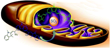
A novel mitochondria-targeted ratiometric fluorescent probe was developed via condensation of carbazole with two indolium units, which could realize the detection of bisulfite in a PBS buffer solution. The probe exhibited good mitochondrial location ability and could selectively respond to bisulfite among other sulfur-containing species in living cells.
Sulfur dioxide (SO2) has been conventionally regarded as an environmental pollutant for several decades. In physiological systems, SO2 mainly exists in sulfite and bisulfite (SO32−/HSO3−), which are widely used as anti-oxidant and antibacterial agents in the food industry.1 Toxicological and epidemiological studies suggest that SO2 and its derivatives are closely associated with various symptoms and diseases including many respiratory problems, neurological disorders and cardiovascular diseases at high concentrations.2 However, recent studies indicated that SO2 could be generated endogenously from sulfur-containing amino acids (cysteine or glutathione) through biosynthetic pathways, which is important in maintaining the biological sulfur balance.3 More recent work have demonstrated that SO2 and its derivatives at low concentrations (less than 450 mM) have vasorelaxing, anti-hypertensive and anti-atherogenic effects and are regarded as novel messengers in cardiovascular system.4 Because of the important roles they play, developing analytical methods that could realize concentration determination and imaging of SO2 and its hydrated derivatives in living cells are highly needed.
Fluorescent probes have been recognized as efficient molecular tools to monitor and visualize bioactive molecules due to their simplicity, convenience and time-saving.5 In the past few years, many probes have been developed to imaging SO2 derivatives in living cells.6–9 Among them, ratiometric fluorescent probes that can eliminate most or all ambiguities by self-calibration at two emission bands, exhibited better performance.7–10 As we know, SO2 mainly generated in mitochondria, therefore, monitoring of SO2 and its hydrated derivatives in mitochondria is particularly important and helpful to understand the mitochondria-directed cell death process.11 However, such mitochondria-targeted ratiometric probes are rare. Not long ago, Yu and Yuan group reported mitochondria-targeted ratiometric fluorescent probes with good selectivity, respectively.9e,10 Inspired by the work of Yu group, herein, we presented another novel carbazole–indolium based ratiometric probe with good water solubility, which could also selectively located in mitochondria and realized the concentration determination of bisulfite in living cells.
We constructed the probe in the same way as Yu's group adopted. The only difference is that two indolium was introduced for DCI, which may result in better water solubility and mitochondria-targeting ability. The probe could be easily synthesized with indolium and carbazolyl dialdehyde through one step condensation reaction, which was further characterized by 1H NMR, 13C NMR and HRMS (Scheme 1).
 |
| Scheme 1 Synthesis route for DCI. | |
With the probe in hand, we first evaluated whether DCI could exhibit optical response towards bisulfite. To a solution of 10 μM DCI in PBS (pH 7.4, 10 mM) was added 100 μM HSO3−, we found that the absorption peak at 508 nm was reduced and a new absorption peak at 295 nm was found (Fig. S1†), accompany by a colour change from light yellow to colourless. On the other hand, in the absence of HSO3−, DCI showed a strong fluorescent emission at 628 nm; with the addition of HSO3− (100 μM) to the solution of 10 μM DCI, the emission at 628 nm would be quenched completely and a new emission would arise at 507 nm (Fig. S2†). In other words, it could realize ratiometric determination of SO2 derivatives. Compared with the probe CZ-Id,9e DCI exhibited similar ratiometric change but with less time to complete the detection (Fig. S3†). Meanwhile, other bio-relevant ions and common sulfur-containing compounds were also investigated to further prove the high selectivity of DCI. As shown in Fig. 1, the addition of 100 equiv. representative anions (CO32−, N3−, AcO−, PO43−, NO2−, F−, Cl−, Br−, I−, SCN−, S2O32−, C2O42−, CN−, N3− and B4O72−) did not cause significantly change of the fluorescence of DCI. Other sulfur-containing compounds (Hcy, Cys and GSH) also could not change the fluorescence emission of the probe. However, once DCI was treated by 10 equiv. HSO3− or SO32−, it exhibited a dramatic ratiometric fluorescence change. These results suggested that DCI could be potential used in imaging of bisulfite in living cells without the interference of Cys and other sulfur-containing compounds.
 |
| Fig. 1 Fluorescence responses of 10 μM DCI toward NaHSO3 (100 μM), Na2SO3 (100 μM), S2− (100 μM), CN− (200 μM), other biological thiols (Cys, Hcy and GSH; 1 mM) and various anions (1 mM). Black bar: DCI + various species. Red bar: DCI + various species + bisulfite. Y-axis represent the fluorescent intensity ratio (I507nm/I625nm), data were acquired in PBS buffer (pH 7.4, 10 mM). λex = 350 nm. Slit: 5 nm/5 nm. | |
Then, fluorescence titration experiments were conducted and the results demonstrated that the emission intensity ratios of the DCI could be amplified evidently upon the addition of HSO3−. As shown in Fig. 2, a linearly proportional increment of emission ratio change to the concentration of HSO3− in the range of 0–40 μM was disclosed, which can be used for the quantification of bisulfite. Quantitative analysis of this approach showed a good limit of detection (LOD), which was calculated to be 30 μM toward bisulfite (S/N = 3, R = 0.999, Fig. S4†). The kinetic study indicated that the sensing process could be completed within 2 min, suggesting the high reactivity of the probe (Fig. S5†).
 |
| Fig. 2 Fluorescence titration spectra of DCI (10 μM) upon the addition of HSO3− (0, 2, 5, 10, 15, 20, 30, 40, 50, 60, 80, 100, 150, 300 μM) λex = 350 nm, slit: 5 nm/5 nm. | |
The sensing mechanism was then investigated. Since two reaction sites existed in DCI, we expected that the double addition product would be formed. As can be seen from the 1H NMR of DCI in the absence and presence of bisulfite, the proton shift of a and b in the double bond between indolium unit and carbazole disappeared, while two new proton shift at 5.15 and 4.63 ppm were observed (Fig. S6†). The addition mechanism undergoes a 1, 4-addition reaction just like Feng group reported.7g Meanwhile, the characteristic peak at 732.2186 (m/z) in HRMS spectra was corresponded to the addition product DCI-SO2 (calcd 732.2178), which further confirmed the double-addition reaction mechanism (Fig. S7†). Additionally, HPLC analysis was performed and further proved the transformation from DCI to DCI-SO2 (Fig. S8†).
A standard MTT assay in A549 cells indicated that more than 90% of cells still remained alive even though 6 μM DCI was internalized for 24 h (Fig. S9†), demonstrating the acceptable cytotoxicity of probe. As reported in the literature, the indolium moiety not only can improve the water solubility of the probe but act as a mitochondria-targeted carrier. Since DCI have two indolium units, we expect it will exhibit good mitochondria-targeting ability. To prove this, DCI as well as Mito Tracker Deep Red (MT Deep Red) were employed for the co-localization studies. As depicted in Fig. 3, the fluorescence signals in DCI-loaded HeLa cells overlayed well with the fluorescence of MT Deep Red (570–620 nm). And the Pearson's co-localization coefficient was calculated to be 0.98, demonstrating that DCI could be site-specifically internalized in mitochondria in living cells (Scheme 2).
 |
| Fig. 3 HeLa cells were stained with DCI (4 μM) and MT Deep Red (1 μM) for 30 min. (a): DCI; (b): MT Deep Red; (c): merged images of (a) and (b), (d): bright field of (a); (e): co-localization image across of HeLa Cells. Ex@488 nm for the green channel (540–680 nm), and Ex@638 nm for MT Deep Red (700–780 nm). | |
 |
| Scheme 2 The reaction mechanism. | |
In light of the excellent cellular penetrability and biocompatibility of DCI, further biological application imaging of bisulfite in living cells was then expanded. We incubated the DCI with HeLa cells at 37 °C for 30 min, fluorescence was clearly observed through green channels (Fig. 4b). Ratio images were obtained with images collected from green (450–530 nm) and blue (600–700 nm) channels, and the fluorescence ratio (Fgreen/Fblue) was calculated to be less than 1.5 (Fig. 4m). Upon addition of HSO3− (20 μM) to DCI-loaded HeLa cells (incubated for another 15 min), the fluorescence intensity of green channel decreased significantly along with slight increment of signals in the blue channel (Fig. 4e and f), and fluorescence ratio increase to 30 (Fig. 4m). On the other hand, N-ethylmaleimide (NEM), which was reported to serves as an intracellular thiol scavenger, was adopted in a control experiment.11,12 The HeLa cells were pre-incubated with NEM which prior to DCI staining for 30 min, followed by treatment HSO3−. We could observe the strong signals in green channels, and the fluorescence ratio (Fgreen/Fblue) was the same as only DCI-load cells (Fig. 4k). From these experiments we can conclude that the observed fluorescence ratio increase is related to existence of HSO3−. Furthermore, fluorescence signal of the NEM pre-treated cells is similarly to that not treated with bisulfite, indicating the decrement of fluorescence signals in green channels is resulted in bisulfite rather than other intracellular thiols within cells.
 |
| Fig. 4 (a) Fluorescence imaging of HeLa cells incubated with DCI (4 μM) from the blue channel; (b) fluorescence imaging of (a) from the green channel; (c) overlay of (a) and (b); (d) bright field of (a). (e) Fluorescence imaging of HeLa cells incubated with DCI (4 μM) for 30 min and further incubated with SO2 donor (60 μM) for 15 min from the blue channel; (f) fluorescence imaging of (e) from the green channel; (g) overlap of (e) and (f); (h) bright field of (e). (i) Fluorescence image of HeLa cells treated with NEM (1 mM) for 15 min, DCI (4 μM) for 30 min, and then incubated with synthetic SO2 donor (60 μM) for 15 min from the blue channel; (j) fluorescence imaging of (i) from the green channel; (k) overlap of (i) and (j); (l) bright field of (i). Ex@405 nm for the blue channel (425–520 nm), Ex@488 nm for the green channel (540–680 nm); (m) statistical analysis were performed with Student's t-test (n = 10 fields of cells). ***P < 0.001, **P < 0.01. | |
Conclusions
In summary, we have developed a mitochondria-targeted fluorescent probe (DCI), which exhibited highly sensitive and selective ability for the sensing of bisulfite/sulfite via ratiometric fluorescence detection. With two indolium moiety was incorporated in the structure, DCI exhibited good water solubility and mitochondria-targeting ability, which was proved by co-localization experiments with MT Deep Red. Meanwhile, DCI was successfully applied in the imaging of bisulfite in living cells.
Preparation and characterization of DCI
3,6-Diformyl-9-methyl carbazole and 1,2,3,3-tetramethyl-3H-indol-1-ium iodide were synthesized according to the literature.13
A mixture of 3,6-diformyl-9-ethyl carbazole (119 mg, 0.5 mmol), 1,2,3,3-tetramethyl-3H-indolium iodide (332 mg, 1.1 mmol), piperidine (1 drop) in ethanol (15 mL) was heated to reflux and stirred overnight. The reaction mixture was cooled to room temperature and filtered. The solid was rinsed with 20 mL of cool methanol and dried over vacuum oven overnight to give 100 mg of desired product as a red solid in 30% yield.
1H NMR (400 MHz, DMSO) δ 9.27 (s, 2H), 8.61 (d, J = 16.1 Hz, 2H), 8.44 (d, J = 8.5 Hz, 2H), 8.01–7.81 (m, 6H), 7.77 (d, J = 16.3 Hz, 2H), 7.67–7.48 (m, 4H), 4.19 (s, 6H), 4.04 (s, 3H), 1.89 (d, J = 47.6 Hz, 12H). 13C NMR (100 MHz, CD3OD, δ) 181.8, 154.8, 145.1, 143.8, 142.4, 129.4, 127.6125.2, 123.6, 123.3, 115.3, 111.7, 111.0, 52.4, 35.2, 30.6, 26.2. MS (ESI) m/z for C39H39IN3, 676.2189, found, 677.2267 (M + H).
Acknowledgements
This work was financially supported by 2010 Science and Technology Support Project on Modernization of Traditional Chinese Medicine (No. 2010-5012) by Guizhou Province of China, and the National Science Foundation of China 81302639. We want to thanks Dr Kun Li (College of Chemistry, Sichuan University) for his kindly advices of the experimental.
Notes and references
- D. H. Daniels, F. L. Joe Jr, C. R. Warner, S. D. Longfellow, T. Fazio and G. W. Diachenko, Food Addit. Contam., 1992, 9, 283–289 CrossRef CAS PubMed.
-
(a) S. Iwasawa, Y. Kikuchi, Y. Nishiwaki, M. Nakano, T. Michikawa and T. Tsuboi, J. Occup. Health, 2009, 51, 38–47 CrossRef CAS PubMed;
(b) L. Migliore and F. Coppede, Mutat. Res., 2009, 674, 73–84 CrossRef CAS PubMed;
(c) N. Sang, Y. Yun, G. Yao, H. Li, L. Guo and G. Li, Toxicol. Sci., 2011, 124, 400–413 CrossRef CAS PubMed;
(d) S. Iwasawa, Y. Kikuchi, Y. Nishiwaki, M. Nakano, T. Michikawa, T. Tsuboi, S. Tanaka, T. Uemura, A. Ishigami and H. Nakashima, J. Occup. Health, 2009, 51, 38–47 CrossRef CAS PubMed;
(e) N. Sang, Y. Yun, H. Li, L. Hou, M. Han and G. Li, Toxicol. Sci., 2010, 114, 226–236 CrossRef CAS PubMed.
- M. H. Stipanuk and I. Ueki, J. Inherited Metab. Dis., 2011, 34, 17–32 CrossRef CAS PubMed.
-
(a) X.-B. Wang, H.-F. Jin, C.-S. Tang and J.-B. Du, Clin. Exp. Pharmacol. Physiol., 2010, 37, 745–752 CrossRef CAS PubMed;
(b) Y. Liang, D. Liu, T. Ochs, C. Tang, S. Chen, S. Zhang, B. Geng, H. Jin and J. Du, Lab. Invest., 2011, 91, 12–23 CrossRef CAS PubMed;
(c) J. Li, R. Li and Z. Meng, Eur. J. Pharmacol., 2010, 645, 143–150 CrossRef CAS PubMed;
(d) H. F. Jin, Y. Wang, X. B. Wang, Y. Sun, C. S. Tang and J. B. Du, Nitric Oxide-Biol. Chem., 2013, 32, 56–61 CrossRef CAS PubMed;
(e) X.-B. Wang, H.-F. Jin, C.-S. Tang and J.-B. Du, Eur. J. Pharmacol., 2011, 670, 1–6 CrossRef CAS PubMed;
(f) L.-M. Luo, S. Chen, H.-F. Jin, C.-S. Tang and J.-B. Du, Biochem. Biophys. Res. Commun., 2011, 415, 61–67 CrossRef CAS PubMed.
-
(a) M.-Y. Wu, T. He, K. Li, M.-B. Wu, Z. Huang and X.-Q. Yu, Analyst, 2013, 138, 3018–3025 RSC;
(b) N. Bones, V. Leen and W. Dehaen, Chem. Soc. Rev., 2012, 41, 1130–1172 RSC;
(c) L. Yuan, W. Lin, K. Zheng, L. He and W. Huang, Chem. Soc. Rev., 2013, 42, 622–661 RSC.
-
(a) M.-Y. Wu, K. Li, C.-Y. Li, J.-T. Hou and X.-Q. Yu, Chem. Commun., 2014, 50, 183–185 RSC;
(b) H.-Y. Tian, J.-H. Qian, Q. Sun, C.-J. Jiang, R.-S. Zhang and W.-B. Zhang, Analyst, 2014, 139, 3373–3377 RSC;
(c) X.-F. Yang, M. Zhao and G. Wang, Sens. Actuators, B, 2011, 152, 8–13 CrossRef CAS;
(d) G.-Y. Li, Y. Chen, J.-Q. Wang, Q. Lin, J. Zhao, L.-N. Jia and H. Chao, Chem. Sci., 2013, 4, 4426–4433 RSC;
(e) L. Tan, W.-Y. Lin, S.-S. Zhu, L. Yuan and K.-B. Zheng, Org. Biomol. Chem., 2014, 12, 4637–4643 RSC.
-
(a) Y.-Q. Sun, J. Liu, J.-Y. Zhang, T. Yang and W. Guo, Chem. Commun., 2013, 49, 2637–2639 RSC;
(b) K. Chen, Y. Guo, Z. Lu, B. Yang and Z. Shi, Chin. J. Chem., 2010, 28, 55–60 CrossRef CAS;
(c) J.-H. Song, D. Zhang, Y.-Q. Liu, Y.-F. Zhao and Y. Ye, New J. Chem., 2015, 39, 6284–6288 RSC;
(d) Y.-Q. Sun, P. Wang, J. Liu, J. Zhang and W. Guo, Analyst, 2012, 137, 3430–3433 RSC;
(e) L.-H. Geng, X.-F. Yang, Y.-G. Zhong, Z. Li and H. Li, Dyes Pigm., 2015, 120, 213–219 CrossRef CAS;
(f) Y. Yang, F. Huo, J. Zhang, Z. Xie, J. Chao, C. Yin, H. Tong, D. Liu, S. Jin, F. Cheng and X. Yan, Sens. Actuators, B, 2012, 665, 166–167 Search PubMed;
(g) Q. Zhang, Y. Zhang, S.-S. Ding, H.-Y. Zhang and G.-Q. Feng, Sens. Actuators, B, 2015, 211, 377–384 CrossRef CAS.
-
(a) G. Wang, H. Qi and X.-F. Yang, Luminescence, 2012, 28, 97–101 CrossRef PubMed;
(b) X.-H. Cheng, H.-Z. Jia, J. Feng, J.-G. Qin and Z. Li, Sens. Actuators, B, 2013, 184, 274–280 CrossRef CAS;
(c) C.-C. Wang, S. Feng, L.-Y. Wu, S.-Y. Yan, C. Zhong, P. Guo, R. Huang, X.-C. Weng and X. Zhou, Sens. Actuators, B, 2014, 190, 792–799 CrossRef CAS;
(d) M.-J. Peng, X.-F. Yang, B. Yin, Y. Guo, F. Suzenet, D. En, J. Li, C.-W. Li and Y.-W. Duan, Chem.–Asian J., 2014, 9, 1817–1822 CrossRef CAS PubMed;
(e) W.-Q. Chen, Q. Fang, D.-L. Yang, H.-Y. Zhang, X.-Z. Song and J. W. Fole, Anal. Chem., 2015, 87, 609–616 CrossRef CAS PubMed;
(f) X.-H. Cheng, H.-Z. Jia, J. Feng, J.-G. Qin and Z. Li, J. Mater. Chem. B, 2013, 1, 4110–4114 RSC;
(g) M.-T. Sun, H. Yu, K. Zhang, Y.-J. Zhang, Y.-H. Yan, D.-J. Huang and S.-H. Wang, Anal. Chem., 2014, 86, 9381–9385 CrossRef CAS PubMed;
(h) C.-Y. Liu, L.-Y. Liu, X. Li, C.-X. Shao, X. Huang, B.-C. Zhu and X.-L. Zhang, RSC Adv., 2014, 4, 33507–33513 RSC.
-
(a) X.-J. Liu, Q.-W. Yang, W.-Q. Chen, L.-N. Mo, S. Chen, J. Kang and X.-Z. Song, Org. Biomol. Chem., 2015, 13, 8663–8668 RSC;
(b) W.-Q. Chen, X.-J. Liu, S. Chen, X.-Z. Song and J. Kang, RSC Adv., 2015, 5, 25409–25415 RSC;
(c) L.-M. Zhu, J.-C. Xu, Z. Sun, B.-Q. Fu, C.-Q. Qin, L.-T. Zeng and X.-C. Hu, Chem. Commun., 2015, 51, 1154–1156 RSC;
(d) Z.-X. Liu, S.-J. Guo, J.-Y. Piao, X. Zhou and X. Wu, RSC Adv., 2014, 4, 54554–54557 RSC;
(e) Y. Liu, K. Li, M.-Y. Wu, Y.-H. Liu, Y.-M. Xie and X.-Q. Yu, Chem. Commun., 2015, 51, 10236–10239 RSC;
(f) J. Yang, K. Li, J.-T. Hou, L.-L. Li, C.-Y. Lu, Y.-M. Xie, X. Wang and X.-Q. Yu. ACS Sens., DOI:10.1021/acssensors.5b00165.
- W. Xu, C. L. Teoh, J. Peng, D. Su, L. Yuan and Y.-T. Chang, Biomaterials, 2015, 56, 1–9 CrossRef CAS PubMed.
- S.-X. Du, H.-F. Jin, D.-F. Bu, X. Zhao, B. Geng, C.-S. Tang and J.-B. Du, Acta Pharmacol. Sin., 2008, 29, 923–930 CrossRef CAS PubMed.
- S. R. Malwal, D. Sriram, P. Yogeeswari, V. B. Konkimalla and H. Chakrapani, J. Med. Chem., 2012, 55, 553–557 CrossRef CAS PubMed.
-
(a) B. Jin, X. Zhang, W. Zheng, X.-J. Liu, C. Qi, F.-Y. Wang and D.-H. Shangguan, Anal. Chem., 2014, 86, 943–952 CrossRef CAS PubMed;
(b) E. A. Owens, N. Bruschi, J. G. Tawney and M. Henary, Dyes Pigm., 2015, 113, 27–37 CrossRef CAS.
Footnote |
† Electronic supplementary information (ESI) available: The NMR and HRMS spectra of the probe, details information for experiments. See DOI: 10.1039/c5ra27805h |
|
This journal is © The Royal Society of Chemistry 2016 |
Click here to see how this site uses Cookies. View our privacy policy here.