DOI:
10.1039/C5RA27605E
(Paper)
RSC Adv., 2016,
6, 15831-15837
Chain-like Fe3O4@resorcinol-formaldehyde resins–Ag composite microstructures: facile construction and applications in antibacterial and catalytic fields†
Received
24th December 2015
, Accepted 26th January 2016
First published on 28th January 2016
Abstract
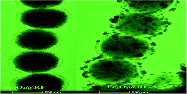
A facile two-step route is designed to synthesize chain-like Fe3O4@resorcinol-formaldehyde resins–Ag composite microstructures (denoted as Fe3O4@RF–Ag). Chain-like Fe3O4@RF microstructures are firstly obtained by the in situ polymerization of RF on Fe3O4 microspheres under the assistance of an external magnetic field; and then, Ag nanoparticles are strewn on the surface of the RF via in situ reduction by hydrazine hydrate. The as-obtained chain-like Fe3O4@RF–Ag microstructures are characterized by powder X-ray diffraction (XRD), scanning electron microscopy (SEM), (high-resolution) transmission electron microscopy (HRTEM/TEM) and energy dispersive spectrometry (EDS). Experiments indicate that the as-prepared chain-like Fe3O4@RF–Ag microstructures can not only serve as outstanding antibacterial agents against bacteria including Staphylococcus aureus and Escherichia coli, but also as highly efficient catalysts for the hydrogenation of various compounds, including 4-nitrophenol, methylene blue and Rhodamine B. Also, the as-prepared chain-like Fe3O4@RF–Ag microstructures can be conveniently regenerated under extra magnetic field due to the presence of Fe3O4 and they show good cyclic stability, which is important for practical applications.
1. Introduction
One-dimensional (1D) magnetic nanostructures have been attracting increasing attention in recent years because of their extensive potential applications in fields, including quantum computing, targeted delivery, cell control, magnetic resonance imaging (MRI), micromechanical sensors and biomolecule desorption.1–9 To obtain magnetic 1D nanomaterials, several synthetic methods were developed in the past years, including template synthesis,10,11 self-assembly,12,13 and magnetic field-induced (MFI) assembly.14–16 Due to dipole-directed assembly, magnetic nanoparticles easily form a chain-like 1D structure under an external magnetic field. Thus, MFI assembly is considered as a comparatively facile and economical route for fabricating magnetic 1D nanostructures. Once the external magnetic field is removed, however, this ordered magnetic 1D structure can hardly be maintained due to the weak or ignorable anisotropic dipolar interaction between the building blocks.17 Therefore, how to immobilize the magnetic 1D nanochains is a challenge. At present, a “glue” strategy is successfully employed for the stabilization of magnetic 1D nanochains. Many materials have been exploited as the “glue”, including carbon, silica, metals and polymers.18–24
Recently, multifunctional nanostructures formed by various components with different properties have been paid much attention, especially the combination of magnetic properties and other functions.25,26 For instance, Fe3O4/P(MAA–DVB)/TiO2 nanochains were successfully prepared via a polymerization and hydrolysis-hydrothermal route.27 Fe3O4/C@ZnO core–shell microrods were obtained through a facile two-step method.28 The as-produced nanochains and microrods had magnetic and semiconducting performances and could be used as photocatalysts for water treatment.27,28 Moreover, Yang et al. synthesized Fe3O4@PZS–Pd nanochains, which were employed as catalytically active magnetic stirring bars.29 Due to the presence of magnetic Fe3O4 nanoparticles, importantly, the above multifunctional nanostructures could be readily recycled under an extra magnetic field, which is favourable for practical applications.
As one typical noble metal nanocrystal, nanostructured Ag particles always receive much interest due to their unique properties and extensive applications. It has been discovered that Ag nanoparticles bear excellent catalytic activity and strong antibacterial ability.30–35 However, the aggregation of Ag nanoparticles and high cost limit their wide practical applications.36–38 In the current work, we constructed Ag nanoparticle-strewn Fe3O4@resorcinol-formaldehyde resin chain-like microstructures, denoted as Fe3O4@RF–Ag, through a two-step route. Firstly, under an external magnetic field RF was in situ polymerized on Fe3O4 microspheres to form chain-like Fe3O4@RF microstructures; and then, chain-like Fe3O4@RF–Ag microstructures were obtained through the reduction of AgNO3 in the system with Fe3O4@RF. Experiments showed that the as-obtained chain-like Fe3O4@RF–Ag microstructures had strong antibacterial ability to Staphylococcus aureus (S. aureus) and Escherichia coli (E. coli), and outstanding catalytic activity for the hydrogenation of organic small molecules such as 4-nitrophenol (4-NP), methylene blue (MB), and Rhodamine B (RdB). Compared with the reported literature, several features can be found in the present work: (1) RF is used as the “glue” for the formation of magnetic nanochains; (2) Ag nanoparticles can be homogeneously dispersed on the surfaces of Fe3O4@RF microstructures due to the interaction between the functional groups of RF and Ag nanoparticles, which prevents the aggregation of Ag nanoparticles; (3) the loaded amount of Ag nanoparticles on the chain-like microstructures can be controlled; and (4) the catalyst and antibacterial reagent can be easily recycled under the extra magnetic field, which can efficiently reduce the cost in practical applications.
2. Experimental
All reagents and chemicals are analytically pure, bought from Shanghai Chemical Company and used without further purification.
2.1 Preparation of chain-like Fe3O4@RF microstructures
Magnetic Fe3O4 microspheres were firstly prepared by a modified hydrothermal method. Typically, 1.0 g FeCl3·6H2O and 0.3 g trisodium citrate dihydrate were first dissolved in 30 mL ethylene glycol under vigorous stirring. Then 1.8 g urea was added with stirring for 30 min, and the mixture was sealed in a Teflon-lined stainless-steel autoclave of 50 mL capacity and heated at 200 °C for 12 h. After cooling down to room temperature naturally, the black product was separated magnetically and washed with ethanol and deionized water several times, and finally dried under vacuum at 60 °C for 6 h. To obtain chain-like Fe3O4@RF microstructures, 0.1 g Fe3O4 nanospheres was added into a 250 mL three-necked round-bottom flask with 100 mL mixed solvent of absolute ethanol (30 mL) and deionized water (70 mL). After ultrasonication for 10 min, 0.3 mL 25 wt% ammonia solution and 0.1 g resorcinol were in turn introduced into the above system. The mixed system was mechanically stirred at 35 °C for 30 min. Then, 0.14 mL formaldehyde solution was added dropwise under continuous stirring. After stirring at 35 °C for another 6 h, the system was naturally cooled down to room temperature and aged overnight without stirring. Finally, the precipitate was collected by a magnet, washed with distilled water and ethanol several times, and air-dried at 60 °C overnight.
2.2 Formation of chain-like Fe3O4@RF–Ag microstructures
To prepare chain-like Fe3O4@RF microstructures with different amounts of Ag nanoparticles, 50 mg of the as-prepared Fe3O4@RF was firstly added into 100 mL AgNO3 ethanol solutions with concentrations of 1.43, 4.29 and 7.14 mmol L−1, respectively, under vigorous stirring. Then, 1 mL 2 mol L−1 N2H4·H2O was injected into the above system dropwise. After the system was stirring at room temperature for 1 h, the product was collected by a magnet, washed with deionized water and ethanol several times, and dried under vacuum. The as-prepared products were labeled as Fe3O4@RF–Ag-1, Fe3O4@RF–Ag-2 and Fe3O4@RF–Ag-3 in turn.
Scheme 1 depicts the formation process of the chain-like Fe3O4@RF–Ag microstructures.
 |
| Scheme 1 Preparation strategy of the chain-like Fe3O4@RFs–Ag core–shell microstructures. | |
2.3 Characterization
X-ray powder diffraction (XRD) patterns were measured on a Shimadzu XRD-6000 X-ray diffractometer equipped with Cu Kα radiation (λ = 0.154060 nm), employing a scanning rate of 0.02° s−1. TEM images were obtained on a FEI Tecnai G2 20 transmission electron microscope, employing an accelerating voltage of 200 kV. FESEM images and energy dispersive spectrometry (EDS) of the product were taken on a Hitachi S-4800 field emission scanning electron microscope, employing an accelerating voltage of 5 kV and 15 kV, respectively. The room temperature hysteresis loops of the products were measured using a superconducting quantum interference device (SQUID) operating at room temperature (300 K) with an applied field up to 1.0 T.
2.4 Performance tests
2.4.1 Antibacterial activity of Fe3O4@RF–Ag. To investigate the antibacterial activity of the as-obtained chain-like Fe3O4@RF–Ag microstructures, 10.0 mg of Fe3O4@RF–Ag microstructures was dispersed in 20.0 mL of nutrient solution with 1% trypton, 0.5% yeast strain and 1% sodium chloride. The pH of the nutrient solution was adjusted to 7.0 by NaOH solution. Then, various Fe3O4@RF–Ag suspensions with a concentration of 250, 125, 62.5, 31.2, 15.6, 7.8, 3.9, 1.95, and 0.98 μg mL−1 obtained by the half-dilution method were introduced into different Erlenmeyer flasks. Finally, 100 μL S. aureus (or E. coli) solution with a concentration of 107 cfu mL−1 (cfu: colony-forming units) was introduced into each of the flasks and shaken to mix the suspension. All the flasks were incubated at 37 °C for 24 h.To evaluate the antibacterial activity of the chain-like Fe3O4@RF–Ag microstructures, the optical density (OD) value of 100 μL suspension was measured on a Thermo Fisher Multiskan GO-1510 spectrophotometer at 540 nm via ELIASA.
2.4.2 Catalytic activity of Fe3O4@RF–Ag. To study the catalytic activity of the as-prepared chain-like Fe3O4@RF–Ag microstructures, the reductions of some organic small molecules including 4-nitrophenol (4-NP), methylene blue and Rhodamine B in excess NaBH4 solution were chosen as the model reactions. In a typical procedure, appropriate amounts of organic compounds and the catalysts were firstly mixed in a small amount of distilled water. Then, a certain volume of NaBH4 solution was introduced into the above system to form a 3 mL solution. Here, the concentrations of the organic compound, NaBH4 and the catalyst were 0.5 × 10⁻⁴ mol L−1, 2.0 × 10⁻² mol L−1 and 10 mg L−1, respectively. The reducing reaction processes were monitored with a Metash 6100 UV-vis spectrophotometer (Shanghai) in the spectral range from 250 to 550 nm. The catalyst was recycled using a magnet, washed 3 times with NaBH4 solution and dried under vacuum at 60 °C for reuse.
3. Results and discussion
3.1 Morphology and structure characterization
Fig. 1a depicts a FESEM image of Fe3O4 prepared by solvothermal technology. The microspheres with an average diameter of ∼300 nm were made of abundant smaller nanoparticles and their surfaces were not smooth, which was proven by TEM observations (Fig. 1b). After the RF polymerization took place in the system containing Fe3O4 microspheres under the external magnetic field, a great deal of chain-like structures assembled by microspheres was obtained (Fig. 1c). The typical TEM image shown in Fig. 1d clearly displays the core–shell structure with an average shell thickness of ∼40 nm. This uncovers that the formation of the chain-like Fe3O4@RF structures is attributed to the immobilization of the RF resin. After N2H4·H2O was dropped into the AgNO3 solution containing the chain-like Fe3O4@RF microstructures, chain-like Fe3O4@RF–Ag microstructures were finally obtained. Fig. 1e exhibits a representative FESEM image of the final product gained from the system with 1.43 mmol L−1 AgNO3 (labeled as Fe3O4@RF–Ag-2). Ag nanoparticles with a mean size of ∼20 nm are distributed on the surfaces of the chain-like Fe3O4@RF microstructures. A typical TEM image of Fe3O4@RF–Ag-2 is depicted in Fig. 1f. Compared with Fig. 1d, the Ag nanoparticles have been successfully anchored on the surfaces of the chain-like microstructures, which is in agreement with the FESEM result. The inset shown in Fig. 1f depicts a HRTEM image of a nanoparticle. The clear crystal fringes indicate the good crystallinity of the nanoparticles. The d-spacing between neighbouring planes is measured to be 0.24 nm, which is very close to 0.23587 nm of the (111) plane of Ag.
 |
| Fig. 1 FESEM and TEM images of various products: (a and b) Fe3O4 microspheres, (c and d) Fe3O4@RF microstructures and (e and f) Fe3O4@RF–Ag-2 microstructures. The inset in (f) is a HRTEM image of Ag nanoparticles. | |
The formation of Fe3O4@RF–Ag microstructures is also proven by XRD diffraction. Fig. 2 compares the XRD patterns of the products before and after Ag nanoparticles being strewn on the surfaces of Fe3O4@RF. By comparison with the data of JCPDS card files no. 89-2355, only diffraction peaks of Fe3O4 are detected before the Ag nanoparticles are strewn. After strewing the Ag nanoparticles, the diffraction peaks belonging to Ag can be also found at 38.1, 44.2, 64.5 and 77.4° besides those of Fe3O4. Based on the calculations of the (311) plane of Fe3O4 and the (111) plane of Ag, their sizes are in turn 12.3 nm and 24.6 nm. The small particle size of Fe3O4 proves the results of FESEM and TEM observations: big Fe3O4 microspheres were made of abundant Fe3O4 nanoparticles. Moreover, the magnetic property studies show that the as-obtained Fe3O4 microspheres were superparamagnetic. After forming chain-like Fe3O4@RF and Fe3O4@RF–Ag-2 microstructures, the saturation magnetization reduced in turn (Fig. 3).
 |
| Fig. 2 XRD patterns of Fe3O4@RF (a), and Fe3O4@RF–Ag-2 (b). | |
 |
| Fig. 3 The room temperature hysteresis loops of Fe3O4 microspheres, Fe3O4@RF and Fe3O4@RF–Ag-2. | |
It was found that changing the concentration of the AgNO3 solution could affect the loading amount of Ag nanoparticles on the surfaces of the chain-like Fe3O4@RF microstructures. Fig. 4 depicts FESEM images of the products obtained from the AgNO3 solutions with concentrations of 1.43 and 7.14 mmol L−1. Comparing with Fig. 1e, one can readily see the density increase of Ag nanoparticles on the surfaces of Fe3O4@RF with the increase of the concentration of the AgNO3 solution. The above Ag amount increase is also confirmed by the EDS analyses of the products. As shown in Fig. 4, Ag peaks increase with the increase of AgNO3 concentration from 1.43 mmol L−1 to 7.14 mmol L−1. The inset of Fig. 5 lists the content of all components for the three composites. The loading amounts of Ag nanoparticles (atom%) are in turn 2.81 for Fe3O4@RF–Ag-1, 7.44 for Fe3O4@RF–Ag-2 and 12.5 for Fe3O4@RF–Ag-3.
 |
| Fig. 4 FESEM images of Fe3O4@RF–Ag-1 (a), and Fe3O4@RF–Ag-3 (b), obtained from AgNO3 solutions with concentrations of 1.43 and 7.14 mmol L−1, respectively. | |
 |
| Fig. 5 EDS analyses of Fe3O4@RF–Ag-1, Fe3O4@RF–Ag-2 and Fe3O4@RF–Ag-3. | |
3.2 Antibacterial activity
It was found that the as-obtained chain-like Fe3O4@RF–Ag microstructures could strongly restrain the propagation of some bacteria such as S. aureus (Gram-positive) and E. coli (Gram-negative). Fig. 6 shows the OD value changes of S. aureus and E. coli solutions with time before and after introducing 125 μg mL−1 Fe3O4@RF–Ag-2. The same change trends can be found in two bacterial solutions: the OD values rapidly rise with duration before introducing Fe3O4@RF–Ag-2 microstructures; after Fe3O4@RF–Ag-2 microstructures are added the OD values firstly decrease, and then only slightly increase within 6 h. The above experimental phenomena show that the propagation of S. aureus/E. coli is restrained after Fe3O4@RF–Ag-2 microstructures are introduced into the system. However, Fe3O4@RF microstructures do not possess the above ability (Fig. 6), implying that the Ag nanoparticles were the active material to restrain the propagation of bacteria. Li et al. considered that Ag nanoparticles could damage the structure of bacterial cell membranes and depress the activity of some membranous enzymes, which causes bacteria to die eventually.39 Fig. S1† depicts the concentration influences of Fe3O4@RF–Ag-2 on the propagation of S. aureus and E. coli. When no Fe3O4@RF–Ag-2 existed in the nutrient solutions containing the two bacteria, both bacteria rapidly propagated after incubation at 37 °C for 24 h. Since abundant bacteria existed in the systems, high OD values were detected. Meanwhile, the system became yellow for S. aureus (inset in Fig. S1a†) and brown for E. coli (inset in Fig. S1b†). With the concentration increase of the chain-like microstructures, the OD values of the two bacteria gradually decreased. Simultaneously, the colors of the systems gradually became pale and closer to the color of the pure nutrient solution (inset given in Fig. S1†). Obviously, increasing the concentration of Fe3O4@RF–Ag-2 is favourable for restraining the propagation of bacteria. In addition, the present antibacterial agent could be easily regenerated owing to its magnetism, which is important in practical applications for reducing the cost. Fig. S2a† exhibits the antibacterial efficiency change of the present antibacterial agent to S. aureus with the cycle time. After cycling for 5 times the antibacterial efficiency still reaches ∼83%. Furthermore, experiments also confirmed that with the loading amount increase of Ag nanoparticles the antibacterial ability of the antibacterial agent could be enhanced (see Fig. S2b†). The above facts indicate that the chain-like Fe3O4@RF–Ag microstructures obtained by the present experimental route are promising antibacterial agents, and have potential applications in many fields relating to the routine lives of human beings.
 |
| Fig. 6 The OD value changes of S. aureus (black lines) and E. coli (red lines) after incubation at 37 °C for various durations in the absence/presence of Fe3O4@RF–Ag-2 microstructures of 125 μg mL−1. | |
3.3 Catalytic activity for the reduction of organic small molecules
Furthermore, the as-obtained chain-like Fe3O4@RF–Ag microstructures also presented excellent catalytic activity for the reduction of some organic small molecules such as 4-nitrophenol (4-NP), methylene blue (MB) and Rhodamine B (RdB). Fig. S3a† shows the UV-vis absorption spectra of the 4-NP–NaBH4 system in the presence of 10 mg L−1 Fe3O4@RF–Ag-2 at various reaction durations. The strong absorption peak at 400 nm, which originates from the intermediate formed by 4-NP and NaBH4,40,41 gradually decreases with the prolonging of the reaction duration. Simultaneously, a new peak centered at ∼300 nm appears, which belongs to the characteristic absorption peak of 4-aminophenol (4-AP). After reacting for 6 min, the peak at 400 nm disappears, indicating the completion of the reduction reaction of 4-NP. Experiments also showed that the loading amount of Ag nanoparticles could markedly affect the reductive rate of 4-NP. As shown in Fig. 7a, when Fe3O4@RF was added into the 4-NP–NaBH4 system, no reaction was initiated. While Fe3O4@RF–Ag-1 and Fe3O4@RF–Ag-3 nanochains with the same concentration were separately selected as the catalyst, it took 10 min and 2 min in turn. Obviously, increasing the loading amount of Ag nanoparticles on the surfaces of the chain-like microstructures can promote the reduction of 4-NP. Since excess NaBH4 was used, the reduction of 4-NP to 4-AP could be reasonably assumed to follow pseudo-first-order kinetics with regard to 4-NP. The corresponding kinetic equation can be described as follows:
where C and C0 are the instantaneous and initial concentration of 4-NP, respectively, and k and t in turn represent the rate constant and the reaction time. Fig. 7b depicts the linear relation between ln(C/C0) and the reaction time in the presences of various catalysts. The corresponding rate constants are calculated to be 0.317 min−1 for Fe3O4@RF–Ag-1, 0.676 min−1 for Fe3O4@RF–Ag-2 and 0.749 min−1 for Fe3O4@RF–Ag-3, respectively. Moreover, compared with some previous reports (Table 1), the present Fe3O4@RF–Ag catalyst also presented better catalytic activity for the reduction of 4-NP. More importantly, due to the magnetism of the chain-like microstructures, the present catalyst could be easily reused. As seen from Fig. S3b,† the catalytic efficiency of the catalyst hardly decreases after 5 runs. Similar experimental phenomena were also found in the reductions of MB and RdB.
 |
| Fig. 7 (a) Conversion curves of 4-NP to 4-AP and (b) the linear relationship between ln(C/C0) and the reaction time in the presence of 10 mg L−1 various catalysts. | |
Table 1 Comparison of the catalytic capacities of various catalysts reported in the literature for the reduction of 4-NP to 4-AP by NaBH4
Catalyst |
Rate constant (min−1) |
Reference |
Fe3O4@RF–Ag-1 nanochains |
0.317 |
This work |
Fe3O4@RF–Ag-2 nanochains |
0.676 |
This work |
Fe3O4@RF–Ag-3 nanochains |
0.749 |
This work |
PPy/Pd nanocapsules |
0.532 |
42 |
Polygonal Au nanoparticles |
0.660 |
43 |
Ag nanodendrites |
0.149 |
44 |
Hierarchical Ag microstructures |
0.251 |
45 |
Fig. S4† describes the UV-vis absorption spectra of the MB–NaBH4 and RdB–NaBH4 system in the presence of 10 mg L−1 Fe3O4@RF–Ag-2 at various reaction durations. With the extension of the reaction time, the absorption peaks of the two dyes gradually decreased. Fig. 8 shows the correlation curves of the MB/RdB concentration vs. time in the presence of 10 mg L−1 various catalysts. When no Ag was loaded on the surfaces of the chain-like microstructures, the reduction reactions did not take place. With the increase of the Ag loading amount from 2.81 to 7.44 and to 12.5 atom%, it took 8 min, 6 min and 4 min for the complete reduction of MB, and 8 min, 6 min and 3 min for the complete reduction of RdB in turn. Nevertheless, a slight difference can be found during the reductions of the above two dyes. In the initial stage, the reduction of RdB is slow. Then, the reaction is accelerated. This fact implies the presence of an inducing period in the reduction of RdB. However, no inducing periods are found in the reductions of 4-NP and MB.
 |
| Fig. 8 The correlation curves of the dye concentration vs. time in the presence of 10 mg L−1 various catalysts: (a) MB and (b) RdB. | |
As mentioned previously, the reduction of organic small molecules could not be induced when no Ag was loaded on the surfaces of the chain-like microstructures, indicating that the above reductive reactions were promoted only by Ag nanoparticles. Generally, it is believed that the catalyst acts a medium for the electron transfer from BH4− ions to organic small molecules. In the current work, the possible reactive mechanism is described as follows: BH4− ions firstly reacted with water to free H atoms/H2 molecules. The produced H atoms/H2 molecules were adsorbed on the Ag nanoparticles. At the same time, organic small molecules were also adsorbed on the surfaces of the Ag nanoparticles. Thus, organic small molecules were reduced at last.
4. Conclusions
In summary, chain-like Fe3O4@RF–Ag microstructures have been successfully obtained by the self-assembly of Fe3O4@RF under an external magnetic field and subsequent in situ deposition of Ag nanoparticles on the surfaces of the Fe3O4@RF microstructures. Experiments showed that the loading amount of the Ag nanoparticles on the surfaces of the chain-like Fe3O4@RF microstructures could be tuned by changing the initial concentration of AgNO3. It was found that the as-obtained chain-like Fe3O4@RF–Ag presented excellent antibacterial capacity to bacteria such as S. aureus (Gram-positive) and E. coli (Gram-negative). With increasing the amount of the antibacterial agent, the ability to restrain the propagation of S. aureus and E. coli was obviously promoted. Furthermore, the as-obtained Fe3O4@RF–Ag chain-like microstructures also displayed outstanding catalytic activity for the reduction of some organic small molecules such as 4-nitrophenol, methylene blue and Rhodamine B. In the presence of 10 mg L−1 catalyst, the rate constants were in turn 0.317 min−1 for Fe3O4@RF–Ag-1, 0.676 min−1 for Fe3O4@RF–Ag-2 and 0.749 min−1 for Fe3O4@RF–Ag-3. More importantly, the present antibacterial agent and catalyst could be easily regenerated under an extra magnetic field, and kept good stability. After cycling for 5 times, the antibacterial efficiency was still ∼83% and the catalytic efficiency hardly decreased (for Fe3O4@RF–Ag-2), indicating that the as-obtained chain-like Fe3O4@RF–Ag microstructures are promising multifunctional materials in future practical applications.
Acknowledgements
The authors thank the National Natural Science Foundation of China (21171005 and 21571005) for the fund support.
Notes and references
- D. Gatteschi and R. Sessoli, Angew. Chem., Int. Ed., 2003, 42, 268 CrossRef CAS PubMed.
- L. Zhang, T. Petit, K. E. Peyer and B. J. Nelson, Nanomedicine: Nanotechnology, Biology and Medicine, 2012, 8, 1074 CrossRef CAS PubMed.
- M. F. Contreras, R. Sougrat, A. Zaher, T. Ravasi and J. Kosel, Int. J. Nanomed., 2015, 10, 2141 CrossRef CAS PubMed.
- A. Hultgren, M. Tanase, E. J. Felton, K. Bhadriraju, A. K. Salem, C. S. Chen and D. H. Reich, Biotechnol. Prog., 2005, 21, 509 CrossRef CAS PubMed.
- J. H. Park, G. V. Maltzahn, L. L. Zhang, M. P. Schwartz, E. Ruoslahti, S. N. Bhatia and M. J. Sailor, Adv. Mater., 2008, 20, 1630 CrossRef CAS PubMed.
- Z. G. Yue, W. Wei, Z. X. You, Q. Z. Yang, H. Yue, Z. G. Su and G. H. Ma, Adv. Funct. Mater., 2011, 21, 3446 CrossRef CAS.
- F. Johansson, M. Jonsson, K. Alm and M. Kanje, Exp. Cell Res., 2010, 316, 688 CrossRef CAS PubMed.
- C. Goubault, P. Jop, M. Fermigier, J. Baudry, E. Bertrand and J. Bibette, Phys. Rev. Lett., 2003, 91, 260802 CrossRef CAS PubMed.
- B. Özkale, N. Shamsudhin, G. Chatzipirpiridis, M. Hoop, F. Gramm, X. Z. Chen, X. Martí, J. Sort, E. Pellicer and S. Pané, ACS Appl. Mater. Interfaces, 2015, 7, 7389 Search PubMed.
- D. Yang, Y. P. Zhou, X. H. Rui, J. X. Zhu, Z. Y. Lu, E. Fong and Q. Y. Yan, RSC Adv., 2013, 3, 14960 RSC.
- Y. R. Wang, L. Zhang, X. H. Gao, L. Y. Mao, Y. Hu and X. W. Lou, Small, 2014, 10, 2815 CrossRef CAS PubMed.
- J. Y. Gong, S. Z. Li, D. G. Zhang, X. B. Zhang, C. Liu and Z. W. Tong, Chem. Commun., 2010, 46, 3514 RSC.
- J. C. Jia, J. C. Yu, Y. X. J. Wang and K. M. Chan, ACS Appl. Mater. Interfaces, 2010, 2, 2579 CAS.
- J. F. Zhou, L. J. Meng, X. L. Feng, X. K. Zhang and Q. H. Lu, Angew. Chem., Int. Ed., 2010, 49, 8476 CrossRef CAS PubMed.
- Y. Kim, Y. S. Choi, H. J. Lee, H. Yoon, Y. K. Kim and M. Oh, Chem. Commun., 2014, 50, 7617 RSC.
- J. J. Zhou, C. X. Wang, P. Wang, P. B. Messersmith and H. W. Duan, Chem. Mater., 2015, 27, 3071 CrossRef.
- G. J. Cheng, D. Romero, G. T. Fraser and A. R. H. Walker, Langmuir, 2005, 21, 12056 Search PubMed.
- Z. B. Zhang, H. F. Duan, S. H. Li and Y. J. Lin, Langmuir, 2010, 26, 6676 CrossRef CAS PubMed.
- S. F. Situ and A. C. S. Samia, ACS Appl. Mater. Interfaces, 2014, 6, 20154 CAS.
- Y. X. Hu, L. He and Y. D. Yin, Angew. Chem., Int. Ed., 2011, 50, 3747 CrossRef CAS PubMed.
- V. T. Trana, H. J. Zhou, S. Kim, J. Lee, J. Kim, F. M. Zou, J. Kim, J. Y. Park and J. Lee, Sens. Actuators, B, 2014, 203, 817 CrossRef.
- V. T. Tran, H. J. Zhou, S. Lee, S. C. Hong, J. Kim, S. Y. Jeong and J. Lee, ACS Appl. Mater. Interfaces, 2015, 7, 8650 CAS.
- J. F. Zhou, L. J. Meng, X. L. Feng, X. K. Zhang and Q. H. Lu, Angew. Chem., Int. Ed., 2010, 49, 8476 CrossRef CAS PubMed.
- M. H. Rashid, M. Raula and T. K. Mandal, J. Mater. Chem., 2011, 21, 4904 RSC.
- H. Wang, Q. W. Chen, Y. B. Sun and M. Y. He, J. Phys. Chem. C, 2010, 114, 19660 CAS.
- C. M. Li, J. J. Tan, X. L. Fan, B. L. Zhang, H. P. Zhang and Q. Y. Zhang, Ceram. Int., 2015, 41, 3860 CrossRef CAS.
- Y. Liu, L. Zhou, Y. Hu, C. F. Guo, H. S. Qian, F. M. Zhang and X. W. Lou, J. Mater. Chem., 2011, 21, 18359 RSC.
- Y. R. Wang, J. Q. Ning, E. L. Hu, C. C. Zheng, Y. J. Zhong and Y. Hu, J. Alloys Compd., 2015, 637, 301 CrossRef CAS.
- S. L. Yang, C. Y. Cao, Y. B. Sun, P. P. Huang, F. F. Wei and W. G. Song, Angew. Chem., Int. Ed., 2015, 54, 2661 CrossRef CAS PubMed.
- Q. L. Li, S. Mahendra, D. Y. Lyon, L. Brunet, M. V. Liga, D. Li and P. J. J. Alvarez, Water Res., 2008, 42, 4591 CrossRef CAS PubMed.
- A. Kumar, P. K. Vemula, P. M. Ajayan and G. John, Nat. Mater., 2008, 7, 236 CrossRef CAS PubMed.
- C. Damm, H. Münstedt and A. Rösch, J. Mater. Sci., 2007, 42, 6067 CrossRef CAS.
- N. R. Panyala, E. M. Pena-Mendez and J. Havel, J. Appl. Biomed., 2008, 6, 47 Search PubMed.
- D. Lee, R. E. Cohen and M. F. Rubner, Langmuir, 2005, 21, 9651 CrossRef CAS PubMed.
- L. F. Hakim, J. L. Portman, M. D. Casper and A. W. Weimer, Powder Technol., 2005, 160, 149 CrossRef CAS.
- A. Dong, Y. Sun, S. Lan, Q. Wang, Q. Cai, X. Z. Qi, Y. L. Zhang, G. Gao and F. Q. Liu, ACS Appl. Mater. Interfaces, 2013, 5, 8125 CAS.
- H. C. Dong, J. Y. Huang, R. R. Koepsel, P. L. Ye, A. J. Russell and K. Matyjaszewski, Biomacromolecules, 2011, 12, 1305 CrossRef CAS PubMed.
- D. Wang and D. Astruc, Chem. Rev., 2014, 114, 6949 CrossRef CAS PubMed.
- W. R. Li, X. B. Xie, Q. S. Shi, H. Y. Zeng, Y. S. Ouyang and Y. B. Chen, Appl. Microbiol. Biotechnol., 2010, 85, 1115 CrossRef CAS PubMed.
- F. Guo, Y. H. Ni, Y. Ma, N. N. Xiang and C. Liu, New J. Chem., 2014, 38, 5324 RSC.
- H. S. Zheng, Y. H. Ni, N. N. Xiang, X. Ma and F. Y. Wan, Mater. Chem. Phys., 2015, 158, 82 CrossRef CAS.
- Y. Xue, X. F. Lu, X. J. Bian, J. Y. Lei and C. Wang, J. Colloid Interface Sci., 2012, 379, 89 CrossRef CAS PubMed.
- M. H. Rashid and T. K. Mandal, Adv. Funct. Mater., 2008, 18, 2261 CrossRef CAS.
- W. Zhang, F. T. Tan, W. Wang, X. L. Qiu, X. L. Qiao and J. G. Chen, J. Hazard. Mater., 2012, 36, 217 Search PubMed.
- S. S. Gu, W. Wang, F. T. Tan, J. Gu, X. L. Qiao and J. G. Chen, Mater. Res. Bull., 2014, 49, 138 CrossRef CAS.
Footnote |
† Electronic supplementary information (ESI) available. See DOI: 10.1039/c5ra27605e |
|
This journal is © The Royal Society of Chemistry 2016 |
Click here to see how this site uses Cookies. View our privacy policy here.