
Morphological changes in carbon nanohorns under stress: a combined Raman spectroscopy and TEM study
Miriam Peña-Álvareza,
Elena del Corroab,
Fernando Langac,
Valentín G. Baonzaa and
Mercedes Taravillo*a
aMALTA-Consolider Team, Department of Physical Chemistry I, Chemistry Faculty, University Complutense of Madrid, 28040 Madrid, Spain. E-mail: mtaravil@ucm.es
bJ. Heyrovsky Institute of Physical Chemistry of the Academy of Sciences of the Czech Republic, v.v.i., Dolejskova 3, 182 23 Prague 8, Czech Republic
cUniversity of Castilla-La Mancha, Instituto de Nanociencia, Nanotecnología y Materiales Moleculares (INAMOL), 45071, Toledo, Spain
First published on 12th May 2016
Abstract
In this work, we present the first study of highly compressed carbon nanohorns (CNHs). The experiments were performed in a sapphire anvil cell and the morphological changes induced in the CNHs samples were monitored simultaneously by Raman spectroscopy and subsequently by transmission electron microscopy. CNHs samples subjected to a maximum stress of 8 GPa in a single direct compression cycle showed broadened Raman spectra, corresponding to carbonaceous regions with graphite-like structures, surrounded by debundled dahlia-like structures. However, samples subjected to a moderate stress single cycle (2 GPa) exhibited morphological changes from dahlia-like to bud-like structures. Finally, consecutive moderate stress cycles led to the aggregation of such bud spheres towards the formation of a laminar material with horn-like structures at the edges; a very promising configuration for targeted functionalization. This study demonstrates the advantages of using stress for pretreating CNHs samples for subsequent reactivity and functionalization studies.
Introduction
Since the discovery of carbon nanotubes (CNTs) in 1991,1 much research has been done to develop more efficient and cleaner synthetic routes. In 1999, Ijima et al., synthesized for the first time a related material: single walled carbon nanohorns (SWCNHs), which can be now obtained without the need of any catalyst.2 This fact turns this new material into an excellent candidate to work with clean and pure carbon materials, so overcoming some of the disadvantages of CNTs. SWCNHs are single graphitic sheets wrapped into nano-sized tubules of 2–3 nm diameter, closed with cone caps of average cone angle ∼20°. Their tubular shape, together with their cone tip, provides CNHs with a selective reactivity towards either the tip or the tube walls, connecting each region to certain functional groups. One key advantage is that, combined with other carbon materials such as fullerenes, functionalization reactions can be conducted in solution under mild conditions, in contrast to what occurs in CNTs.3 Depending on the conditions under which they were produced, SWCNHs aggregate with different shapes; four different types have been identified: dahlia-like, bud-like, seed-like and petal-like.CNTs are very stiff materials, with Young modulus of about 1 TPa,4,5 whose response to high pressure has been already explored.6,7 Concerning morphological changes under stress, first, along their radial direction, CNTs collapse at certain pressure values, depending on their diameter and number of walls;8–12 and second, high stress provides a mechanical alternative to debundle CNTs.6 So far, little is known about the response of CNHs to stress and possible debundling effects. Just, a few previous works13,14 showed how repeated hydrostatic compressions of SWCNHs up to 50 MPa, generated high bulk density nanocarbon material which could be used as methane storage with high capacity; due to the generation of two types of channel with different geometry, intratubular and interstitial.
As in other graphitic materials, Raman spectroscopy is among the best diagnostic tools for providing information about the mechanical properties,7,15–17 and the stress induced morphological changes.13 Additionally, molecular dynamics18–20 studies carried out on graphene, have demonstrated how the mechanical properties of a graphene layer strongly depend on its morphology and defects distribution. The Raman spectra of CNHs appear quite similar to those of their CNTs analogues, but showing broader features. Due to their conical shape, CNHs present larger diameter distribution which implies a wider G band distribution;21 and the coexistence of pentagonal and heptagonal rings into the hexagonal carbon network also justifies the observed broadening of the D band.22 However, in contrast to what is observed in the Raman spectra of CNTs,23 radial breathing modes are absent in CNHs, probably because of their distorted symmetry.
In this work, we study the changes induced by stress in several CNHs samples by combining in situ Raman spectroscopy and Transmission Electronic Microscopy (TEM) on the recovered samples. We observe that the application of a single high stress cycle generates carbonaceous regions with graphite like structure, within a sample with broken dahlias morphology. However, after consecutive moderate stress-cycles over the same CNHs sample, a layered material with graphite-like surfaces and horns remaining on the edges is formed. These observations provide an important reference for characterizing CNHs in future applications and open new avenues for stress-controlled pretreatment of CNHs that may develop into tailored and targeted functionalization reactions.
Materials and methods
In this work we use dahlia-like aggregates of CNHs of 90% purity. They were produced by CO2 laser ablation of graphite in the absence of any metal catalyst under an argon atmosphere (760 Torr) at room temperature, as described elsewhere.2Raman spectroscopy
Two Raman setups were used in this work. The first is based on an ISA HR460 monochromator using a 600 grooves per mm grating and liquid nitrogen cooled CCD detector (ISA CCD3000, 1024 × 256 pixels). The sample was excited at 532 nm using a Spectra Physics solid-state laser and the scattered light was collected using a 10× Mitutoyo long-working distance objective coupled to a 10× Navitar zoom. The second Raman setup is a confocal BWTEK Voyage™ BWS435-532SY spectrometer coupled to an Olympus BX51 microscope; we used a 532.0 nm solid-state laser as excitation line and a 20× long-working distance microscope objective. The CCD detector is based on a 2048 × 122 pixels back-thinned Hamamatsu S10141-1107S chip, thermoelectrically cooled to 253 K. All the spectra were excited at laser powers of about 0.16 mW to avoid sample heating and damaging. Typical sampling areas were about 1–2 μm in diameter. Spectra were measured at spectral resolutions of about 2–3 cm−1 and calibrated with a standard Ne-emission lamp.TEM characterization
Recovered specimens after stress were analyzed with a transmission electron microscope (JEOL JEM 2100 operated at 200 kV), and a high resolution transmission electron microscope (JEOL JEM-3000F, operated at 300 kV) from the Spanish CNME facility.24High stress experiments
These experiments were conducted using a Sapphire Anvil Cell (SAC) described elsewhere.25 The CNHs samples were supported on Inconel gaskets (not drilled), so that after the stress treatment the samples could be recovered to perform successful TEM experiments. Using this configuration non-hydrostatic stresses are generated and unwanted interactions between pressure-transmitting media and the CNHs samples are discarded. This configuration requires using the Raman shifts of the sapphire anvil bands (A1g and Eg modes at 417 and 750 cm−1, respectively26,27) as secondary standards to estimate the local effective stress, σeff, acting on the sample.28,29 Two kinds of stress experiments were performed. First, a single stress cycle up to 8 GPa was applied on a given sample, in which Raman spectra were collected at increasing small stress steps. Second, consecutive lower stress cycles (2 GPa, 20 cycles maximum) were applied over CNHs samples and Raman spectra were registered at the maximum stress and on the recovered sample. Both kinds of experiments were run at least two times to confirm the reproducibility of the measurements.Results and discussion
In Fig. 1 we present the Raman spectrum of pristine SWCNHs at room conditions. It exhibits the typical sp2 defected carbon materials first-order features: the D (∼1350 cm−1) and the G (∼1589 cm−1) bands.30,31 In CNHs the D band is originated from two kind of topological distortions relative to a perfect sp2 graphene hexagonal lattice: (1) layer folding, similar to a armchair-like edge in CNTs,32 and (2) pentagons to generate the characteristic conical-like shape and the CNH's apex.22 The later topological D band appears about 50 cm−1 downshifted from the usually present D band;22 thus deriving, at low resolution conditions, in a unresolved broad (Full Width at Half Maximum (FWHM) ∼ 60 cm−1) D band. The G band also presents a FWHM (ca. 55 cm−1) larger than graphite and graphene at room conditions33,34 and it is centered at a slightly higher frequency.31 Such differences with respect to other pristine sp2 carbon materials,35 can be very likely attributed to remaining amorphous carbon (a-C) resulting from the CNHs synthesis. In fact, CNHs have a small percentage of a-C, which shows a G band around 1510 cm−1,30,31,36 that contributes to additional broadening in both G and D profiles. The Raman spectrum of a-C also presents less intense Raman features around 1350 and 2750 cm−1,37 that cannot be distinguished in the spectrum of pristine CNHs but that may appear under compression. CNHs' first-order spectrum is also formed by the D′ band, around 1615 cm−1, as a shoulder of the G band,15 with an intensity proportional to that of the D band.38 Regarding the second-order Raman spectra, the most intense contribution is the characteristic 2D band (2678 cm−1), which varies with the ABAB stacking of graphene layers in graphite39 and the graphitic crystal size.40 Additional Raman contributions like the D + D′′ band (2451 cm−1) and the D + D′ band (2799 cm−1),41 which are also activated by disorder and showing intensities proportional to that of the D band, are observed in the Raman spectra of compressed CNHs.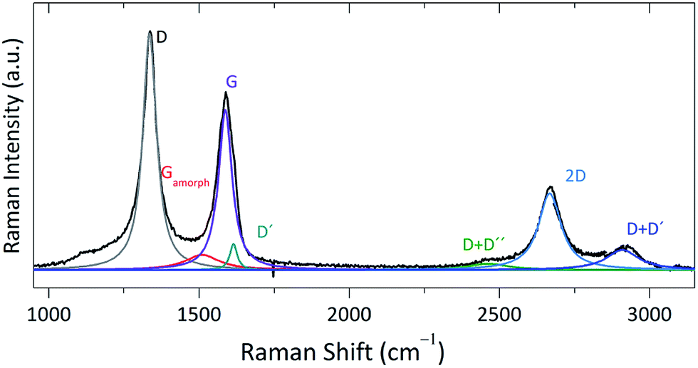 | ||
| Fig. 1 Raman spectrum of pristine CNHs. Colored Lorentzian curves correspond with the different contributions of the spectrum mentioned in the text. | ||
Morphological changes under high stress
Fig. 2 shows the evolution of the Raman spectrum of CNHs with increasing stress. As expected from previous carbon materials studies,6,34 a general upshift with stress is observed. As already commented in the introduction, CNTs under compression undergo a flattening of their circular section8–10 at a stress threshold that varies depending on the diameter and the number of walls in the tube.11,12 Specifically, the flattening stress barrier is lower for single-walled tubes with larger diameter (0.8 GPa for 1.6 nm diameter tubes).8 Therefore, we may expect flattening of the horns in our CNHs samples (2–3 nm) at stresses below 0.5 GPa; this provides a stress limit beyond which additional compression would lead to the formation of graphite-like carbon.42 Unfortunately, the stress value estimated for a similar transition in CNHs is below the detection limit in our experimental setup, and the evolution of the Raman frequencies with stress follows a linear behavior over the whole stress range monitored in this work. Moreover, the Raman spectrum of the recovered sample is presented in red in Fig. 2; it shows an upshift of the main Raman features, giving a clear indication of a debundling process of the CNHs sample, similar to that already described in the literature for CNTs6 and confirmed by the TEM images discussed below. | ||
| Fig. 2 Raman spectra of CNHs with increasing stress. (a) Fundamental bands in the spectral range 1200–1800 cm−1 and (b) combination bands and overtones from 2300 to 3500 cm−1. In (a) the Lorentzian functions representing the most relevant bands are plotted in different colors. | ||
An increase of the stress on the sample induces a broadening of the whole Raman spectrum. From a spectral profile analysis using Lorentzian contributions, we estimate that such broadening is more than 3 times larger for the D band than for the other first-order bands (G and D′), see Fig. 3a. On the other hand, a considerable broadening of the second-order spectrum occurs with increasing stress, and above ca. 6 GPa an unexpected vanishing of this spectral range takes place. As mentioned above, a-C presents broad contributions overlapping with the D, 2D and D + D′ bands of carbon materials,37 which are not visible in the Raman spectrum of pristine CNHs but may become more intense with the stress treatment, thus causing the broadening of the spectrum in the corresponding spectral regions. Moreover, vanishing of the combination bands and overtones is also characteristic of the formation of amorphous carbon material, so our findings are indicative of a drastic morphological transition in the compressed sample.
 | ||
| Fig. 3 (a) FWHM of the D, G and D′ bands as a function of applied stress. Intensity ratios (b) IG/ID and (c) IG/ID′ and IG/Ia-CG as a function of stress. | ||
The evolution of the intensity of the Raman bands induced by the stress is plotted in Fig. 3b–c, related to the G band intensity. With stress the intensity of the D′ band and the G band of a-C increases, a fact that at a first view could be simply related with an increasing generation of defects towards the sample destruction. However, interestingly, the D band decreases in intensity to become approximately equal to that of the G band, thus discarding this possibility. The intensity ratio G/D has been related with the relative amount of double (C![[double bond, length as m-dash]](https://www.rsc.org/images/entities/char_e001.gif) C) and single (C–C) bonds. In other words, such ratio establishes some sort of hybridization scale for the material under study, being infinite for HOPG graphite and zero for diamond.43,44 Our results reveal an increase in CC bonds when the CNHs samples are subjected to large compression. Having into account the evolution with stress of the Raman spectra, as measured by the FWHMs and relative band intensities, we conclude that CNHs turn into a carbonaceous sp2 structure upon compression. Such modification appears to be only partially reversible, since the intensity of the second-order Raman spectrum after releasing the stress is not fully recovered.
C) and single (C–C) bonds. In other words, such ratio establishes some sort of hybridization scale for the material under study, being infinite for HOPG graphite and zero for diamond.43,44 Our results reveal an increase in CC bonds when the CNHs samples are subjected to large compression. Having into account the evolution with stress of the Raman spectra, as measured by the FWHMs and relative band intensities, we conclude that CNHs turn into a carbonaceous sp2 structure upon compression. Such modification appears to be only partially reversible, since the intensity of the second-order Raman spectrum after releasing the stress is not fully recovered.
To give additional support to this conclusion we conducted series of TEM experiments on the recovered samples. In Fig. 4 and 5 we show selected TEM images (under different magnifications) of both pristine CNHs and the corresponding recovered sample after an 8 GPa stress cycle, respectively. The TEM images of the pristine sample correspond to CNHs aggregates exhibiting dahlia-like morphology.45 The TEM images of the recovered samples resembles that of amorphous carbon (Fig. 5a–b) at low resolution, but at the highest magnification a clear graphitic-like structure is distinguished (Fig. 5c). Furthermore, a remarkable observation is that while the interlayer spacing between single CNHs is about 0.4 nm in pristine CNHs, which is 18% larger than that of graphite,46,47 the interlayer distance deduced from Fig. 5c is closer to 0.34 nm, which exactly corresponds to the typical van der Waals interlayer distance in graphite. This observation justifies the relative increase observed in the IG/ID ratio deduced from the Raman spectra. In other regions of the recovered sample (Fig. 5d–f) we observe that the CNHs still remain as aggregates, but they have lost their initial dahlia-like shape. These regions consist of aggregates of debundled CNHs, in good agreement with the remaining upshift observed in the Raman spectrum of the recovered sample (notice that the aggregates have some of their original arrangement, and there are more individual horns which look more parallel to each other).48 Both kind of TEM images, those corresponding to sp2 amorphous carbon and those to debundled aggregates, confirms our previous discussion on the Raman spectrum of the recovered samples (bottom red spectrum in Fig. 2): the G band of amorphous sp2 carbon contributes, but the G band of the CNHs still predominates.
 | ||
| Fig. 4 TEM images of pristine CNHs samples at different magnifications: dahlia-like aggregates. | ||
 | ||
| Fig. 5 TEM images at different magnifications of the CNHs sample recovered after a 8 GPa stress cycle, in two different regions, A (a–c) and B (d–f), with graphite-like amorphous carbon and broken dahlias morphologies, respectively. | ||
Thus, the application of stress promotes a morphological change in CNHs that generates carbonaceous regions with graphite like structure within regions of broken dahlia-like morphology. We believe that these two different responses to stress depending on the region are simply due to different CNHs concentrations in the sample. When the CNHs sample is low concentrated and has the purest quality (low a-C content), the applied stress is essentially invested in debundling the dahlia-like structures; however, in more concentrated samples, with larger amount of a-C, the formation of graphite like lumps upon stress is observed, as if a-C were acting as a seed for generating such kind of morphology.
Moderate compression/decompression cycles on CNHs
We also subjected different CNHs samples to several moderate-stress cycles (maximum stress ∼ 2 GPa), in order to analyze a possible high bulk density material formation like that obtained by other authors elsewhere,13,14 after consecutive 50 MPa hydrostatic pressure cycles. In Fig. 6 we compare the Raman spectra of the recovered samples after consecutive compression–decompression cycles with the Raman spectrum of pristine CNHs. In contrast to the increase in the a-C content observed after a higher stress treatment, when the samples are subjected to several moderate-stress cycles the graphitic crystallinity remains, as proven by the Raman spectra of the recovered sample where the second order features remain visible and the G band of the amorphous carbon does not increase. In contrast with the observations of Bekyarova et al.,13,14 broadening of the Raman spectrum is barely observed; furthermore, we see in Fig. 7a that the FWHM of the G band remains almost unaltered after the stress cycles (in fact, it is slightly narrowed). Moreover, the FWHM of the D band also narrows in about 30 cm−1, with respect to that of pristine CNHs, in a sample recovered after 20 stress cycles. This result, together with the slight narrowing of the G band, is indicative of the disappearance of the pentagonal defects and the conical shapes contributions to the spectrum; a conclusion also confirmed by TEM experiments, as will be discussed below. Additionally we observe in Fig. 7b that the frequency of the G band of CNHs decreases in about 10 wavenumbers upon stress cycling, approaching the reference value of the G band of highly oriented pyrolytic graphite (HOPG).33,49 Regarding the intensities variations we observe in Fig. 7c that the ratio IG/ID decreases from 0.85 to 0.60 after consecutive stress cycles, thus indicating significant changes in the ratio double (CC) to single (C–C) bonding in the sample.43,44
 | ||
| Fig. 6 Raman spectra of a CNHs sample recovered after different consecutive stress cycles up to ca. 2 GPa. Labels indicate the maximum cycle number reached for each sample. The red spectrum at the bottom corresponds to that of the pristine CNHs sample. | ||
 | ||
| Fig. 7 Characteristics of the D and G bands in recovered CNHs samples as a function of the number of consecutive stress cycles: (a) FWHM, (b) Raman shift and (c) intensity ratio (IG/ID). | ||
The above findings are supported by the TEM and HRTEM images of the recovered samples shown in Fig. 8. After one stress cycle, a transition from dahlia-like to bud-like carbon nanohorns takes place (Fig. 8a–e).45,49 In this new morphology the horns do not protrude out from aggregates' surface, in contrast to what occurs in the dahlia-like arrangement. Samples consisting of variable CNHs concentrations were subjected to a single stress cycle, and in all cases a certain amount of isolated bud-like aggregates appeared, more extensively in low-concentrated CNHs samples. Thus, it seems that for a single moderate stress cycle bud-like aggregates tend to isolate instead of forming larger protuberances. However, after several consecutive stress cycles (up to six) we observed that the bud-like spheres tend to aggregate (Fig. 8f–h) towards the creation of graphite-like surfaces with horns at the edges (Fig. 8i–l). After six or more stress cycles, we observe the formation of a layered material (Fig. 8m–o), similar to that obtained after a high temperature treatment of CNHs,49 but with a larger degree of disorder.
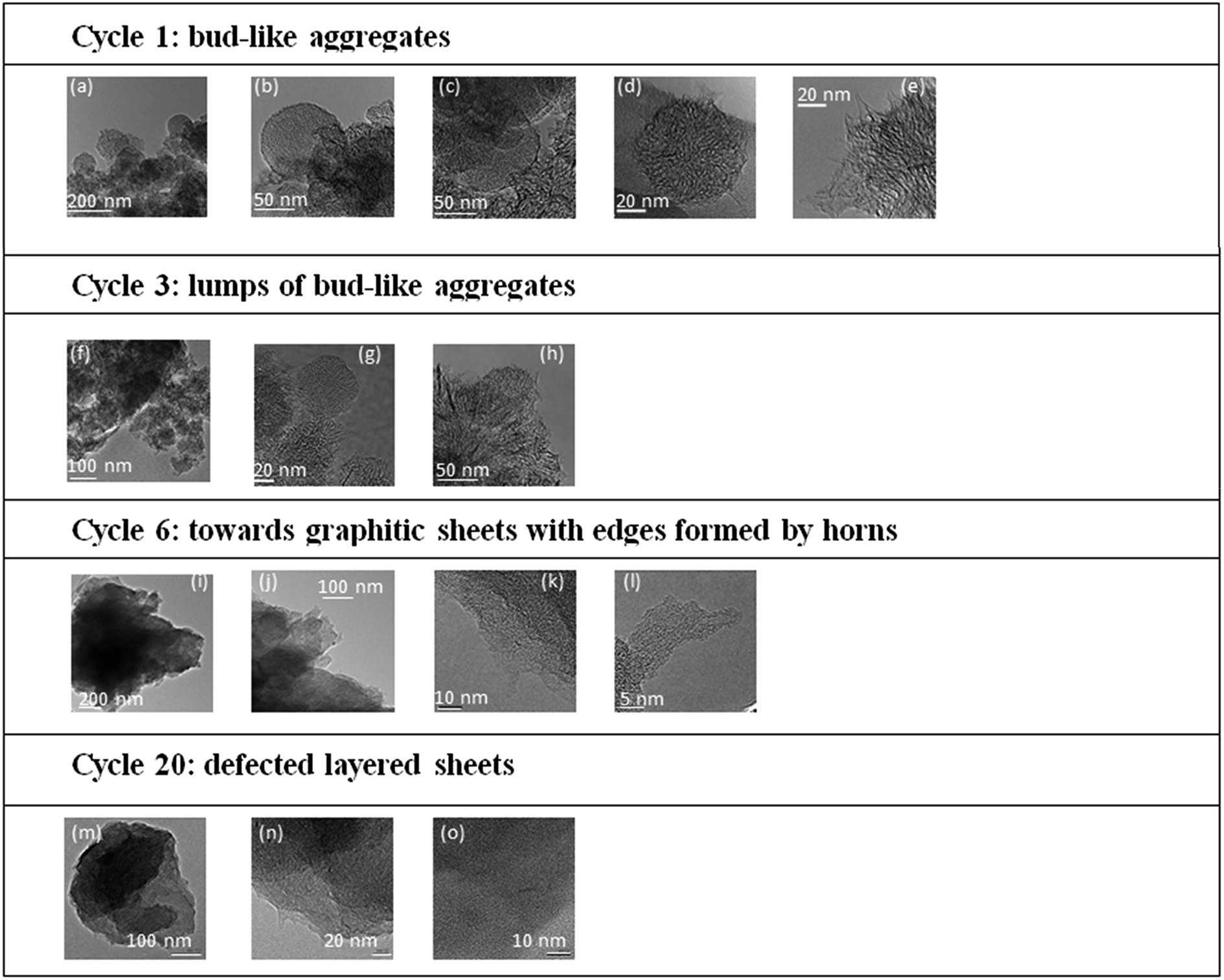 | ||
| Fig. 8 Selected TEM images of the CNHs samples recovered after a certain number of stress cycles. | ||
Conclusions
SWCNHs samples have been subjected to several high stress treatments and characterized by combined TEM and Raman spectroscopy in order to evaluate morphological changes upon compression. After a single high stress cycle of ∼8 GPa, we observed the appearance of two different formations depending on the initial quantity of CNHs in the pristine sample: debundled dahlias and graphite like lumps (turning into amorphous sp2 carbon). In contrast, after single lower stress cycle of ∼2 GPa the transition from dahlia to bud-like sphere is observed. Consecutive 2 GPa cycles leads to the aggregation of the bud-like spheres towards the formation of a layered material (with sp2 graphitic crystallinity) in whose edges, remnant horn structures can be found. All these findings demonstrate the advantages of using high to moderate stress for pretreating CNHs samples, thus increasing their subsequent reactivity and, moreover, opening new possibilities for a tailored and targeted functionalization. Finally, it would be interesting to study the effect of such treatments in their electronic properties.Acknowledgements
We thank Prof. Masako Yudasaka from Nanotube Research Center, National Institute of Advanced Industrial and Technology, Higashi, Japan for a critical reading of the manuscript. We also thank Prof. Iijima (Meijo University) for providing us with the Carbon Nanohorn sample. This work has been supported by MINECO through the projects CSD2007-00045, CTQ2012-38599-C02-02 and CTQ2013-48252-P. We thank the National Center for Electron Microscopy at Universidad Complutense de Madrid for facilities. MPA is grateful to the Spanish Ministerio de Educación, Cultura y Deporte for an FPU grant.References
- S. Iijima, Nature, 1991, 354, 56–58 CrossRef CAS.
- K. Ajima, M. Yudasaka, K. Suenaga, D. Kasuya, T. Azami and S. Iijima, Adv. Mater., 2004, 16, 307–401 CrossRef.
- M. Vizuete, M. J. Gómez-Escalonilla, J. L. G. Fierro, M. Yudasaka, S. Iijima, M. Vartanian, J. Iehl, J. F. Nierengarten and F. Langa, Chem. Commun., 2011, 47, 12771–12773 RSC.
- J. P. Lu, Phys. Rev. Lett., 1997, 79, 1297–1300 CrossRef CAS.
- M. Meo and M. Rossi, Compos. Sci. Technol., 2006, 66, 1597–1605 CrossRef CAS.
- E. Del Corro, J. González, M. Taravillo, E. Flahaut and V. G. Baonza, Nano Lett., 2008, 8, 2215–2218 CrossRef CAS PubMed.
- A. J. Ghandour, I. F. Crowe, J. E. Proctor, Y. W. Sun, M. P. Halsall, I. Hernandez, A. Sapelkin and D. J. Dunstan, Phys. Rev. B, 2013, 87, 085416 CrossRef.
- J. A. Elliott, J. K. W. Sandler, A. H. Windle, R. J. Young and M. S. P. Shaffer, Phys. Rev. Lett., 2004, 92, 095501 CrossRef PubMed.
- D. Y. Sun, D. J. Shu, M. Ji, F. Liu, M. Wang and X. G. Gong, Phys. Rev. B, 2004, 70, 165417 CrossRef.
- M.-F. Yu, T. Kowalewski and R. S. Ruoff, Phys. Rev. Lett., 2001, 86, 87–90 CrossRef CAS PubMed.
- A. L. Aguiar, A. San-Miguel, E. B. Barros, M. Kalbac, D. Machon, Y. A. Kim, H. Muramatsu, M. Endo and A. G. Souza Filho, Phys. Rev. B, 2012, 86, 195410 CrossRef.
- A. L. Aguiar, R. B. Capaz, A. G. Souza Filho and A. San-Miguel, J. Phys. Chem. C, 2012, 116, 22637–22645 CAS.
- E. Bekyarova, K. Murata, M. Yudasaka, D. Kasuya, S. Iijima, H. Tanaka, H. Kahoh and K. Kaneko, J. Phys. Chem. B, 2003, 107, 4681–4684 CrossRef CAS.
- E. Bekyarova, K. Kaneko, M. Yudasaka, K. Murata, D. Kasuya and S. Iijima, Adv. Mater., 2002, 14, 973–975 CrossRef CAS.
- M. Peña-Álvarez, E. del Corro, V. G. Baonza and M. Taravillo, J. Phys. Chem. C, 2014, 118, 25132–25140 Search PubMed.
- E. Del Corro, M. Taravillo and V. G. Baonza, Phys. Rev. B, 2012, 85, 033407 CrossRef.
- U. D. Venkateswaran, A. M. Rao, E. Richter, M. Menon, A. Rinzler, R. E. Smalley and P. C. Eklund, Phys. Rev. B, 1999, 59, 10928–10934 CrossRef CAS.
- S. Wang, B. Yang, J. Yuan, Y. Si and H. Chen, Sci. Rep., 2015, 5, 14957 CrossRef CAS PubMed.
- B. Yang, S. Wang, Y. Guo, J. Yuan, Y. Si, S. Zhanga and H. Chen, RSC Adv., 2014, 4, 54677–54683 RSC.
- S. Wang, B. Yang, S. Zhang, J. Yuan, Y. Si and H. Chen, ChemPhysChem, 2014, 15, 2749–2755 CrossRef CAS PubMed.
- A. Jorio, A. G. Souza Filho, G. Dresselhaus, M. S. Dresselhaus, A. K. Swan, M. S. Ünlü, B. B. Goldberg, M. A. Pimenta, J. H. Hafner, C. M. Lieber and R. Saito, Phys. Rev. B, 2002, 65, 155412 CrossRef.
- K. I. Sasaki, Y. Sekine, K. Tateno and H. Gotoh, Phys. Rev. Lett., 2013, 111, 116801 CrossRef PubMed.
- M. S. Dresselhaus, G. Dresselhaus, R. Saito and A. Jorio, Phys. Rep., 2005, 409, 47–99 CrossRef.
- http://www.cnme.es.
- V. G. Baonza, M. Taravillo, A. Arencibia, M. Cáceres and J. Núñez, J. Raman Spectrosc., 2003, 34, 264–270 CrossRef CAS.
- G. H. Watson Jr, W. B. Daniels and C. S. Wang, J. Appl. Phys., 1981, 52, 956–958 CrossRef.
- J. A. Xu, E. Huang, J. F. Lin and L. Y. Xu, Am. Mineral., 1995, 80, 1157–1165 CAS.
- P. Loubeyre, F. Occelli and R. LeToullec, Nature, 2002, 416, 613–617 CrossRef CAS PubMed.
- B. J. Baer, M. E. Chang and W. J. Evans, J. Appl. Phys., 2008, 104, 034504 CrossRef.
- T. Yamaguchi, S. Bandow and S. Iijima, Chem. Phys. Lett., 2004, 389, 181–185 CrossRef CAS.
- A. C. Ferrari and J. Robertson, Phys. Rev. B, 2000, 61, 14095–14107 CrossRef CAS.
- L. G. Cançado, M. A. Pimenta, B. R. A. Neves, M. S. S Dantas and A. Jorio, Phys. Rev. Lett., 2004, 93, 247201 CrossRef PubMed.
- A. C. Ferrari, J. C. Meyer, V. Scardaci, C. Casiraghi, M. Lazzeri, F. Mauri, S. Piscanec, D. Jiang, K. S. Novoselov, S. Roth and A. K. Geim, Phys. Rev. Lett., 2006, 97, 187401 CrossRef CAS PubMed.
- E. del Corro, A. Otero de la Roza, M. Taravillo and V. G. Baonza, Carbon, 2012, 50, 4600–4606 CrossRef CAS.
- T. C. Hirschmann, P. T. Araujo, H. Muramatsu, J. F. Rodriguez-Nieva, M. Seifert, K. Nielsch, Y. Ahm Kim and M. S. Dresselhaus, ACS Nano, 2014, 8, 1330–1341 CrossRef CAS PubMed.
- R. Saito, A. Jorio, A. G. S. Filho, A. Grueneis, M. A. Pimenta, D. Dresselhaus and M. S. Dresselhaus, Phys. B, 2002, 323, 100–106 CrossRef CAS.
- J. Hong, M. K. Park, E. J. Lee, D. Lee, D. S. Hwang and S. Ryu, Sci. Rep., 2013, 3, 2700 Search PubMed.
- E. Del Corro, M. Taravillo and V. G. Baonza, J. Raman Spectrosc., 2014, 45, 476–480 CrossRef.
- N. Larouche and B. L. Stansfield, Carbon, 2010, 48, 620–629 CrossRef CAS.
- R. J. Nemanich and S. A. Dolin, Phys. Rev. B, 1979, 20, 392–401 CrossRef CAS.
- A. C. Ferrari and D. M. Basko, Nat. Nanotechnol., 2013, 8, 235–246 CrossRef CAS PubMed.
- F. Colonna, A. Fasolino and E. J. Meijer, Phys. Rev. B, 2013, 88, 165416 CrossRef.
- S. Utsumi, H. Honda, Y. Hattori, H. Kanoh, K. Takahashi, H. Sakai, M. Abe, M. Yudasaka, S. Iijima and K. Kaneko, J. Phys. Chem. C, 2007, 111, 5572–5575 CAS.
- R. Yuge, S. Bandow, K. Nakahara, M. Yudasaka, K. Toyama, T. Yamaguchi, S. Iijima and T. Manako, Carbon, 2014, 75, 322–326 CrossRef CAS.
- S. Iijima, M. Yudasaka, R. Yamada, S. Bandow, K. Suenaga, F. Kokai and K. Takahashi, Chem. Phys. Lett., 1999, 309, 165–170 CrossRef CAS.
- S. Bandow, F. Kokai, K. Takahashi, M. Yudasaka, L. C. Qin and S. Iijima, Chem. Phys. Lett., 2000, 321, 514–519 CrossRef CAS.
- K. Yoshizawa, T. Yumura, T. Yamabe and S. Bandow, J. Am. Chem. Soc., 2000, 122, 11871–11875 CrossRef CAS.
- M. Zhang, M. Yudasaka, J. Miyawaki, J. Fan and S. Iijima, J. Phys. Chem. B, 2005, 109, 22201–22204 CrossRef CAS PubMed.
- D. Kasuya, M. Yudasaka, K. Takahashi, F. Kokai and S. Iijima, J. Phys. Chem. B, 2002, 106, 4947–4951 CrossRef CAS.
| This journal is © The Royal Society of Chemistry 2016 |