DOI:
10.1039/C5RA27072C
(Paper)
RSC Adv., 2016,
6, 32115-32123
Targeting CD44, ABCG2 and CD133 markers using aptamers: in silico analysis of CD133 extracellular domain 2 and its aptamer†
Received
17th December 2015
, Accepted 16th March 2016
First published on 21st March 2016
Abstract
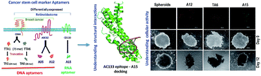
The application of nucleic acid aptamers for the diagnosis and therapy of cancer stem cells (CSCs) is expanding. The current study truncated and probed various existing aptamers against CSC markers CD44, ABCG2 and CD133 in retinoblastoma (RB) primary cells, cell lines, a breast cancer cell line and MCF7-sphere. Truncated CD44 aptamer retained its specific binding to cancer cells, ABCG2+ve MCF7-spheres and CD133+ve RB cells. Similarly, ABCG2 and CD133 aptamers showed higher affinity to ABCG2+ve, CD133+ve cells than the negative population and cell lines. All aptamers appreciably reduced viability of up to 50% and 32% of the primary RB tumor cells and cell lines, respectively. Colony formation of MCF7, RB cell lines and MCF7-sphere growth were inhibited significantly. Structure prediction, simulation of CD133 extracellular domain 2 (ExD2) and A15 followed by docking to comprehend the potential interaction revealed hydrogen bonds and non bonded interactions between them. This information could be used to improve the A15 aptamer to gain more interactions with CD133. Thus approaches undertaken here can be applied universally for cell-specific targeting, and the aptamers studied against CSC markers deserve further in vivo studies.
Introduction
Many solid tumors contain cancer stem cells (CSCs) that contribute to their aggressiveness and resistance to therapy.1 Numerous CSC markers such as the epithelial cell adhesion molecule (EpCAM), prominin 1 (PROM1 or CD133), ATP-binding cassette sub-family G member 2 (ABCG2) and homing cell adhesion molecule (HCAM or CD44) have been identified in many solid epithelial cancers. These surface antigens are common in many epithelial cancers and childhood eye tumors including retinoblastoma (RB).2–7 The CSC markers identified in RB include CD24, EpCAM, ABCG2, MCM2, CD44, CD133, CD90, ALDH1, CD166, SCA-I, and p63.5,8–11
CSCs can be targeted using antibodies, ankyrins, affibodies and aptamers.12–16 Aptamers are new class of oligonucleotides that bind to a specific target. Cell based systemic evolution of ligands by exponential enrichment (cell-SELEX) is used for developing aptamers for specific cell types.17 Aptamers possess several advantages such as reduced immuno-reactivity, being free from contaminations, smaller size, high specificity, ease of synthesis and eligibility for xeno-modifications that prevent serum nuclease degradation.18–20 Aptamers have a wide application in biosensing and in theranostics.21 Macugen, was the first aptamer approved by the FDA for the treatment of age-related macular degeneration22 and recently an aptamer based diagnostic platform for analysis of mycotoxins in grain became available for commercial use. Many aptamer conjugates are also in preclinical trials.23,24
The proteins that attributes to cancer stemness are located in different regions of the cell, cytoplasm, nucleus and the cell surface. The cell surface proteins are of interest due to ease of targeting, which could aid in receptor mediated internalization. Aptamers were developed for binding and targeting CSC markers such as EpCAM, CD133, ABCG2 and CD44 using SELEX technology. Both RNA and DNA aptamers targeting EpCAM were developed and studied for therapy and imaging in cell culture and in animal models.25–27 In the current study, we sought to analyze the aptamers targeting CD44, ABCG2 and CD133. The CD44 thio-aptamer first isolated was against the HA-binding domain of CD44 and studied in vitro against ovarian cancer cell line.28 Later, RNA aptamer targeting CD44 and DNA aptamer against CD44v10 were developed; the DNA aptamer was able to inhibit the breast cancer cell migration.29,30 ABCG2 targeted DNA aptamers were isolated using ABCG2 expressing stable cell line and MCF7-spheres.31 Similarly, an RNA aptamer against CD133 that binds to the AC133 epitope was developed and shown to penetrate hepatospheres.32
CD133 is considered an important stem cell and CSC marker and understanding its AC133 epitope structure could generate data for future studies.7,33,34 The AC133 epitope resides in the extracellular domain 2 of the CD133 protein and its presence marks the stemness property. Upon differentiation this epitope is lost due to changes in the tertiary structure. The A15-CD133 RNA aptamer was selected against AC133 epitope, thus in silico analysis can predict the interacting residues of the CD133 extracellular domain 2 (ExD2) with A15 aptamer. We used bioinformatics tools to perform molecular dynamics, energy minimization of structures and docking. Unavailability to model single stranded DNA (ssDNA) sequences restricted our interest to study the DNA aptamer protein interaction.
Thus in the current study, we truncated CD44 thio-aptamer, utilized ABCG2 and CD133 aptamers mentioned above.28,31 We investigated their functional role using breast cancer cell line, MCF7-spheres (MCF7), RB cell lines (Y79, WERI-Rb1), RB primary tumors, non-malignant retinal cells. Additionally, we assessed their specificity using ABCG2+ve/−ve MCF7-sphere and CD133+ve/−ve RB/normal retina cells respectively. We envisage that truncated CD44 aptamer and deciphering interacting residues of CD133 and its aptamer using computational modelling studies would have better theranostic value.
Results
Expression of CD44 variants in RB
CD44 is cell surface marker involved in cell–cell interaction and expressed as alternate splice variants in cancer conditions. The expression of CD44 (CD44+/CD24−) marks the CSC in breast cancer population and the alternately spliced variants are well studied in breast cancer.35,36 Expression of CD44 overlapping with other CSC markers in RB primary tumor cells were showed by flow cytometry technique.9 The arrangement of CD44 exons and the splice variants of CD44 generated are show as insert in Fig. 1A.
 |
| Fig. 1 The expression of CD44 variants in RB primary tumor. (A) CD44 and its variants generated by alternate splicing mechanism. CD44s (standard form) carries exon S1–5 and S7–10, while the exon v1–10 are designated variant exons. The CD44s with insertion of exon 6 is represented as CD44v6, similarly for other variants. (B) Graph showing the relative fold change in the CD44 variant mRNAs in RB tumors and cell lines, WERI-Rb1 and Y79 normalized with adult cadaveric non-malignant retina by SYBR green based qPCR method. | |
We sought to analyze the CD44 standard form (CD44s) and its splice variants as data is lacking in RB tumors. CD44 variants mRNA levels were analyzed by qPCR using the variant primers that are designed between the exonic regions.35 The expression of CD44 was limited and varied between tumor samples and mostly found to be differential in the RB (50%). The expression levels of the CD44v6, CD44v8, CD44v9 and CD44-T were around two fold upregulated in 50% of the cases, whereas the CD44v10 was downregulated (Fig. 1B). Though 50% of cases showed CD44-S standard form, the expression levels were low in order to be considered for targeting. The presence of the variant forms needs further validation using variant specific antibodies especially for the CD44v8 and CD44v9 respectively. The clinicopathological correlation with the expression status of the CD44 variants in RB tumors show increased expression of variants in non-invasive tumors, while the expression decreased as the choroid invasion increased >3 mm.37 The CD44s (standard form) did not show significant change in expression with choroid invasion. The pathological finding and the changes in the expression levels are presented with upregulation or downregulation sign (0.8 > fold as upregulation and −0.8 < fold as downregulation; values between are considered insignificant) in Table S3.† Thus the information on CD44v forms in RB helps to use it for targeting.
Truncation of CD44 aptamer: cellular uptake of CD133, ABCG2 and truncated CD44 aptamer
The CD44 thio-aptamers TTA1 and TTA6 (Fig. S2A†) were shown to bind the ovarian cancer cell lines, while the later aptamer had better as studied for affinity using HA binding domain of CD44 using binding assays.28 Hence we analyzed the binding of TTA6 full length aptamer onto WERI-Rb1, MCF7 and RB primary tumor cells. TTA6 binding on different RB tumor cells (RB1 and RB2) are shown in Fig. S2B.† Further truncations of the CD44 aptamer were performed for the TTA1 and TTA6 to obtain smaller aptamers with lower molecular weight. The truncated TTA6 and TTA1 are referred as TA6 and TA1 respectively. The aptamer truncation is based on the secondary structure, trimming of the selection sequence and their secondary structures predicted by Mfold are given in Fig. S2C.†
Apart from the CD44 aptamers (TA1 and TA6), CD133 and ABCG2 aptamers were also utilized as these markers were expressed by CSCs of different cancer types and by RB and breast cancer studied currently. The CD133 aptamer (A15 aptamer) binds to the CD133 extracellular domain 2 that harbor the AC133 epitope, identified during the undifferentiated state.32 Similarly, the aptamers that were targeted to the ABCG2 protein were isolated using cell-SELEX by utilizing the ABCG2 over expressing BHK cells (A12 aptamer), MCF7-sphere cultures that overexpresses CD44 and ABCG2 (A35 aptamer) were also utilized in this study.31 The A12, A35, TA1 and TA6 are DNA aptamers while the A15 is an RNA aptamer (Fig. 2A).
 |
| Fig. 2 Truncated CD44 aptamer and CSC marker aptamer uptake on cancer cell lines. (A) Schematic representation of the cancer type studied vs. the CSC markers and their respective aptamers. Aptamer truncation and the type of aptamer (DNA/RNA) are represented in the scheme. (B) FACS analysis and scatter plots of CD133-A15APT uptake at the concentration of 500 nM to RB cell lines, Y79, WERI-Rb1 and MIOM1 (non-malignant retinal) cell line. (C) The scatter plots showing the uptake of ABCG2 (A12 and A35) APT and CD44 truncated APTs (TA1 and TA6) binding to MIOM1, MCF7 and WERI-Rb1 cells with the percentage positive cells indicated on lower right quadrant. Experiments were repeated minimum thrice and the scatter plot shows results from representative experiment. | |
All the aptamers were studied for cell uptake using primary RB cells, non-cancerous retinal cell line, MIOM1, RB cell line, WERI-Rb1 (positive for CD133 and ABCG2) and breast cancer cell line, MCF7 (positive for CD44 and ABCG2). The CD133 – A15 aptamer showed specific binding and uptake by the RB cell lines and not by the MIOM1 (Fig. 2B). The A12, A35 and TA6 showed consistent binding and uptake by cancer cell lines, sparing MIOM1. MIOM1, the control cell line did not uptake any of these aptamers. The truncated TA1 aptamer showed reduced binding than TA6 and it was insignificant on the cell lines (Fig. 2C).
The aptamers were further studied for their ability to penetrate the MCF7-sphere or spheroids generated from MCF7 cells. Generating spheroids were not possible for RB cells as they grow as clusters/chains in nature. Aptamers were tested for their ability to penetrate the intact MCF7 spheroids by incubating with the aptamer for 2 h followed by fluorescent microscopic analysis. All aptamers showed good penetration except for TA1 and they accrued at specified sites (Fig. 3A), while TA6 showed more diffused staining. The TA1 aptamer showed moderate penetration with most of the aptamers bound to spheroid surface. The degree of penetration observed was in the order of TA6 > A35 > A12 > A15 > TA1. Further CSC marker mediated uptake of aptamer was confirmed by using the ABCG2+ve/−ve cells isolated from MCF7-spheres. The A12 and A35 aptamers were more specific to ABCG2+ve and showed very less uptake on to ABCG2−ve cells. Thus enrichment of cells led to higher uptake of all aptamers including the TA1 that showed poor uptake by the monolayer or MCF7-sphere cells (Fig. 3B).
 |
| Fig. 3 Cellular uptake of APT on ABCG2+ve-MCF7-spheroids and CD133+ve RB cells. (A) MCF7 cells were grown on agarose coated plates for more than 15 days. The spheroids were incubated with 500 nM of fluorescently labelled CSC marker aptamers and imaged under Axiovision fluorescent microscope (5× objective, scale bar = 200 μm). Control MCF7-spheroid (i) A12 (ii) A35 (iii) TA1 (iv) TA6 (v) and A15 (vi). (B) Scheme showing the monolayer vs. spheroid culture and the ABCG2+ve cell isolation. The monolayer cells, MCF7-spheroids, ABCG2+ve and −ve cells were analyzed for CSC marker aptamer uptake. Graph shown below represents the percentage cells positive for the uptake of the ABCG2 (A12 and A35) aptamer and CD44 truncated aptamers (TA1 and TA6)-cy3 labelled aptamers and CD133-A15 aptamer is labelled with FITC. (C) Schematic representation of isolation of CD133+ve and CD133−ve cells from the RB tumor and non malignant cadaveric retina. Graph below shows the efficiency of the CSC marker aptamer staining of the isolated CD133+ve/−ve cells. | |
Cellular uptake of CSC marker aptamers by primary RB and CD133+ve RB cells
The CSC marker aptamers were studied for uptake in the RB primary tumor cells as well in non-malignant, normal cadaveric retina cells. The scatter plot showing the aptamer uptake with the percentage positivity or mean fluorescence intensity labeled in the same quadrant is shown in Fig. S2A and B.† The tumor cells showed very high uptake of all the aptamers except the TA1 which showed lower binding even in the cell lines, could be that upon truncation, the aptamer affinity for the CD44 has reduced further. But the TA6 uptake was equally maintained with no compromise in its uptake. We tested two cadaveric retinas for the aptamer uptake and both showed very less binding to the aptamers which signifies their specificity for cancer cells.
From the FACS analysis, the average binding of the A12 and A35 aptamer were found to be 56.21 ± 2.59% and 58.072 ± 5.11% respectively. The average binding of the TA1 and TA6 aptamer were 4.296 ± 0.69% and 68.496 ± 0.57% respectively. The tumor samples showed very good binding for A15 aptamer, having maximum binding of 95.1%. Interestingly all aptamers when subjected to binding with normal retinas, showed very less binding similar to that of MIOM1 cells (Table S4†). Aptamers were further tested using primary RB cells, CD133 marker enriched/depleted RB primary tumors and normal retina cells. RB primary cells were chosen due to innate expression of the stem cell antigens. The AC133 epitope specific monoclonal antibody (Miltenyi biotech, UK) was used for the isolation of the CD133+ve/−ve cells (Fig. 3C). Both the CD133+ve and negative cells subjected for the aptamer uptake study exhibited higher uptake of aptamer by CD133+ve cells than the negative cells. The CD133+ve RB cells had differences of ∼300 mean fluorescence intensity (MFI) than the negative RB cells for the A15 aptamer uptake, while the normal retina isolated cells showed ∼130 MFI difference. The enhancement in binding of the aptamer in the normal retina CD133+ve could be due to the innate CD133, also the negative cells too showed uptake of CD133 aptamer, which could be due to innate basal CD133 expression in normal retina. The A12, A35 and TA6 aptamers uptake on the CD133+ve and CD133−ve normal retina cells did not show any greater difference, the MFI were closer in both the uptake population. However, the TA1 aptamer binding was not enhanced even in the CD133+ve cells confirming its specificity for CD44/ABCG2 receptors (Fig. 3C).
Functional activity of the CSC marker aptamers
The functional activity to inhibit the cell proliferation was tested using RB cells, cell lines and breast cancer cell line by MTT assay. RB primary tumor cells showed upto 55% of cell death upon CSC maker aptamers treatment. The A15 aptamer showed significant inhibition of cell proliferation of Y79 and RB primary cells. Similarly, TA6 aptamer showed inhibition on WERI-Rb1, MCF7 and RB cells. In general results clearly indicate that CSC marker aptamers act better on primary RB cells that express CSC markers and uptake aptamer at higher rate. The functional activity of these aptamers were more pronounced in the primary cells, as they expressed the CSC markers higher in amount than the cell lines (Fig. 4A).
 |
| Fig. 4 Functional activity of the CSC marker aptamers. (A) Graph showing the percentage cell viability of the MIOM1, MCF7, Y79, WERI-Rb1 and RB primary cells upon treatment with the cancer stem cell marker aptamers. The aptamers were treated at a concentration of 500 nM for 48 h and assessed for their functional activity by MTT assay. (B) Graph showing the total area of growth calculated from the colony formation assay of MCF7, Y79 and WERI-Rb1 cells upon treatment with 500 nM of the aptamers for the period of 10 days in MCF7 and Y79 and 12 days in WERI-Rb1. The error bars represents the standard error between the duplicate experiment, the * indicates P < 0.05 and ** indicates P < 0.001 significantly different from the untreated group (representative images are shown in section C). (C) Colony formation of MCF7, Y79 and WERI-Rb1 cells were treated with 500 nM of the APTs and imaged upon visible colonies formation (MCF7 – colonies were stained, Y79 and WERI-Rb1 – cell clusters formed were imaged) in the untreated set showing reduction in size or number of colonies upon treatment with the aptamers. (D) MCF7 spheroids imaged before and after 12 days of treatment with APTs showing reduction in spheroid size and disintegration of spheroids post aptamer treatment in the below panel. The size ratios normalized to the untreated spheroids from day 0 to day 12 are represented below. | |
Further we tested the aptamers for their activity to inhibit cell proliferation/growth over period of 10 days using colony formation assay. This could in turn detail the ability of aptamer to reduce the proliferation of the cancer cells. The aptamers tested over seven to ten days of treatment on MCF7, Y79 and WERI-Rb1 cells resulted in decreased growth area for most of the aptamers (Fig. 4B). A15 aptamer showed the best inhibiting characteristics (colony formation as well growth inhibition). The representative panels in Fig. 4C shows the colony formation (MCF7) and cluster/colony formation from Y79 and WERI-Rb1 cells and the aptamer activity. Also the aptamers were tested on spheroids, 3D cellular architecture similar to in vivo condition. The intactness and size of spheroids were analyzed between the day 0 and day 12 after aptamer treatment. The size ratios of day 12 to day 0 normalized to untreated spheroids showed that the TA6 and A15 had best activity in reducing the spheroid size followed by A35 and A12 with no change in TA1 treatment. Aptamer treatment had led to disintegration of the spheroids except for the A35, while there was only size difference (Fig. 4D).
CD133 protein and aptamer interaction: molecular dynamics and docking studies
CD133 ExD2 harboring the AC133 epitope predicted to interact with the A15 aptamer was analyzed using molecular dynamics and docking studies. Molecular simulation on the CD133 ExD2 and A15 aptamer were performed to attain a stable conformation under dynamic nature. The structural stability assessed by RMSD trajectory for CD133 ExD2 exhibited initial fluctuations followed by stable conformation attained. Fluctuations were observed upto 5 Å until 5 ns and stable conformation was gained after 7.5 ns till 15 ns. Similarly, the A15 aptamer RMSD plot attained stability after 4 ns with a deviation within 1 Å (Fig. S3A and B†). Molecular dynamics studies were performed to obtain optimal structure with lowest potential energy highlighted with red circles (Fig. S3C and S3D†). The optimized structure of protein was superimposed with structure obtained from i-TASSER (Fig. 5A) and A15 aptamer (Fig. 5B) obtained based on the lowest potential energy conformations sampled during the simulation process were further utilized for docking study.
 |
| Fig. 5 Structure prediction, molecular dynamics and docking of CD133 ExD2 with A15 aptamer. (A) Initial predicted structure (RED) super imposed to the final structure (lowest potential energy) derived from molecular dynamics (CYAN). (B) Structure showing the lowest potential energy conformation of A15 aptamer derived from molecular dynamics simulation. (C) The cartoon view of the docked complex of aptamer and the CD133 ExD2 protein. (D) NucPlot results showing the predicted interacting residues between the aptamer and the protein. The legend showing the details of the bonds and the chemical groups are given beneath. | |
The docking studies using Hex Dock server returned 100 docked conformations, among which the best conformation was auto selected by the server (Fig. 5C). NucPlot tool generates a 2D plot of the interactions by analyzing the best docked conformation. This revealed the A15 aptamer to form two hydrogen bonds with Glu1 and Asp264 of CD133 ExD2 region. Moreover, the aptamer also showed non-bonded interactions with Thr229, Gly232, His236, Thr261, Thr257, Ala256, Asp260, Ser79, Thr75 and Leu267 residues of CD133 ExD2 region (Fig. 5D). The predicted interacting sites were further utilized for future studies.
Discussion
The CD44 variant exons spliced with the standard exons gets translated and assembles on membrane. The variant region forms part of the extracellular region and hence targeting such chimeric regions that does not exist in normal condition is of interest under diseased condition.38 CSC expressing CD44 or cancer cells expressing CD44v and other stem cell markers are potential targets.39–42 The expression of CD44 variants in RB is not yet available and the current study illustrated for the first time that variant forms are expressed in RB. While the CD44v are present in the primary tumor samples analyzed the cell lines did not show upregulation of CD44 variants could be due to changes acquired during continuous line generation.
The information on the presence of CD44v forms is currently utilized for targeting using aptamer based approach. Aptamers (DNA or RNA) are selected to bind specifically the target proteins or cells. CSC aptamers have already been reported for EpCAM, CD133, CD44 and ABCG2. EpCAM aptamer has been extensively studied, but there is lack of information on the utility of aptamers targeting CD44, ABCG2 and CD133 in RB. We further utilized breast cancer cell line and MCF7-sphere models that express high levels of CD44 and ABCG2. We illustrated that aptamers could be truncated, specifically target cancer cells for inhibiting the cellular activity to bring down cell viability. The CSC markers and their respective aptamers studied are represented in Fig. 2A.
The CD44 aptamer earlier published was 73 nucleotides in size and carried modifications such as “thio” for stability.28 Truncating aptamers will bring down its molecular weight desirable for its clearance from the systemic circulation while applying for imaging purposes. But this would be undesirable when used for therapeutic purpose as the half-life decreases, efficacy would decrease too. Nevertheless, synthesis would become easier and immunogenicity can be brought down upon truncation. Hence we opted for truncating the CD44 aptamer. Of the two truncation performed from aptamers that binds the CD44 hyaluronic acid (HA) domain, one aptamer (TA6) retained its binding while in the other aptamer (TA1) binding and uptake was greatly reduced. But the TA1 aptamer showed uptake on the ABCG2+ve MCF7-spheres and CD133+ve RB cells. Thus TA1 aptamer retained binding efficiency to stem cell marker expressing population. The ABCG2 (A12) and MCF7-mammosphere (A35) aptamers were also actively taken up by RB and breast cancer cells sparing the normal retinal cell line, MIOM1. Though cell lines were used as models, the RB primary tumor cells had acted as better systems as they expresses higher levels of stem cell markers.5,8,43
Spheroid cultures express the CSC markers at higher levels44 and so we used it as an in vitro model to confirm the binding of all the aptamers and ability to penetrate to cells. Our results confirmed the specificity of CD44 aptamers post-truncation in MCF7 and WERI-Rb1 cell lines and spheroid models. Also, these aptamers were tested for functional activities such as inhibition of cellular activity or colony forming ability for the first time. Earlier studies, explored the use of EpCAM, CD133 RNA aptamers in various cell lines and spheroid cultures of hepatocellular carcinoma respectively.32,45 Our results confirms that the CD133 and CD44 aptamers have potential use for CSC targeting as they disintegrated the 3D cell culture model and lead to cell death. It will be of greater interest to study the mechanism adopted by these aptamers.
CD133 being a marker for stem cells and cancer stemness, understanding its structure details will help drug discovery. But the crystal structure of CD133 protein is not yet available. To study the interaction between the residues within the CD133 extracellular domain 2 (ExD2) that harbours the AC133 epitope, modelling and structure prediction of the CD133 protein is necessary. AC133 epitope maintains the undifferentiated state of the cells. The re-folding of protein results in disappearance of the AC133 epitope leads to transformation and masking of the cancer stem cell property.46 The A15 aptamer selected against the CD133 recombinant protein binds to the AC133 epitope bearing cells and that the binding abolishes with the transformation of cells. Thus taken together the CD133 ExD2 harbouring AC133 epitope was studied for its interaction with the A15 aptamer. Current study revealed the interacting residues of CD133 protein with A15 aptamer aided in understanding the functional activity of the aptamer. Also these residues could be used as targets for anti-cancer small molecules.
Experimental
Materials and methods
Cell lines and cell culture.
RB cell lines, Y79 and WERI-Rb1 and breast cancer cell line MCF7 were obtained from the cell bank, RIKEN BioResource Center (Ibaraki, Japan). A non-cancerous Müller glial, MIOM1 cell line was gifted by Dr Limb. MCF7 and MIOM1 was cultured in DMEM media with 10% FBS (Gibco, Life technologies, Bangalore, India). Y79 and WERI-Rb1 cells were maintained in RPMI-1640 media with 10% FBS. All cell lines were maintained with 1% pen–strep and incubated at 37 °C in a 5% CO2 incubator. Mammospheres or MCF7 spheroids were grown by culturing the cells on uncoated plates or defined cell numbers seeded in 96 well plates coated with agarose for 15–18 days. The cells were given intermittent media change and used for evaluating the cellular uptake of the aptamers. For establishing the spheroid cultures, MCF7 monolayer cultures were trypsinised, counted, 2.5 × 10⁴ cells seeded per well of agarose coated 96 well plates. The cultures were given media changes on alternate days and cultured for 15 days. Roswell Park Memorial Institute (RPMI) 1640 media, Dulbecco's modification of eagle's medium (DMEM), heat-inactivated Fetal Bovine Serum (FBS), poly-L-lysine (PLL) were purchased from Sigma Aldrich (St. Louis, MO). The study adheres to the declaration of the Helsinki.
Sample collection and RB primary tumor cells.
RB primary tumors samples collected from enucleated eye balls as part of therapy were utilized for research purpose. A written general consent was obtained from the parents/guardians of the patient undergoing enucleation. Cornea removed normal retinas from cadaveric donors received from C U shah eye bank, Sankara Nethralaya was used anonymously in the study as control retina sample. The study was conducted after obtaining the approval from the Ethics Sub-Committee (Institutional Review Board) of Sankara Nethralaya eye hospital [Ethical clearance no. DBT-318-2012-P].
RNA extraction and qPCR analysis.
The total RNA was isolated from RB cell lines, non-malignant cadaveric retina and RB tumor cells by Trizol method. 500 ng to 1 μg of total RNA was transcribed into cDNA using oligo-dT and random hexamers. The quantitative PCR was performed in Applied Biosystem 7500 by using Sybr-green (Thermoscientific, Mumbai, India). The CD44 variant (CD44v) specific primers were utilized from earlier publication.36 The primers used are listed in the Table S1† and they are designed between the exonic regions to specifically analyze the splice variants. Relative quantification of CD44 variants (control retina to tumor retina) was determined using the formula 2−ΔΔCt and fold change or relative expression normalized to the beta-2-microglobulin (β2M) endogenous control were used for plotting the graphs. Experiments were performed in triplicate for the same samples.47
CSC marker aptamer and truncation of CD44 aptamer.
DNA aptamers targeting CD44, ABCG2 and RNA aptamer targeting CD133 were included in the study. The CD44 thio-aptamers (73 nucleotide) were truncated further to harbor the loop and the few nucleotides from the stem region, so as to maintain the predicted secondary structure. The thio-aptamers, TTA1 and TTA6 against CD44 (Fig. S1A†) shown to bind to the ovarian cancer cells,28 were truncated to generate TA1 and TA6 (24 and 28 nucleotide length) used in the study (Fig. S1B†). The ABCG2 DNA aptamers (A12, A35) and CD133 RNA aptamer (A15) from previous studies were also utilized in this study.31,32 The aptamer sequences used are listed in the Table S2.†
Cellular uptake of the CSC marker aptamers.
The uptake of fluorescently labeled aptamers by the target cells was studied by flow cytometry and fluorescent microscopy. 2 × 10⁵ MCF7, MIOM1, Y79 and WERI-Rb1 cells were incubated with 50, 250, 500 nM aptamers for 2 h in 1× binding buffer.25 After incubation, the cells were washed with 1× PBS and acquired by flow cytometer. Similarly uptake studies were performed in the primary RB tumor samples and normal retina that were stored frozen. Tissues were cut, minced, homogenously suspended, washed with PBS and used for the study. The spheroids were checked for the viability using propidium iodide staining and analyzed for cellular uptake of aptamers. The aptamer uptake was acquired using fluorescent microscope at 5× objective (Z-stacking was performed) to study the penetration of aptamers in the core of spheroid. Further the MCF7-sphere cell uptake of aptamer was studied by flow cytometry after disrupting the spheroids into single cell suspension, following the protocol for cellular uptake of aptamer.
Isolation of CD133+ve/−ve and ABCG2+ve/−ve cells and aptamer uptake study.
CD133+ve cell isolation was performed from the RB primary tumor sample and normal retina using CELLection™ Pan Mouse IgG Kit (Invitrogen life science, India) following the manufacturer's instructions. Similarly ABCG2+ve cells were isolated from the MCF7-spheres. Cell suspensions were incubated with AC133 epitope binding anti-CD133 antibody (Miltenyi biotech, UK) or anti-ABCG2 antibody (Sigma Aldrich, India) for 4 h at RT, followed by incubation with washed Dynabeads (Invitrogen, Bangalore, India) for 4 h, separation using magnetic stand yields negative cells and further incubation with DNAse I enzyme releases the positive cells for the antigens desired, from the beads. All the cells were washed twice with 1× PBS and incubated with the CSC marker aptamers (A12, A35, TA1, TA6 and A15).
Cellular activity of the CSC marker aptamers.
Cell proliferation was studied by MTT assay of aptamer treated cells. 6000 cells were seeded a day before treatment. The cells were treated with 500 nM concentration of aptamer for 48 h. Post treatment cells were incubated with 100 μl of media containing 10 μl of 5 mg ml−1 MTT and incubated for 3–4 h at 37 °C. Then MTT was removed, 100 μl of DMSO was added and absorbance was measured at 570 nm. Colony formation assay was performed on MCF7, Y79 and WERI-Rb1 cells. MCF7 cells were seeded at a density of 5000 cells per well of six well plates and treated 24 h post seeding with 500 nM of each aptamer (day 0) and incubated until visible colonies developed in untreated plate. Y79 and WERI-Rb1 cells were seeded as single cells on day 0 and treated with 500 nM of aptamer until larger colony/cluster of cells were grown. Colony formation assays were performed twice, total area of growth calculated using imageJ software using particle analysis tool and the mean ± SEM was plotted as graph. MTT assays were performed in triplicate thrice respectively and the mean ± SD was plotted as graph.
Structure prediction and molecular dynamics of CD133 protein and A15 aptamer.
The extracellular domain 2 of the CD133 (ExD2) protein was modeled by fold recognition techniques using I-TASSER48 as there were no structural homologue from the BLAST. The structure with the highest C-score was refined using modeller9v7 software.49 The three dimensional structure of aptamer was predicted using MC Fold-MC Sym50 pipeline of tools. The MC fold predicts the secondary structure and the MC Sym predicts the three dimensional coordinates utilizing the secondary structure with template from nucleotide cyclic motifs (NCM).
Molecular dynamics51 simulations were performed using GROMACS 4.5 (ref. 52) in which the system was prepared by solvating the protein structure in SPCE model and A15 aptamer in TIP3P water models with OPLS as force field. The charge of the system was neutralized by adding one Cl-ion for the protein system and 14Na+ ions for the A15 aptamer. The neutralized systems were further energy minimized using steepest decent algorithm which was followed by equilibration by NVT and NPT ensembles. The simulation was carried out for 15 nanoseconds (ns) for the protein and 5 ns for the RNA, and the coordinates were sampled at every two pico seconds (ps) for both the systems. Further, the root mean square deviations (RMSD) trajectories of the molecular backbones of sampled coordinates were plotted against the time duration to observe the dynamics of the molecules. Similarly, the potential energy of all the evolved structural conformation was also calculated and plotted against time duration.
Docking.
The interaction between the protein CD133 and A15 RNA aptamer was predicted by performing docking simulation using Hex Dock (http://hexserver.loria.fr) which calculates the docking poses on the shape complementarities using fast Fourier transformation. Based on HexDock scoring function, the best binding pose was chosen and was subjected to Nuc Plot53 analysis to visualize the interactions between the docked complex.
Conclusions
The two novel aptamers derived post truncation of the CD44 aptamers, without altering the specificity towards CD44 positive cells. Truncated aptamers, ABCG2 aptamer and CD133 aptamer showed functional activity in primary RB tumors. For the first time we showed the binding and functional activity of the CSC marker aptamers in RB cell lines and primary tumors. CD133 aptamer interactions with the extracellular domain 2 possessing the AC133 epitope were confirmed using in silico analysis. Thus the CSC marker aptamers studied by our group can be further modified with stability incorporating nucleotides, chimerized with drugs or peptides and can be tested in RB and other cancer types in vivo and for imaging.
Acknowledgements
We acknowledge the Department of Biotechnology (Indo Australian Biotechnology grant Locked nucleic acid modified aptamer-siRNA chimeric conjugates for improved chemosensitivity and eradication of cancer stem cells) BT/Indo-Aus/06/08/2011 in part from the Programme support on Retinoblastoma, BT/01/CEIB/11/V/16 from the Department of Biotechnology. Vision Research Foundation, core lab facility is acknowledged for their flow cytometry and fluorescent microscopy. Deakin University is acknowledged for the PhD scholarship for Nithya Subramanian.
Notes and references
- J. E. Visvader and G. J. Lindeman, Nat. Rev. Cancer, 2008, 8, 755–768 CrossRef CAS PubMed.
- C. Guo, H. Liu, B. H. Zhang, R. M. Cadaneanu, A. M. Mayle and I. P. Garraway, PLoS One, 2012, 7, e34219 CAS.
- F. Kroepil, A. Dulian, D. Vallbohmer, H. Geddert, A. Krieg, C. Vay, S. A. Topp, J. S. am Esch, S. E. Baldus, O. Gires, W. T. Knoefel and N. H. Stoecklein, BMC Res. Notes, 2013, 6, 253 CrossRef PubMed.
- E. Mato, C. Gonzalez, A. Moral, J. I. Perez, O. Bell, E. Lerma and A. de Leiva, J. Mol. Endocrinol., 2014, 52, 289–300 CrossRef CAS PubMed.
- A. Mohan, M. Kandalam, H. L. Ramkumar, L. Gopal and S. Krishnakumar, Br. J. Ophthalmol., 2006, 90, 889–893 CrossRef CAS PubMed.
- F. Prall, C. Maletzki and M. Linnebacher, Histopathology, 2010, 56, 553–555 CrossRef PubMed.
- X. Wei, J. Wang, J. He, B. Ma and J. Chen, Int. J. Clin. Exp. Med., 2014, 7, 2453–2462 CAS.
- M. Adithi, V. Nalini, M. Kandalam and S. Krishnakumar, Exp. Eye Res., 2008, 87, 312–318 CrossRef CAS PubMed.
- M. M. Balla, G. K. Vemuganti, C. Kannabiran, S. G. Honavar and R. Murthy, Invest. Ophthalmol. Visual Sci., 2009, 50, 1506–1514 Search PubMed.
- G. M. Seigel, L. M. Campbell, M. Narayan and F. Gonzalez-Fernandez, Mol. Vision, 2005, 11, 729–737 CAS.
- G. M. Seigel, A. S. Hackam, A. Ganguly, L. M. Mandell and F. Gonzalez-Fernandez, Mol. Vision, 2007, 13, 823–832 CAS.
- K. Kasuya, M. Shimazu, M. Suzuki, T. Itoi, T. Aoki and A. Tsuchida, Int. J. Mol. Med., 2010, 25, 209–215 CAS.
- P. Martin-Killias, N. Stefan, S. Rothschild, A. Pluckthun and U. Zangemeister-Wittke, Clin. Cancer Res., 2011, 17, 100–110 CrossRef PubMed.
- A. V. Salnikov, A. Groth, A. Apel, G. Kallifatidis, B. M. Beckermann, A. Khamidjanov, E. Ryschich, M. W. Buchler, I. Herr and G. Moldenhauer, J. Cell. Mol. Med., 2009, 13, 4023–4033 CrossRef PubMed.
- J. Wang, K. Sefah, M. B. Altman, T. Chen, M. You, Z. Zhao, C. Z. Huang and W. Tan, Chem.–Asian J., 2013, 8, 2417–2422 CrossRef CAS PubMed.
- C. Yang, F. Xiong, J. Wang, J. Dou, J. Chen, D. Chen, Y. Zhang, S. Luo and N. Gu, Nanomedicine, 2014, 9, 45–60 CrossRef CAS PubMed.
- K. Sefah, K. M. Bae, J. A. Phillips, D. W. Siemann, Z. Su, S. McClellan, J. Vieweg and W. Tan, Int. J. Cancer, 2013, 132, 2578–2588 CrossRef CAS PubMed.
- K. Hagiwara, H. Fujita, Y. Kasahara, Y. Irisawa, S. Obika and M. Kuwahara, Mol. BioSyst., 2015, 11, 71–76 RSC.
- S. Shigdar, J. Macdonald, M. O'Connor, T. Wang, D. Xiang, H. Al Shamaileh, L. Qiao, M. Wei, S. F. Zhou, Y. Zhu, L. Kong, S. Bhattacharya, C. Li and W. Duan, Sensors, 2013, 13, 13624–13637 CrossRef CAS PubMed.
- A. I. Taylor, S. Arangundy-Franklin and P. Holliger, Curr. Opin. Chem. Biol., 2014, 22, 79–84 CrossRef CAS PubMed.
- D. Xiang, S. Shigdar, G. Qiao, T. Wang, A. Z. Kouzani, S.-F. Zhou, L. Kong, Y. Li, C. Pu and W. Duan, Theranostics, 2014, 5, 23–42 CrossRef PubMed.
- P. Ray and R. R. White, Pharmaceuticals, 2010, 3, 1761–1778 CrossRef CAS.
- J. R. Kanwar, J. S. Shankaranarayanan, S. Gurudevan and R. K. Kanwar, Drug Discovery Today, 2014, 19, 1309–1321 CrossRef CAS PubMed.
- X. Ni, M. Castanares, A. Mukherjee and S. E. Lupold, Curr. Med. Chem., 2011, 18, 4206–4214 CrossRef CAS PubMed.
- N. Subramanian, V. Raghunathan, J. R. Kanwar, R. K. Kanwar, S. V. Elchuri, V. Khetan and S. Krishnakumar, Mol. Vision, 2012, 18, 2783–2795 CAS.
- L. Li, D. Xiang, S. Shigdar, W. Yang, Q. Li, J. Lin, K. Liu and W. Duan, Int. J. Nanomed., 2014, 9, 1083–1096 CrossRef PubMed.
- N. Subramanian, J. R. Kanwar, P. Athalya, N. Janakiraman, V. Khetan, R. K. Kanwar, S. Eluchuri and S. Krishnakumar, J. Biomed. Sci., 2015, 22, 4 CrossRef CAS PubMed.
- A. Somasunderam, V. Thiviyanathan, T. Tanaka, X. Li, M. Neerathilingam, G. L. Lokesh, A. Mann, Y. Peng, M. Ferrari, J. Klostergaard and D. G. Gorenstein, Biochemistry, 2010, 49, 9106–9112 CrossRef CAS PubMed.
- N. Ababneh, W. Alshaer, O. Allozi, A. Mahafzah, M. El-Khateeb, H. Hillaireau, M. Noiray, E. Fattal and S. Ismail, Nucleic Acid Ther., 2013, 23, 401–407 CrossRef CAS PubMed.
- J. Iida, R. Clancy, J. Dorchak, R. I. Somiari, S. Somiari, M. L. Cutler, R. J. Mural and C. D. Shriver, PLoS One, 2014, 9, e88712 Search PubMed.
- K. Palaniyandi, B. A. Pockaj, S. J. Gendler and X. B. Chang, J. Cancer Sci. Ther., 2012, 4, 214–222 CrossRef CAS PubMed.
- S. Shigdar, L. Qiao, S. F. Zhou, D. Xiang, T. Wang, Y. Li, L. Y. Lim, L. Kong, L. Li and W. Duan, Cancer Lett., 2013, 330, 84–95 CrossRef CAS PubMed.
- S. T. Kim, I. Sohn, I. G. Do, J. Jang, S. H. Kim, I. H. Jung, J. O. Park, Y. S. Park, A. Talasaz, J. Lee and H. C. Kim, Cancer Genomics Proteomics, 2014, 11, 259–266 Search PubMed.
- I. Saricanbaz, E. Karahacioglu, O. Ekinci, H. Bora, D. Kilic and M. Akmansu, Asian Pac. J. Cancer Prev., 2014, 15, 8215–8219 CrossRef PubMed.
- S. A. Khan, A. C. Cook, M. Kappil, U. Gunthert, A. F. Chambers, A. B. Tuck and D. T. Denhardt, Clin. Exp. Metastasis, 2005, 22, 663–673 CrossRef CAS PubMed.
- B. Krishnamachary, M. F. Penet, S. Nimmagadda, Y. Mironchik, V. Raman, M. Solaiyappan, G. L. Semenza, M. G. Pomper and Z. M. Bhujwalla, PLoS One, 2012, 7, e44078 CAS.
- X. Sastre, G. L. Chantada, F. Doz, M. W. Wilson, M. T. de Davila, C. Rodriguez-Galindo, M. Chintagumpala and P. Chevez-Barrios, Arch. Pathol. Lab. Med., 2009, 133, 1199–1202 Search PubMed.
- M. Uhl-Steidl, E. Muller-Holzner, A. G. Zeimet, G. R. Adolf, G. Daxenbichler, C. Marth and O. Dapunt, Oncology, 1995, 52, 400–406 CrossRef CAS PubMed.
- A. P. Megaptche, U. Erb, M. W. Buchler and M. Zoller, Immunol. Cell Biol., 2014, 92, 709–720 CrossRef CAS PubMed.
- J. L. Lee, M. J. Wang, P. R. Sudhir, G. D. Chen, C. W. Chi and J. Y. Chen, Cancer Res., 2007, 67, 2089–2097 CrossRef CAS PubMed.
- M. Yoshikawa, K. Tsuchihashi, T. Ishimoto, T. Yae, T. Motohara, E. Sugihara, N. Onishi, T. Masuko, K. Yoshizawa, S. Kawashiri, M. Mukai, S. Asoda, H. Kawana, T. Nakagawa, H. Saya and O. Nagano, Cancer Res., 2013, 73, 1855–1866 CrossRef CAS PubMed.
- J. Zeilstra, S. P. Joosten, H. van Andel, C. Tolg, A. Berns, M. Snoek, M. van de Wetering, M. Spaargaren, H. Clevers and S. T. Pals, Oncogene, 2014, 33, 665–670 CrossRef CAS PubMed.
- M. Mitra, M. Kandalam, A. Harilal, R. S. Verma, U. M. Krishnan, S. Swaminathan and S. Krishnakumar, Mol. Vision, 2012, 18, 290–308 CAS.
- J. Liao, F. Qian, N. Tchabo, P. Mhawech-Fauceglia, A. Beck, Z. Qian, X. Wang, W. J. Huss, S. B. Lele, C. D. Morrison and K. Odunsi, PLoS One, 2014, 9, e84941 Search PubMed.
- S. Shigdar, J. Lin, Y. Yu, M. Pastuovic, M. Wei and W. Duan, Cancer Sci., 2011, 102, 991–998 CrossRef CAS PubMed.
- K. Kemper, M. R. Sprick, M. de Bree, A. Scopelliti, L. Vermeulen, M. Hoek, J. Zeilstra, S. T. Pals, H. Mehmet, G. Stassi and J. P. Medema, Cancer Res., 2010, 70, 719–729 CrossRef CAS PubMed.
- N. Subramanian, S. Navaneethakrishnan, J. Biswas, R. K. Kanwar, J. R. Kanwar and S. Krishnakumar, PLoS One, 2013, 8, e70422 CAS.
- Y. Zhang, BMC Bioinf., 2008, 9, 40 CrossRef PubMed.
- A. Sali and T. L. Blundell, J. Mol. Biol., 1993, 234, 779–815 CrossRef CAS PubMed.
- M. Parisien and F. Major, Nature, 2008, 452, 51–55 CrossRef CAS PubMed.
- T. Ito, B. Zimdahl and T. Reya, Cancer Cell, 2012, 21, 140–142 CrossRef CAS PubMed.
- S. Pronk, S. Pall, R. Schulz, P. Larsson, P. Bjelkmar, R. Apostolov, M. R. Shirts, J. C. Smith, P. M. Kasson, D. van der Spoel, B. Hess and E. Lindahl, Bioinformatics, 2013, 29, 845–854 CrossRef CAS PubMed.
- N. M. Luscombe, R. A. Laskowski and J. M. Thornton, Nucleic Acids Res., 1997, 25, 4940–4945 CrossRef CAS PubMed.
Footnotes |
† Electronic supplementary information (ESI) available. See DOI: 10.1039/c5ra27072c |
‡ Current address: MRC Laboratory of Molecular Biology [LMB], Cambridge, CB2 0QH, UK. |
|
This journal is © The Royal Society of Chemistry 2016 |
Click here to see how this site uses Cookies. View our privacy policy here.