
Multiferroic PVDF–Fe3O4 hybrid films with reduced graphene oxide and ZnO nanofillers
Ehab H. Abdelhamida,
O. D. Jayakumar*b,
Vasundhara Kotarib,
Balaji P. Mandalb,
Rekha Raoc,
Vaman M. Naikd,
Ratna Naik*a and
A. K. Tyagib
aDepartment of Physics and Astronomy, Wayne State University, Detroit, MI, USA-48201. E-mail: rnaik@wayne.edu; Fax: +1-313-577-3932; Tel: +1-313-577-2104
bChemistry Division, Bhabha Atomic Research Centre, Mumbai, India-400085. E-mail: ddjaya@barc.gov.in; Fax: +91-22-25505151; Tel: +91-22-25595330
cSolid State Physics Division, Bhabha Atomic Research Centre, Mumbai, India-400085
dDepartment of Natural Sciences, University of Michigan-Dearborn, MI, USA-48128
First published on 9th February 2016
Abstract
Flexible and self-standing polyvinylidene fluoride (PVDF) films loaded with nanofillers, reduced graphene oxide (RGO), zinc oxide (ZnO) and magnetic iron oxide (Fe3O4) nanoparticles, were prepared by a solvent casting method. The crystallinity, morphology and structure of these films were studied using XRD, SEM, FTIR and Raman spectroscopy. FTIR studies reveal a higher percentage of polar ferroelectric β-phase (∼80%) in both pristine PVDF and PVDF–RGO films, whereas the addition of nanofillers, Fe3O4 and ZnO, resulted in a reduced amount of β-phase (∼50%) in the films. Of all the films studied, PVDF–RGO shows an enhanced dielectric constant as well as maximum electric polarization. On the other hand, Fe3O4 loaded–PVDF composite films exhibit reduced values of both dielectric constant and electric polarization. A weak magneto-dielectric behavior is observed in Fe3O4 loaded PVDF nanocomposite films at room temperature with a coupling constant ∼0.04%.
Introduction
Magnetoelectric multiferroic systems, which show simultaneous ferroelectric and ferromagnetic orders,1,2 have attracted much interest.3–8 As of today, single phase multiferroic materials are sorted into two categories:9 type (I) multiferroics, in which room temperature coupling is either weak or non-existent,5,10 and type (II) multiferroics in which coupling usually exists at very low temperatures,11,12 making them inaccessible to room temperature device applications. Motivated by their potential diverse applications, researchers aim to achieve room temperature magnetoelectric coupling by synthesizing multi-phase multiferroics,13–17 in which magnetoelectric coupling is achieved by having ferroelectric and ferromagnetic phases in proximity, inducing strain on one another.9Among ferroelectric materials that are used in extrinsic multiferroic systems is polyvinylidene fluoride (PVDF), a thermoplastic polymer with semi crystalline nature.18 Research work on PVDF is usually aimed at increasing the quantity of the ferroelectric β-phase19,20 and to improve its dielectric properties through the addition of nanofillers.21,22 In particular, a significant number of reports have been dedicated to studying PVDF/zinc oxide (PVDF/ZnO),23–29 which has been shown to enhance the optical and dielectric properties of PVDF.30–32 Another well studied nanofiller is the reduced graphene oxide (RGO), due to its high theoretical specific surface area (2600 m2 g−1), high electric and thermal conductivity.33 An earlier study on RGO–loaded PVDF has shown an increase in its dielectric constant.34
In this work, in addition to using RGO and ZnO to improve the dielectric properties and ferroelectric response of PVDF, magnetic Fe3O4 nanoparticles are added as fillers to introduce ferromagnetism and induce magneto-dielectric coupling in composite films. By adding Fe3O4, we aim to utilize the microscopic magnetostriction caused by applying an external magnetic field to create piezoelectricity in PVDF β-phase.
Experimental
Synthesis
Fe3O4 nanoparticles were synthesized by co-precipitation method in an inert (Ar) atmosphere. Ammonium hydroxide solution was added dropwise to iron(III) chloride and iron(II) chloride solution (till the pH was 12) taken in a round bottomed flask in 2![[thin space (1/6-em)]](https://www.rsc.org/images/entities/char_2009.gif) :1 molar ratio and heated and stirred at 50 °C for 1 h. The precipitate was cooled to room temperature and separated by centrifugation followed by drying. ZnO nanorods of micrometer size dimensions were synthesized by a solvothermal method using zinc acetate dehydrate and tri-n-octylamine by heating them at 320 °C for 2 h followed by washing and drying. Commercially available graphene oxide (GO) was reduced to obtain reduced graphene oxide (RGO) by dispersing GO in dimethyl formamide (DMF) and hydrazine hydrate by heating at 150 °C for 1 h. In a typical synthesis to produce RGO, 100 mg of GO was dispersed in 20 mL DMF and 2 μL hydrazine hydrate by ultrasonication for 30 minutes followed by heating at 150 °C for 1 h and centrifuging the product and drying it. Composite films were prepared by mixing the required amount (5 wt%) of fillers (Fe3O4, RGO and ZnO) with PVDF dissolved in dimethyl formamide at room temperature. This mixture was poured on a clean glass side and heated at 80 °C to ensure the evaporation of DMF to obtain ∼50 μm thick self-standing flexible films of PVDF, PVDF–RGO, PVDF–RGO–Fe3O4 and PVDF–RGO–Fe3O4–ZnO.
:1 molar ratio and heated and stirred at 50 °C for 1 h. The precipitate was cooled to room temperature and separated by centrifugation followed by drying. ZnO nanorods of micrometer size dimensions were synthesized by a solvothermal method using zinc acetate dehydrate and tri-n-octylamine by heating them at 320 °C for 2 h followed by washing and drying. Commercially available graphene oxide (GO) was reduced to obtain reduced graphene oxide (RGO) by dispersing GO in dimethyl formamide (DMF) and hydrazine hydrate by heating at 150 °C for 1 h. In a typical synthesis to produce RGO, 100 mg of GO was dispersed in 20 mL DMF and 2 μL hydrazine hydrate by ultrasonication for 30 minutes followed by heating at 150 °C for 1 h and centrifuging the product and drying it. Composite films were prepared by mixing the required amount (5 wt%) of fillers (Fe3O4, RGO and ZnO) with PVDF dissolved in dimethyl formamide at room temperature. This mixture was poured on a clean glass side and heated at 80 °C to ensure the evaporation of DMF to obtain ∼50 μm thick self-standing flexible films of PVDF, PVDF–RGO, PVDF–RGO–Fe3O4 and PVDF–RGO–Fe3O4–ZnO.
Characterization methods
X-ray diffraction (XRD) measurements were performed using a Rigaku Minflex-600 diffractometer with Cu K-α (λ = 1.54 Å) X-rays to determine the phase purity of the samples. The morphology of the samples was investigated using JSM-6510-LV-LGS SEM and JEOL 2010 TEM instruments. Fourier transform infrared (FTIR) spectra were measured using a Perkin-Elmer SpectrumTwo FTIR spectrometer in the total attenuated reflection geometry. Raman spectra were collected with a Renishaw inVia Raman microscope spectrometer using a 785 nm line from a diode laser and a 50× objective. The resolution of the spectrometer using 1200 grooves per mm grating was ∼3.5 cm−1. The laser power at the sample was limited to 1–2 mW to avoid the phase transformation due to heating of the films. The dielectric (DE) measurements of the films were carried out at room temperature in a parallel plate capacitor configuration using an Agilent 4284A LCR meter in the frequency range of 1–106 Hz at 1 volt excitation. The ferroelectric (FE) measurements were performed using a commercial FE measurement instrument (aixACCT: TF-2000) at 100 Hz under a field of 60 kV cm−1.Results and discussion
X-ray diffraction and electron microscopy
Fig. 1 shows X-ray diffraction (XRD) patterns of pure PVDF and the composite films studied. For comparison, we also show the XRD pattern of a commercial PVDF film consisting of predominantly α-phase (nonpolar) with characteristic (100), (020), (110) and (021) peaks at 2θ = 17.6°, 18.4°, 19.9°, and 26.6°, respectively. The PVDF composite films prepared for this study show a broad peak around 2θ = 20.6°, indicative of polar β-phase (200) and (110) peaks. However, the XRD peaks in these films are broad implying the presence of a semi crystalline mixture of α-, β- and γ-phases. Further, the XRD intensity is dictated by the crystallinity and texture structure of the films and thus quantitative assessment of the phase composition of the films is difficult. Addition of RGO, Fe3O4 and ZnO to PVDF does not alter the gross crystal structure compared to that of pure PVDF while composition of α-, β- and γ-phases in the composite films could be different. As the concentration of both RGO and Fe3O4 is only 5 wt% in the composite films, their corresponding peaks in the XRD pattern are very weak and just lead to some peak broadening. However, PVDF–RGO–Fe3O4–ZnO film shows strong peaks (indicated by asterisks in Fig. 1) due to highly crystalline nature of ZnO nanorods. | ||
| Fig. 1 XRD patterns of PVDF (commercial), PVDF, and PVDF composite films. | ||
Fig. 2(a–c) shows the SEM images of graphene oxide (GO), reduced graphene oxide (RGO) and ZnO nanorods, including the TEM image 2(e) and SAED 2(f) pattern of ZnO nanorods. From these images, the crystalline nature of ZnO nanorods with ∼10 μm in length is evident. Fig. 2(d) shows the TEM image Fe3O4 nanoparticles and the corresponding SAED pattern (g) is shown as an inset. The estimated average particle size of Fe3O4 nanoparticles is ∼10–12 nm and that they are crystalline in nature. Low resolution scanning electron microscopy (SEM) images (not shown here) of the composite films show that PVDF film to be smooth and dense with no visible voids, whereas nanoparticle-loaded PVDF composite films have rough surfaces with granular particles distributed nearly homogenously in the film.
 | ||
| Fig. 2 SEM of (a) GO, (b) RGO (G), (c) ZnO; TEM of (d) Fe3O4, (e) ZnO, and SAED of (f) ZnO, and (g) Fe3O4. | ||
Infrared and Raman spectroscopy
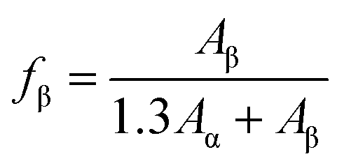
In order to quantitatively estimate the α- and β-phases in the films, we have used their Raman and infrared spectra. Fig. 3(a) shows the Raman spectra of the composite films along with that of a commercial PVDF film consisting of predominantly α-phase. The Raman spectra of PVDF films loaded with nanofillers are dominated by D and G bands of RGO, and hence the weak bands arising from PVDF are barely discernible. However, the Raman spectra of pure PVDF film prepared for this study and the commercial PVDF film show many distinct bands and clear difference between them. The Raman bands observed at 795 cm−1 and 839 cm−1 in pure PVDF films correspond to α- and β-phases and are often are used to identify the phases of PVDF.35 Clearly, the commercial PVDF film is dominated by the characteristic α-phase band at 795 cm−1 and a weak band at 839 cm−1 corresponding to the β-phase. But the opposite trend is seen in the pure PVDF film. We have estimated the amount of α- and β-phases in the two PVDF films by fitting the Raman spectral profile in the 700–950 cm−1 region with Lorentzian–Gaussian line profiles. Fig. 3(b) shows the fitted peaks and profiles. Using the integrated area of 795 and 839 cm−1 peaks, we estimate the amount of α- and β-phase in the pure PVDF film to be ≈18% and ≈82% (see Table 1), whereas in the commercial PVDF film it is found to be ≈74% and ≈26%. Thus, the solvent cast PVDF film consists of predominantly β-phase with a smaller fraction of α-phase. As the Raman spectra of PVDF–RGO, PVDF–RGO–Fe3O4 and PVDF–RGO–Fe3O4–ZnO are dominated by D and G bands of RGO, we could not estimate the amount α- and β-phases using the phase sensitive Raman modes. These samples are dark colored and tend to burn when incident laser power is increased in an attempt to obtain good Raman spectra in the region not masked by the strong and broad bands arising from RGO. We have used infrared spectroscopy studies to estimate the fractions of α- and β-phases in these films, as discussed below.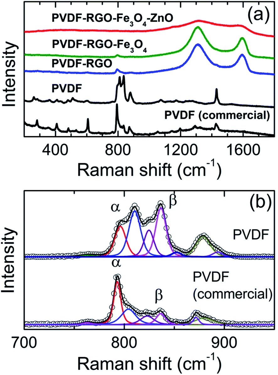 | ||
| Fig. 3 (a) Raman spectra of PVDF (commercial), PVDF, and PVDF composite films, (b) Lorentzian–Gaussian peak fits to Raman spectral profile of PVDF films. | ||
| Film | Fraction of β-phase (%) | |
|---|---|---|
| From IR | From Raman | |
| PVDF-commercial | 27 | 26 |
| PVDF | 81 | 82 |
| PVDF–RGO | 77 | — |
| PVDF–ZnO (spectrum not shown) | 58 | — |
| PVDF–RGO–Fe3O4 | 53 | — |
| PVDF–RGO–Fe3O4–ZnO | 49 | — |
Fig. 4 shows the attenuated total reflection FTIR spectra of the composite films in the 650–950 cm−1 regions. PVDF α-phase shows the characteristic peaks at 490, 530, 615, 760, 795 and 975 cm−1 whereas the polar β-phase shows peaks at 440, 510, 745, 840 and 878 cm−1.36–40 The commercial PVDF film consisting of predominantly α-phase shows all the expected characteristic bands of the α-phase. The pure PVDF sample prepared for this study shows mainly the β-phase with a minor fraction of α-phase. The IR bands at 762 and 840 cm−1 are characteristic of α- and β-phases and the absorbances at these wavenumbers have been used in determining the fraction of the β-phase present in the samples.41,42
 | ||
| Fig. 4 FTIR spectra of PVDF (commercial), PVDF, and PVDF composite films. | ||
The relative fraction of the β-phase in a sample containing both phases is given by,41
 | (1) |
Dielectric, ferroelectric and magnetic properties
Fig. 5(A) is a graph of dielectric constant versus frequency for all samples along with that of PVDF–ZnO film. All the films show nearly frequency independent behavior for f > 1 kHz. Both PVDF–RGO and PVDF–ZnO films show an enhancement in dielectric constant (25–30 at 1 Hz), compared to that of pure PVDF (∼6). Since the polymer matrix and the inorganic fillers have different dielectric constant, a large number of charge carriers accumulate on these interfaces leading to strong MWS (Maxwell–Wagner–Sillars) polarization.34 On the other hand, both PVDF–RGO–Fe3O4 and PVDF–RGO–Fe3O4–ZnO nanocomposite films show a reduced dielectric constant (∼12). We attribute this to increased electrical conduction in Fe3O4 aggregates in the PVDF composite films containing Fe3O4. It is interesting to note that PVDF–RGO film which contains a high percentage of β-phase shows a lower dielectric loss (not shown here) and a high dielectric constant compared to pure PVDF and other composite films. This results in a higher electrical polarization as described below. Generally, the dielectric loss arises from several contributions, including direct current conduction, MWS relaxation and the Debye loss factor (δ = δDC + δMW + δD). | ||
| Fig. 5 (A) Dielectric constant vs. frequency plots, (B) P–E curves, (C) M vs. H curves, and (D) MD vs. H curves of (a) PVDF, (b) PVDF–RGO, (c) PVDF–RGO–Fe3O4 and (d) PVDF–RGO–Fe3O4–ZnO, (e) PVDF–ZnO. Inset in (C) shows FC and ZFC curves for PVDF–RGO–Fe3O4–ZnO. | ||
The room temperature FE (P–E) loops of the composite films are shown in Fig. 5(B). A maximum polarization of 0.13 μC cm−2 is observed in PVDF–RGO film compared to PVDF (0.04 μC cm−2), PVDF–RGO–Fe3O4 (0.05 μC cm−2) and PVDF–RGO–Fe3O4–ZnO (0.06 μC cm−2) films at 60 kV cm−1. The polarization seems to be affected by both dielectric constant and the percentage of β-phase in the film. Although the fraction of β-phase is similar (∼80%) in both PVDF and PVDF–RGO films, the increase in polarization in PVDF–RGO seems to be due to the accumulation of the charges at the interface of the conducting and dielectric phases facilitating the heterogeneous polarization in the systems. However, both PVDF–RGO–Fe3O4 and PVDF–RGO–Fe3O4–ZnO films show lower polarization because of significant reduction in β-phase (∼50%) as well as reduced dielectric constant.
The magnetization vs. field (M vs. H) measurements for composite films are shown in Fig. 5(C). While the pure PVDF and PVDF–RGO films show curves characteristic of diamagnetic behavior, PVDF–RGO–Fe3O4 and PVDF–RGO–Fe3O4–ZnO films show superpara-magnetic behavior. The magnetization vs. temperature (ZFC–FC) curves of PVDF–RGO–Fe3O4–ZnO film (inset in Fig. 5(C)), further confirm its superparamagnetic nature. Fig. 5(D) shows the plots of magneto-dielectric coupling constant (MD%) vs. magnetic field at 30 kHz and 300 K. Here, MD = [(εH − ε0)/ε0] × 100%, where εH and ε0 are the dielectric constants with and without the applied magnetic field, H. Clearly, the diamagnetic PVDF and PVDF–RGO samples do not show any change in dielectric constant upon application of magnetic field. On the other hand, the samples loaded with magnetic Fe3O4 show variation in dielectric constant upon application of the magnetic field. The curves can be fitted using a parabolic equation, MD = (εH − ε0)/ε0 = γH2, which originates from the lowest-order coupling terms polarization (P) and magnetization (M) in the free-energy term.4 The MD effect in these samples arises from terms proportional to P2M2 in a symmetry-allowed Ginzburg–Landau free energy, where P and M are electrical polarization and magnetization. The quadratic dependence of MD on magnetization has been observed in other multiferroics.47,48 The samples of PVDF–RGO–Fe3O4 and PVDF–RGO–Fe3O4–ZnO show similar MD% of ∼0.035 and 0.031 respectively at 300 K and 10 kOe.
The origin of magneto-dielectric coupling in these samples could be attributed to the combined effect of magnetostriction in magnetic Fe3O4 causing piezoelectricity in ferroelectric PVDF. Upon application of magnetic field, the magnetic domains of Fe3O4 align in such a way that it can cause stress on neighboring PVDF phase via magnetostriction, which eventually leads to accumulation of some surface charge due to the piezoelectric effect. The elastic interaction between the magnetic (Fe3O4) and ferroelectric (FE) phases strongly depends on the microstructure and coupling interaction between the magnetic and FE interfaces.
Conclusions
In conclusion, PVDF based self-standing, flexible films loaded with RGO, Fe3O4 and ZnO nanofillers were prepared via solvent casting method. Analysis of IR spectra reveal a higher percentage of polar β-phase (∼80%) in both pure PVDF and PVDF–RGO films whereas the addition of nanofillers, Fe3O4 and ZnO, resulted in a reduced amount of β-phase (∼50%) in the films. All nanofiller loaded PVDF films show an increase in the dielectric constant but the electric polarization depends on the percentage of polar ferroelectric β-phase in the films. Fe3O4-loaded hybrid PVDF films exhibit a multiferroic behavior with a magneto-dielectric coupling constant ∼0.04%. These findings suggest that the PVDF–nanofiller composites may be tailored to make them suitable for room temperature magneto-dielectric device applications.Acknowledgements
Ehab H. Abdelhamid is supported by National Science Foundation DMR-1306449.References
- G. Lawes and G. Srinivasan, J. Phys. D: Appl. Phys., 2011, 44, 243001 CrossRef.
- H. Schmid, Ferroelectrics, 1994, 162, 317–338 CrossRef.
- T. Kimura, T. Goto, H. Shintani, K. Ishizaka, T. Arima and Y. Tokura, Nature, 2003, 426, 55–58 CrossRef CAS PubMed.
- B. Ramachandran, A. Dixit, R. Naik, G. Lawes and M. S. Ramachandra Rao, J. Appl. Phys., 2011, 110, 104105 CrossRef.
- A. K. Pradhan, K. Zhang, D. Hunter, J. B. Dadson, G. B. Loiutts, P. Bhattacharya, R. Katiyar, J. Zhang, D. J. Sellmyer, U. N. Roy, Y. Cui and A. Burger, J. Appl. Phys., 2005, 97, 093903 CrossRef.
- W. Eerenstein, N. D. Mathur and J. F. Scott, Nature, 2006, 442, 759–765 CrossRef CAS PubMed.
- L. W. Martin, S. P. Crane, Y. H. Chu, M. B. Holcomb, M. Gajek, M. Huijben, C. H. Yang, N. Balke and R. Ramesh, J. Phys.: Condens. Matter, 2008, 20, 434220 CrossRef.
- J. F. Scott, Nat. Mater., 2007, 6, 256–257 CrossRef CAS PubMed.
- D. Khomskii, Physics, 2009, 20 DOI:10.1103/physics.2.20.
- N. A. Hill and K. M. Rabe, Phys. Rev. B: Condens. Matter Mater. Phys., 1999, 59, 8759–8769 CrossRef CAS.
- I. Cabrera, M. Kenzelmann, G. Lawes, Y. Chen, W. C. Chen, R. Erwin, T. R. Gentile, J. B. Leão, J. W. Lynn, N. Rogado, R. J. Cava and C. Broholm, Phys. Rev. Lett., 2009, 103, 087201 CrossRef CAS PubMed.
- O. Heyer, N. Hollmann, I. Klassen, S. Jodlauk, L. Bohatý, P. Becker, J. A. Mydosh, T. Lorenz and D. Khomskii, J. Phys.: Condens. Matter, 2006, 18, L471 CrossRef CAS.
- K. Raidongia, A. Nag, A. Sundaresan and C. N. R. Rao, Appl. Phys. Lett., 2010, 97, 062904 CrossRef.
- S. Ryu, J. H. Park and H. M. Jang, Appl. Phys. Lett., 2007, 91, 142910 CrossRef.
- J. Ryu, A. V. Carazo, K. Uchino and H.-E. Kim, J. Electroceram., 2001, 7, 17–24 CrossRef CAS.
- O. D. Jayakumar, E. H. Abdelhamid, V. Kotari, B. P. Mandal, R. Rao, Jagannath, V. M. Naik, R. Naik and A. K. Tyagi, Dalton Trans., 2015, 44, 15872–15881 RSC.
- G. Srinivasan, E. T. Rasmussen, A. A. Bush, K. E. Kamentsev, V. F. Meshcheryakov and Y. K. Fetisov, Appl. Phys. A: Mater. Sci. Process., 2003, 78, 721–728 CrossRef.
- J. Liu, X. Lu and C. Wu, Membranes, 2013, 3, 389–405 CrossRef PubMed.
- Y. K. A. Low, L. Y. Tan, L. P. Tan, F. Y. C. Boey and K. W. Ng, J. Appl. Phys., 2013, 128, 2902–2910 CAS.
- P. Thakur, A. Kool, B. Bagchi, S. Das and P. Nandy, Appl. Clay Sci., 2014, 99, 149–159 CrossRef CAS.
- Y. Liu, Y. Sun, F. Zeng and Y. Chen, Int. J. Electrochem. Sci., 2013, 8, 5688–5697 CAS.
- C.-W. Tang, B. Li, L. Sun, B. Lively and W.-H. Zhong, Eur. Polym. J., 2012, 48, 1062–1072 CrossRef CAS.
- P. I. Devi and K. Ramachandran, J. Exp. Nanosci., 2011, 6, 281–293 CrossRef CAS.
- J. S. Dodds, F. N. Meyers and K. J. Loh, IEEE Sens. J., 2012, 12, 1889–1890 CrossRef CAS.
- M. S. Gaur and A. P. Indolia, J. Therm. Anal. Calorim., 2010, 103, 977–985 CrossRef.
- F. Chen, N. Hu, E.-Y. Ding and Z.-Z. Yang, Gaofenzi Cailiao Kexue Yu Gongcheng, 2007, 23, 242–245 CAS.
- A. Chilvery, A. Batra and M. Thomas, Phys. Rev. Res. Int., 2014, 4, 734 Search PubMed.
- G. Wang, Y. Deng, Y. Xiang and L. Guo, Adv. Funct. Mater., 2008, 18, 2584–2592 CrossRef CAS.
- G.-S. Wang, Y.-Y. Wu, X.-J. Zhang, Y. Li, L. Guo and M.-S. Cao, J. Mater. Chem. A, 2014, 2, 8644–8651 CAS.
- A. P. Indolia and M. Gaur, J. Polym. Res., 2013, 20, 1–8 CAS.
- A. P. Indolia and M. Gaur, J. Therm. Anal. Calorim., 2013, 113, 821–830 CrossRef CAS.
- D. Rouxel, B. Vincent, L. Badie, F. D. Dos Santos, E. Lamouroux and Y. Fort, Appl. Surf. Sci., 2013, 279, 204–211 CrossRef.
- D. He, K. Cheng, T. Peng, X. Sun, M. Pan and S. Mu, J. Mater. Chem., 2012, 22, 21298–21304 RSC.
- D. Wang, Y. Bao, J.-W. Zha, J. Zhao, Z.-M. Dang and G.-H. Hu, ACS Appl. Mater. Interfaces, 2012, 4, 6273–6279 CAS.
- S. Satapathy, S. Pawar, P. Gupta and K. Varma, Bull. Mater. Sci., 2011, 34, 727–733 CrossRef CAS.
- M. Kobayashi, K. Tashiro and H. Tadokoro, Macromolecules, 1975, 8, 158–171 CrossRef CAS.
- S. Lanceros-Mendez, J. Mano, A. Costa and V. Schmidt, J. Macromol. Sci., Part B: Phys., 2001, 40, 517–527 CrossRef.
- M. Latour, A. Montaner, M. Galtier and G. Geneves, J. Polym. Sci., Polym. Phys. Ed., 1981, 19, 1121–1129 CrossRef CAS.
- T. Boccaccio, A. Bottino, G. Capannelli and P. Piaggio, J. Membr. Sci., 2002, 210, 315–329 CrossRef CAS.
- Y. Bormashenko, R. Pogreb, O. Stanevsky and E. Bormashenko, Polym. Test., 2004, 23, 791–796 CrossRef CAS.
- R. Gregorio Jr and M. Cestari, J. Polym. Sci., Part B: Polym. Phys., 1994, 32, 859–870 CrossRef.
- S. Osaki and T. Kotaka, Ferroelectrics, 1981, 32, 1–11 CrossRef CAS.
- J. Andrew and D. Clarke, Langmuir, 2008, 24, 8435–8438 CrossRef CAS PubMed.
- B. P. Mandal, K. Vasundhara, E. Abdelhamid, G. Lawes, H. G. Salunke and A. K. Tyagi, J. Phys. Chem. C, 2014, 118, 20819–20825 CAS.
- M. E. Mackay, A. Tuteja, P. M. Duxbury, C. J. Hawker, B. Van Horn, Z. Guan, G. Chen and R. Krishnan, Science, 2006, 311, 1740–1743 CrossRef CAS PubMed.
- S. Chandran, N. Begam, V. Padmanabhan and J. Basu, Nat. Commun., 2014, 5, 3697 Search PubMed.
- D. P. Dutta, B. P. Mandal, R. Naik, G. Lawes and A. K. Tyagi, J. Phys. Chem. C, 2013, 117, 2382–2389 CAS.
- G. Lawes, T. Kimura, C. Varma, M. Subramanian, N. Rogado, R. Cava and A. Ramirez, Prog. Solid State Chem., 2009, 37, 40–54 CrossRef CAS.
| This journal is © The Royal Society of Chemistry 2016 |