DOI:
10.1039/C5RA26866D
(Paper)
RSC Adv., 2016,
6, 42589-42595
Formation of pH-responsive drug-delivery systems by electrospinning of vesicle-templated nanocapsule solutions†
Received
15th December 2015
, Accepted 18th April 2016
First published on 25th April 2016
Abstract
A novel polyethylene oxide (PEO) nanofibrous membrane, which contains chitosan (CS)/sodium alginate (SA) or CS nanocapsules formed by vesicles as a template, has been designed as a pH-responsive drug-delivery system and fabricated via an electrospinning process. Three different vesicle systems, including didodecyldimethylammonium bromide (DDAB), cetyl trimethyl ammonium bromide (CTAB)/sodium dodecyl benzene sulfonate (SDBS) (7/3) and CTAB/SDBS (3/7), were employed as the templates to construct the nanocapsules and 5-fluoro-2,4(1H,3H)pyrimidinedione (5-Fu) was selected as the model drug to be loaded within the drug delivery system. Structural characterization of the composites was obtained by means of zeta potential and a digital microscope image. The pH-responsive behaviors of the nanocapsules made from three different surfactant systems were detected by fluorescence spectroscopy. Drug release from the electrospun nanofibers with nanocapsules made from these different systems was investigated by a UV-visible spectrophotometer. The result showed that these different drug-delivery systems exhibit different release rates and pH-responsive behaviors. They can be good candidates for anticancer therapy in the organism, especially in wound healing dressing used after surgical procedures to improve the therapeutic value and reduce the local toxicity of medicinal drugs in clinical practice.
1. Introduction
In recent years, a great number of controlled drug-delivery systems have been developed to carry drugs released at a desired site and a desired rate to maximize the drug efficacy and minimize its side effects.1–4 The introduction of macromolecular drugs, which are easy to be denatured and thus lose their bioactivity, further increases the need for new controlled delivery systems. In drug-delivery systems, nanofibrous scaffolds often have been applied as drug carriers because of their high surface-to-volume ratio and functional characteristics. As a novel nanofiber-producing technology, electrospinning has gotten attentions due to the electrospun nanofibrous scaffolds' remarkable properties (such as a very large specific surface area and high porosity), which make them excellent candidates for drug carriers.5–7 It has also been found that electrospinning can simulate the natural extracellular matrix (ECM) to produce an similar architecture and size scale.8–10 The diameter of electrospun fibers closely matches the size scale of the ECM, which is ideal for cell attachment, proliferation, and differentiation.11,12 Therefore, electrospun nanofibrous membranes can be used as carriers for various drugs and bioactive molecules13,14 to be applied in wound healing or be implanted in surgery, which offer site-specific delivery of drugs to the body. The electrospun nanofibrous drug-delivery system can also be designed to control the drug release, no matter whether the release mechanism focus in diffusion alone or diffusion and scaffold degradation simultaneously.15 This drug delivery system has attracted attention in the field of clinical therapy for cancer in recent decades.16–19 Cancerous cells cannot be sliced off entirely by the surgical operation and maybe proliferate after surgical procedures. It is very important for anticancer drugs to be released from the wound dressing materials (electrospun nanofibrous drug-delivery system) at a proper time and in appropriate doses, to reduce the amount of cancer cells and prevent proliferation after surgical procedures.20 Furthermore, an effective therapy should combine sufficiently high and sustained drug levels at the injury site with minimal systemic and local toxicity. Therefore, development of delivery systems for the therapy of cancerous cells after surgical procedures must combine abrupt release bioactivity of the anticancer drug, which is incorporated within the scaffolds, with the controlled release of these anticancer drugs according to the time frame of cancer cell regeneration. Different demands for the drug-releasing rate and amount will handicap the drug-loading process, and it is hard to control the loading amount of different processes on the carriers for achieving a desirable ratio, which is important for improving the clinical effect. It is common and useful to improve cancer therapeutic value of medicinal drugs in clinical practice.21–23 Nowadays, great attention has been paid to the research on the preparation of the polymer nanoparticles and liposomes. It is known that chemical conjugation is generally involved in the loading processes, which increases the operational complexity, and the stoichiometry issue is another obstacle to control the dose ratio. So it is very important for us to find a simple and convenient method to obtain a drug delivery system, in which drugs release controllably, for the therapy of cancerous cells after surgical procedures.
Stimuli-responsive polymersomes have emerged for controlled drug delivery in recent years. In particular, pH-responsive polymersomes have attracted most attention for cancer diagnosis and therapy because cancerous tissue regions and intracellular compartments are more acidic than normal tissues.21 Among them, liquid-core (preferably water-filled) polymeric pH-responsive nanocapsules are of particular interest due to their special hollow morphology and potential for the encapsulation of a variety of guest substances within their empty core domain. Compared to nanospheres, liquid-core particles offer several advantages such as the relatively low density, high loading capacity, easy incorporation/encapsulation and subsequently more release of the substance of interest.22–24 The layer-by-layer (LbL) methodology is appreciable in many recent studies, especially including fabricating oil-core nanocapsules efficiently coated by biocompatible polyelectrolytes for controlled drug-release.25,26 And chitosan (CS)/sodium alginate (SA) nanocapsules can be prepared with templating vesicles of didodecyldimethylammonium bromide (DDAB) by a LbL approach.27 It is straightforward that the CS/SA nanocapsules can be used as the pH responsive carries within the electrospun nanofibrous membranes, and applied in cancer diagnosis and therapy. Here, we investigate three different vesicle systems, including single surfactant (DDAB), cationic mixed surfactant systems (CTAB/SDBS, 7/3) and anionic mixed surfactant systems (CTAB/SDBS, 3/7). In this case, the surfactants, which are used to form the vesicle, do not need to be moved from the vesicular template with difficulty. The surfactants are chosen as the component for constructing vesicles and chemosensitizers for overcoming the multi-drug resistance. Furthermore, they can enhance the drug efficacies28 with their anti-cancer activity which have been proven to be exceptionally high.29,30 A schematic drawing of the production of electrospun nanofibers with hollow nanocapsules is presented in Scheme 1. The procedure is divided into two main parts, i.e., preparation of the hollow nanocapsules with layer-by-layer deposition and preparation of the electrospun nanofibrous membranes by electrospinning the template solution. 5-Fu, a hydrophilic anticancer drug, is selected as model drug to be loaded within the drug delivery system. Drug release from the electrospun nanofibrous membranes is detected with fluorescence and UV-visible spectroscopy. Moreover, the pH-responsive behavior of the electrospun nanofibers has been studied by UV-visible spectroscopy.
 |
| | Scheme 1 Schematic of the preparation of vesicle-templated and pH-responsive electrospun nanofibers. | |
It is notable that all materials used in this study have been approved for drug delivery systems in the field of pharmacy.31–34 CS and SA, which came from the natural polysaccharide, were used to prepare the nanocapsules. The synthetic PEO polymers, which can be used in food, cosmetics, personal care products and pharmacy, are electrospun to the nanofibrous membranes as the carrier. Moreover, besides DDAB, CTAB and SDBS, some other surfactants such as Tween 80, Triton X-100, P123 and F127 are all well qualified as both anti-cancer active drugs and chemosensitizers for overcoming the multi-drug resistance in cancer.29,35–37 Therefore, this versatile method can provide production with a series of vesicle template nanocapsules which can be readily adjusted with different surfactants and proportions. Furthermore, the proportion of mixed surfactant systems offers reasonable control over capsule morphology and the shell thickness, which can indicate the release rates and the potential applications.
2. Experimental
2.1. Materials
PEO with a molecular weight of 600 kDa was purchased from Sigma-Aldrich. DDAB (99.0%), CTAB (99.0%) and SDBS (99.0%) were supplied by Sinopharm Chemical Reagent Co. Ltd. Low-molecular-weight SA and low-molecular-weight CS were purchased from Sigma-Aldrich. 5-Fu (99.9%) was purchased from Aladdin Chemistry Co. Ltd. Analytical reagent sodium chloride (NaCl), potassium chloride (KCl), potassium phosphate (KH2PO4) and disodium hydrogen phosphate dodecahydrate (Na2HPO4·12H2O), which were used to prepare the phosphate buffer solutions (PBS), analytical reagent sodium hydroxide (NaOH) and hydrochloric acid (HCl) were all purchased from Guangdong Xilong Chemical Co., Ltd. All samples were used without further purification. All solutions were prepared with deionized water. And chemical formulas for main reagents studied in this paper are displayed in Scheme S1,† which makes this article easy to follow.
2.2. Preparation of vesicle solutions
The vesicle solutions as the template were obtained from dispersions of the surfactant in water. The cationic DDAB vesicle solution was prepared just by dissolving a certain amount of surfactant in water and shaking the suspension for about 5 h. A surfactant stock solution (1000 μL, 10 mM DDAB) was dispersed in the starting volume of 10 mL H2O. A literature method for the preparation of spontaneous vesicle formation from mixed cationic and anionic single chain surfactants was adopted to prepare CTAB/SDBS vesicle solution.38 A bluish turbidity solution was obtained after being stirred, which suggested the formation of CTAB/SDBS vesicles in the mixed solution. All the vesicles above were maintained with bluish turbidity after standing for more than one month, declaring the favorable physical stability of the surfactant-assisted vesicles. A surfactant stock solution (1000 μL, ctotal = 10 mM) was dispersed in the starting volume of 10 mL H2O for the following study work.
2.3. Preparation of vesicle-templated nanocapsules
A LbL method for preparation of nanoparticles in cationic vesicle solution was adopted to prepare two-layer (SA/CS) nanocapsules.27 Stock solutions (1 mg mL−1) of SA and CS were prepared respectively. CS solution was prepared in a 1% (v/v) acetic acid solution, and the final pH was adjusted to 5.5 with 1 M NaOH. Not only the single surfactant (DDAB) but also the mixed surfactant systems (CTAB/SDBS, 7/3) can form the cationic vesicles in the aqueous solution.27,38 The cationic vesicles were coated with alternating layers of negatively charged SA at first and then layers of positively charged CS. A small increment of the added volume of polyelectrolyte gave the reverse surface charge. The amount of polyelectrolyte for the formation of the individual shells was established through titration, adding the polyelectrolyte solution until the isoelectric point was exceeded, which corresponded to a higher instability of the aggregates. Then the two-layer (SA/CS) nanocapsules were obtained in the cationic vesicle solution. The anionic vesicles were formed in the CTAB/SDBS (3/7) aqueous solution. In this case, the anionic vesicles were coated only with a layer of positively charged CS. Similarly, the amount of CS solution which was added into the vesicle solution was also determined by the isoelectric point. Thus, one-layer CS nanocapsules were formed in the anionic vesicle solution. In this study, stability of the aggregates was checked by measuring zeta potential of the surface and the result was over 30 mV than the neutrality. And zeta potential was used to measure the hydrodynamic diameter (DH) of the vesicles, nanocapsules size, polydispersity index (PdI) of these aggregates. And negative staining technique using uranyl acetate was applied to prepared samples for TEM of vesicles, which is in order to support to the data of zeta potential. Anticancer drug 5-Fu was added into the vesicle solution before preparation of nanocapsules. It could be encapsulated into the aqueous core of vesicle to maintain their concentration identical on the inner and outer sides of the nanocapsules after nanocapsules' formation.
2.4. Fabrication of drug delivery fibrous membranes via electrospinning
PEO was added into the prepared nanocapsules solution to get the mixed solution (2 w/v%, w in g and v in mL) for electrospinning. The prepared solutions were stored at room temperature before use. When electrospinning, the prepared PEO/nanocapsule solutions were loaded into a glass syringe with a 5 mL blunt-end tip capillary. The syringe was placed into a syringe pump (TJ-3A/W0109-1B, Baoding Longer Precision Pump Co., Ltd.) that delivered the solution at a controlled flow rate of 0.5 mL h−1. A horizontal electrospinning setup was used in all experiments.15 The high voltage power supply source (HB-Z503-1AC, Tianjin Hengbo Co., Ltd.) maintained a voltage of 17.0 kV between the capillary tip and a grounded metal plate covered in aluminum foil, with an interelectrode distance of 18.0 cm. Dry nanofibers were collected directly from the aluminum-foil-covered plate and stored at room temperature.
2.5. Characterization of nanocapsules and electrospun nanofibrous membranes
An ECLIPSE LV150N Upright Digital Microscope, manufactured by Japan Nikon Ltd., was used to examine the morphologies of the nanocapsules. A Hitachi-7650 digital vacuum Transmission Electron Microscope (TEM), manufactured by Japan Hitachi Ltd., was used to examine the morphologies of the prepared vesicles and electrospun nanofibres in this work. The Fourier transform infrared (FTIR) spectrum of the electrospun nanofibrous membranes and the neat powder was recorded on a Bruker-Tensor 27 FTIR spectrometer. The zeta potential and size of the aggregates was measured by Malvern UK Zetasizer-Nano ZSP commercial instrument operating with a 4 mW He–Ne laser (wavelength at 633 nm). Samples were injected into dedicated disposable capillary cells. All measurements were performed at 25.0 °C.
2.6. pH-responsive behavior of nanocapsules to calcein
The pH-responsive behavior of the formed nanocapsules was characterized by using calcein (a model compound) as the fluorescent marker and cobalt chloride as the quenching agent.39 These two compounds are commercially available and there is no separation of vesicles from bulk media. The fluorescence intensity of the solution was measured by HITACHI F-4500 Fluorescence Spectrophotometer. The entrapment quantity of nanocapsules to calcein was deduced according to the difference of fluorescence intensity before and after quenching. There was a linear relationship between the fluorescence intensity and the concentration, which could be obtained from 0 to 8 mM in a buffer solution of 0.1 M NaOH–KH2PO4 (pH = 6.80).40 Therefore, the concentration of calcein in the bulk solution was kept at 1 mM. As a metal-sensitive probe, calcein can react with Co2+ and produce a complex with lower fluorescence intensity, which is the so-called quenching.41 The quenching ratio (molar ratio) of Co2+ to calcein is 1/1, and the quenching is a time-dependent process.39 Calcein was added into the solution before nanocapsules were formed to maintain their concentration identical on the inner and outer sides of the nanocapsules after nanocapsules' formation, followed by 100 μL cobalt chloride solution (0.1 mM) to quench any external calcein in bulk solution. The fluorescence intensity of calcein was monitored using an excitation wavelength of 495 nm and an emission wavelength of 515 nm. The excitation and emission slits were set at 5 nm.
2.7. Studies of the drug-releasing
The drug-loaded electrospun nanofibrous membranes were incubated in a 700 mL PBS solution (0.2 M, pH = 7.40) at 37.0 °C and gently stirred at 60 cycles/min. At a certain time, 0.5 mL of the buffer was taken out and an equal amount of fresh buffer was added. The release of 5-Fu from the electrospun nanofibrous membranes was monitored by a UV-visible spectrophotometer (Beijing Purkinje General Instruments Ltd., TU-1810) at the wavelength of 265 nm, and converted to the 5-Fu concentration according to the calibration curve of 5-Fu in the same buffer. The accumulative amount of the released 5-Fu was calculated as a function of incubation time.
3. Results and discussion
3.1. Characterization of morphologies for nanocapsules and electrospinning nanofibres
The fabrication of hollow capsules is derived from the vesicle template made of surfactant. To study the preparation of nanocapsules systematically, three different surfactant systems were employed in the study. DDAB and CTAB/SDBS (7/3) vesicles, the cationic surfactant systems, were used as the template for the LbL deposition. The cationic surface of the vesicles was first covered with the anionic polyelectrolyte SA, until reversing the surface charge from positive to negative which was measured by zeta potential. The positively charged CS was added to form the outer layer. CS was then added into the solution, and the amount was controlled by the zeta potential value until the surface charge changed from negative to positive. Thus, the CS/SA hollow capsules with two layers were obtained. A CTAB/SDBS (3/7) vesicle, the anionic surfactant system, was also used as a template for the deposition of cationic polyelectrolyte CS. However, the anionic vesicle's surface was only covered with a layer of cationic polyelectrolyte. The prepared CS hollow capsules were unilaminar, and the derived hollow capsules maintain the shape of the template. The stability and permeability features depend on the polyelectrolyte complex used. As far as the production of the hollow capsules is concerned, their characterization will also be reported. The variation of the surface charge values, the morphologies of the hollow capsules and the electrospinning nanofibres were determined with the zeta potential, digital microscope, and TEM, respectively. They are shown in Fig. 1.
 |
| | Fig. 1 (A) Zeta potential mean values with respect to the amount of alternate adsorption of polyelectrolyte (stock concentration of SA and CS is 1 mg mL−1), data with error bars were calculated from three independent experiments; (B) digital microscope images of capsules with vesicles as template; (C) TEM image of electrospun nanofibers with capsules in different vesicle systems (2 w/v% PEO, electrospinning in 5 mL capillary at flow rate of 0.5 mL h−1, voltage of 17.0 kV, interelectrode distance of 18.0 cm): (1) DDAB surfactant system (1000 μL of 10 mM DDAB dispersed in 10 mL H2O, VSA = 3.0 mL, VCS = 2.0 mL); (2) CTAB/SDBS (7/3) surfactant system (ctotal = 10 mM, 1000 μL of 10 mM surfactant solution dispersed in 10 mL H2O, VSA = 1.0 mL, VCS = 1.5 mL); (3) CTAB/SDBS (3/7) surfactant system (ctotal = 10 mM, 1000 μL of 10 mM surfactant solution dispersed in 10 mL H2O, VCS = 1.0 mL). All the scale bars are 1 μm. | |
From Fig. 1A1, it can be seen that the zeta potential value of the bare DDAB vesicles was 86.8 mV in the solution. The zeta potential value decreased quickly as the increasing amount of SA. With the increased amount of SA which covered the surface of DDAB vesicles, the surface charge reversed to −56.6 mV when added 3 mL SA. Then, the CS solution with the positive charge was added, and the zeta potential value turned to augment. When 2.0 mL of CS solution was added, the zeta potential values increased to 37 mV. After that, it would change slightly with the increasing amount of CS. This implies that the amount of polyelectrolyte deposited onto the vesicle did no longer increase, substantially. The vesicles size, the amount of polyelectrolyte added into the three systems and the resulting nanocapsules size is listed in Table 1. All the nano-sized vesicle systems are with a very low PDI value indicating the excellent monodispersity of these vesicle systems, which is necessary for biomedical needs. And the TEM of different vesicle systems in Fig. S1† shows the same result. It is clear that the amount of deposition onto vesicles is associated with an increase in particle size determined by zeta potential instrument, basically consistent with that determined by digital microscope image. Fig. 1B shows the shape of the obtained nanocapsules is oval. No appreciable difference in the morphology of the nanocapsules is observed when using different surfactant systems as the template. Furthermore, all nanocapsules are nano-sized with a low PDI value suggests the possibility of them to be a drug carrier. From TEM images of the electrospun nanofibers (Fig. 1C), the nanocapsules can be seen clearly within the nanofibers. This implies that the prepared nanocapsules are incorporated into the electrospinning nanofibres successfully.
Table 1 The hydrodynamic diameter of vesicles and the amount of added polyelectrolyte and the average size of nanocapsules
| System |
DDAB |
CTAB/SDBS (7/3) |
CTAB/SDBS (3/7) |
| Data origin: particle size analysis from the zeta potential instrument. Added polyelectrolyte: 1 mg mL−1 SA. Added polyelectrolyte: 1 mg mL−1 CS. And details of each system are same with captions of Fig. 1, one-to-one. |
| Vesicle DH/nma |
105 |
85 |
90 |
| Vesicle PDIa |
0.098 |
0.050 |
0.064 |
| First layer/mL |
3.0b |
1.0b |
1.0c |
| Second layer/mL |
2.0c |
1.5c |
— |
| Nanocap. size/nma |
340 |
310 |
230 |
| Nanocap. PDIa |
0.124 |
0.089 |
0.101 |
3.2. Characterization of Fourier transforms infrared spectroscopy for electrospinning nanofibres
The composition of the electrospun nanofibrous membrane was characterized by FTIR spectroscopy. Fig. 2 illustrates the FTIR spectra of neat 5-Fu, blank PEO nanofibrous membrane and nanofibers loading 5-Fu electrospun with three different surfactant systems respectively. The characteristic peaks of 5-Fu (Fig. 2a) are clued at 1663 cm−1 (overlapped stretching vibration absorption of C![[double bond, length as m-dash]](https://www.rsc.org/images/entities/char_e001.gif) O and CC), 1246 cm−1 (absorption of C–F stretching vibration), 639 cm−1 and 550 cm−1 (out-of-plane bending vibration absorption of C–H in –CFCH–).42,43 All of these peaks can be detected in the composite electrospun nanofibers, which indicate that 5-Fu has been successfully loaded in all these systems. Meanwhile, it is found that these peaks shift slightly from the original powder (Fig. 2a) to different systems in different degree. This may be due to the different electrostatic interactions between the different surfactants and 5-Fu in these systems.
O and CC), 1246 cm−1 (absorption of C–F stretching vibration), 639 cm−1 and 550 cm−1 (out-of-plane bending vibration absorption of C–H in –CFCH–).42,43 All of these peaks can be detected in the composite electrospun nanofibers, which indicate that 5-Fu has been successfully loaded in all these systems. Meanwhile, it is found that these peaks shift slightly from the original powder (Fig. 2a) to different systems in different degree. This may be due to the different electrostatic interactions between the different surfactants and 5-Fu in these systems.
 |
| | Fig. 2 FTIR spectra of (a) 5-Fu neat powder; (b) PEO electrospun nanofibrous blank membrane; (c) electrospun membrane loading 5-Fu with DDAB as template; and (d) electrospun membrane loading 5-Fu with CTAB/SDBS (7/3) as template; (e) electrospun membrane loading 5-Fu with CTAB/SDBS (3/7) as template. Electrostatic spinning parameters and details of each system are same with captions of Fig. 1, one-to-one. | |
3.3. Responsive behavior of nanocapsules
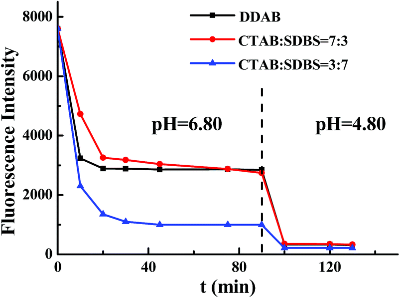
For the possible applications of nanocapsules as controlled release systems, their responsiveness to pH stimuli is a basic requirement. To detect pH-responsive behavior of nanocapsules, the entrapment quantity of the nanocapsules to calcein was employed in our study just as the former study.39 The results are shown in Fig. 3. At first, the same amount of calcein was added into all three vesicle solutions. Though different amount of calcein was entrapped into the nanocapsules, the fluorescence intensity of the three vesicle solutions was kept at the same value. When cobalt chloride was added into the solution, calcein outside of the vesicle was quenched and the fluorescence intensity decreased rapidly for the first 20 minutes. After that, the fluorescence intensity of the solution was almost unchanged with the time for every system. After 90 minutes, the fluorescence intensity of DDAB solution was a little more than that of CTAB/SDBS (7/3) solution, and they were much more than that of the CTAB/SDBS (3/7) solution. When the pH value of the solution was changed from neutral to acidic (pH = 4.80) by adding a certain amount of HCl, an obvious decrease of fluorescence intensity was observed from these three systems. The fluorescence intensity decreased from 2846 to 316 for the DDAB solution, and decreased from 2741 to 331 for the CTAB/SDBS (7/3) solution. While for the CTAB/SDBS (3/7) solution, the fluorescence intensity also changed from 1000 to 215. Thus it proves that all of three nanocapsules are pH-responsive, although there are some differences among them.
 |
| | Fig. 3 Dependence of fluorescence intensity in different vesicle systems on time with two different pH surrounding: pH = 6.80 (0.1 M NaOH–KH2PO4) and pH = 4.80 (adding HCl into 0.1 M NaOH–KH2PO4). ccalcein = 1 mM, quenching ratio (Co2+/calcein) is 1/1, λex = 495 nm, λem = 515 nm, excitation and emission slits at 5 nm and details of each system are same with captions of Fig. 1, one-to-one. | |
3.4. Drug release study of electrospun nanofibrous membrane with different vesicle-template nanocapsules
To explore the pH-responsiveness capacity of electrospun nanofibrous drug-delivery systems with vesicle-templated nanocapsules, four surfactant systems were prepared and investigated in this study. 5-Fu, a hydrophilic anticancer drug, was used as a model drug and loaded into the vesicle, the release profile was determined by UV-Vis characterization. 5-Fu can be dissolved into the free water of the solution and bond water of the inner vesicle. When the vesicle was used as the template, some 5-Fu was encapsulated into the prepared nanocapsules. The release behaviors of 5-Fu from electrospun nanofibrous membrane with different vesicle systems were assessed at 37 °C in PBS of pH at 7.40 and 5.45, respectively. The results are shown in Fig. 4. For the neutral pH solution (pH = 7.40), all drawings in Fig. 4 present a similar tendency, which implies that the drug release for the different vesicle systems is almost same. Initially, for the first 15 min, a rapid release of 5-Fu can be observed in all systems. Subsequently, the release rate reaches a plateau followed up to the cumulative release time of 240 min. There is a little difference among them for the final release rates. When the samples were performed with a pH of 5.45, the release rates increased at different extents for different systems. The detailed results of Fig. 4 are listed in Table 2. It is clear that the release rate when the pH was 7.40 for DDAB system is very similar to that of CTAB/SDBS (7/3) system. This implies that the drug amount encapsulated by the nanocapsules is almost the same for these two systems. For the different release rates between a pH of 7.40 and 5.45, the value of the CTAB/SDBS (7/3) system is higher than that of the DDAB system. It means that pH-responsiveness is more sensitive. This is coincided with the result of the responsive behavior of nanocapsules which is detected in part 3.3. The release rate of pH at 7.40 in CTAB/SDBS (6/4) system is slightly higher than the others, which implies that the amount of drug encapsulated by the nanocapsules is lower. The reason may be due to the amount of the bond water in the vesicle template which was smaller. Meanwhile, the release rate of the CTAB/SDBS (6/4) system is the smallest in the former three systems, which implies that the pH-responsiveness of it is the worst. For the CTAB/SDBS (3/7) system, some drugs were separated out in the solution during electrospinning. Therefore, the release rate and pH-responsiveness was very small. Thus, the system is not stable for being used as a drug-delivery system. Compared to the other systems, the d-value of CTAB/SDBS (7/3) system is higher, coincided with the monodispersity discussed in Part 3.1.
 |
| | Fig. 4 Cumulative 5-Fu mean release profiles from electrospun nanofibrous membranes with different vesicle systems (data with error bars were calculated from three independent experiments): (A) DDAB; (B) CTAB/SDBS (7/3); (C) CTAB/SDBS (6/4); (D) CTAB/SDBS (3/7). Parameters of drug release experiment: 700 mL PBS solution, 37.0 °C, 60 cycles per min, λUV = 265 nm and details of each system are same with captions of Fig. 1, one-to-one. | |
Table 2 Release rate at a pH of 7.40, 5.45 and the d-value between them
| System |
Release rate (pH = 7.40) |
Release rate (pH = 5.45) |
d-valueb |
| Details of each system are same with captions of Fig. 1, one-to-one. The d-value is calculated as the difference value between release rate of pH = 7.40 and release rate of pH = 5.45. |
| DDAB |
75.7% |
93.6% |
17.9% |
| CTAB/SDBS (7/3)a |
76.5% |
96.9% |
20.4% |
| CTAB/SDBS (6/4)a |
79.5% |
92.4% |
12.9% |
| CTAB/SDBS (3/7)a |
62.9% |
67.2% |
4.3% |
The pH responsive character of the nanocapsules can be attributed to the special chemical structure of CS/SA backbone. At lower pH values, protonation can occur at the –NH2 groups of CS leading to the disruption of the hydrogen bonding involving these groups, which consequently facilitates the entrance of the swelling fluid into the nanocapsules to attain higher values of swelling.44 Meanwhile, the reversible protonation and deprotonation of the carboxyl groups on the backbone of make alginate a good candidate for preparing pH-responsive matrices. It is extremely available for the release of the drug within the nanocapsules. The increased hydrophobicity of these CS-based nanocapsules at higher pH values will thus prevent faster swelling in neutral or alkaline medium.45 The CS-based nanocapsules are able to trigger an effective release of 5-Fu in the medium with a low pH, which shows a clear response to the surrounding pH value. Therefore, this pH-responsive property of the drug-delivery system is suitable to be candidates for anticancer therapy in the organism, especially for wound healing dressings used after cancer surgical procedures.
Interestingly, it is very easy and convenient to control the amount of drugs in and out of the nanocapsules by varying the ratio of anionic to cationic surfactant. Meanwhile, these release rates were slow due to the existence of structure of vesicle. Therefore, this new method can offer a desirable amount and ratio of hydrophilic and hydrophobic drugs to meet the requirements of clinical therapy.
4. Conclusions
The drug delivery systems with three different pH-responsive nanocapsules were successfully prepared by electrospinning. The hollow nanocapsules were produced by using an alternating deposition of oppositely charged polyelectrolytes onto vesicles. Three different vesicle systems, DDAB, CTAB/SDBS (7/3) and CTAB/SDBS (3/7), were employed as the template to construct the nanocapsules. 5-Fu, as the model drug, was dissolved into the vesicle solution and loaded into the PEO electrospun nanofibrous membranes successfully. The pH-responsive behaviors of nanocapsules with different vesicle systems were investigated by fluorescence spectroscopy. The results showed that these nanocapsules were all sensitive to the acidic solution due to the –NH2 of CS and the –COOH of SA. It also showed that the pH-responsive behavior of DDAB surfactant system was almost similar with that of the CTAB/SDBS (7/3) mixed surfactant system. Drug released from the electrospun nanofibers with different nanocapsules was also investigated. The results showed different drug-delivery systems with different release rates and pH-responsive behaviors. This kind of drug-delivery system can be good candidates for anticancer therapy in the organism, especially for wound healing dressings after cancer surgical procedures, for improving the therapeutic value of cancer and decreasing the side cytotoxicity of medicinal drugs in clinical practice.
Acknowledgements
This work was supported by Beijing Natural Science Foundation (2142011).
Notes and references
- C. S. Kim, G. Y. Tonga, D. Solfiell and V. M. Rotello, Adv. Drug Delivery Rev., 2013, 65, 93 CrossRef CAS PubMed.
- Y. Engel, J. D. Schiffman, J. M. Goddard and V. M. Rotello, Mater. Today, 2012, 15, 478 CrossRef CAS.
- A. Greiner and J. H. Wendorff, Angew. Chem., Int. Ed., 2007, 46, 5670 CrossRef CAS PubMed.
- T. J. Sill and H. A. von Recum, Biomaterials, 2008, 29, 1989 CrossRef CAS PubMed.
- G. C. Rutledge and S. V. Fridrikh, Adv. Drug Delivery Rev., 2007, 59, 1384 CrossRef CAS PubMed.
- E. R. Kenawy, J. M. Layman, J. R. Watkins, G. L. Bowlin, J. A. Matthews, D. G. Simpson and G. E. Wnek, Biomaterials, 2003, 24, 907 CrossRef CAS.
- J. M. Deitzel, J. Kleinmeyer, D. Harris and N. C. BeckTan, Polymer, 2000, 42, 261 CrossRef.
- K. D. Andrews, J. A. Hunt and R. A. Black, Polym. Int., 2008, 57, 203 CrossRef CAS.
- A. S. Badami, M. R. Kreke, M. S. Thompson, J. S. Riffle and A. S. Goldstein, Biomaterials, 2006, 27, 596 CrossRef CAS PubMed.
- C. A. Bashur, L. A. Dahlgren and A. S. Goldstein, Biomaterials, 2006, 27, 5681 CrossRef CAS PubMed.
- Z. G. Chen, X. M. Mo, C. L. He and H. S. Wang, Carbohydr. Polym., 2008, 72, 410 CrossRef CAS.
- A. I. Teixeira, G. A. Abrams, P. J. Bertics, C. J. Murphy and P. F. Nealey, J. Cell Sci., 2003, 116, 1881 CrossRef CAS PubMed.
- Y. K. Luu, K. Kim, B. S. Hsiao, B. Chu and M. Hadjiargyrou, J. Controlled Release, 2003, 89, 341 CrossRef CAS PubMed.
- C. Huang, S. J. Soenen, E. van Gulck, G. Vanham, J. Rejman, S. Van Calenbergh, C. Vervaet, T. Coenye, H. Verstraelen, M. Temmerman, J. Demeester and S. C. De Smedt, Biomaterials, 2012, 33, 962 CrossRef CAS PubMed.
- W. Li, T. Luo, Y. Shi, Y. J. Yang, X. S. Huang, K. X. Xing, L. F. Liu and M. Q. Wang, Integr. Ferroelectr., 2014, 151, 164 CrossRef CAS.
- T. M. Allen and P. R. Cullis, Science, 2004, 303, 1818 CrossRef CAS PubMed.
- R. Langer and D. A. Tirrell, Nature, 2004, 428, 487 CrossRef CAS PubMed.
- I. Brigger, C. Dubernet and P. Couvreur, Adv. Drug Delivery Rev., 2012, 64, 24 CrossRef.
- M. E. Davis, Z. Chen and D. M. Shin, Nat. Rev. Drug Discovery, 2008, 7, 771 CrossRef CAS PubMed.
- M. Konishi, Y. Tabata, M. Kariya, H. Hosseinkhani, A. Suzuki, K. Fukuhara, M. Mandai, A. Takakura and S. Fujii, J. Controlled Release, 2005, 103, 7 CrossRef CAS PubMed.
- F. H. Meng, Z. Y. Zhong and J. Feijen, Biomacromolecules, 2009, 10, 197 CrossRef CAS PubMed.
- C. Vauthier and K. Bouchemal, Pharm. Res., 2009, 26, 1025 CrossRef CAS PubMed.
- B. H. Tan and K. C. Tam, Adv. Colloid Interface Sci., 2008, 136, 25 CrossRef CAS PubMed.
- M. Motornov, Y. Roiter, I. Tokarev and S. Minko, Prog. Polym. Sci., 2010, 35, 174 CrossRef CAS.
- U. Bazylińska, R. Skrzela, M. Piotrowski, K. Szczepanowicz, P. Warszyński and K. A. Wilk, Bioelectrochemistry, 2010, 87, 147 CrossRef PubMed.
- K. Szczepanowicz, U. Bazylińska, J. Pietkiewicz, L. Szyk-Warszyńska, K. A. Wilk and P. Warszyński, Adv. Colloid Interface Sci., 2011, 222, 678 CrossRef PubMed.
- F. Cuomo, F. Lopez, M. G. Miguel and B. Lindman, Langmuir, 2010, 26, 10555 CrossRef CAS PubMed.
- Q. J. He, Y. Gao, L. X. Zhang, Z. W. Zhang, F. Gao, X. F. Ji, Y. P. Li and J. L. Shi, Biomaterials, 2011, 32, 7711 CrossRef CAS PubMed.
- D. W. Loe and F. J. Sharom, Br. J. Cancer, 1993, 68, 342 CrossRef CAS PubMed.
- E. Ito, K. W. Yip, D. Katz, S. B. Fonseca, D. W. Hedley, S. Chow, G. W. Xu, T. E. Wood, C. Bastianutto, A. D. Schimmer, S. O. Kelley and F. F. Liu, Mol. Pharmacol., 2009, 76, 969 CrossRef CAS PubMed.
- K. Aiedeh, E. Gianasi, I. Orienti and V. Zecchi, J. Microencapsulation, 1997, 14, 567 CrossRef CAS PubMed.
- S. Miyazaki, H. Yamaguchi, M. Takada, W. M. Hou, Y. Takeichi and H. Yasubuchi, Acta Pharm. Nord., 1990, 2, 401 CAS.
- F. Gu, B. Amsden and R. Neufeld, J. Controlled Release, 2004, 96, 463 CrossRef CAS PubMed.
- V. Gopishetty, Y. Roiter, I. Tokarev and S. Minko, Adv. Mater., 2008, 20, 4588 CrossRef CAS.
- H. Riehm and J. L. Biedler, Cancer Res., 1972, 32, 1195 CAS.
- Q. J. He, J. L. Shi, F. Chen, M. Zhu and L. X. Zhang, Biomaterials, 2010, 31, 3335 CrossRef CAS PubMed.
- W. Zhang, Y. A. Shi, Y. Z. Chen, J. A. Ye, X. Y. Sha and X. L. Fang, Biomaterials, 2011, 32, 2894 CrossRef CAS PubMed.
- E. W. Kaler, A. K. Murthy, B. E. Rodriguez and J. A. N. Zasadzinski, Science, 1989, 245, 1371 CAS.
- L. Wei, Y. J. Yang, L. Tian, J. L. Zhang and B. X. Han, Phys. Chem. Chem. Phys., 2014, 16, 3640 RSC.
- L. M. Zhai, J. P. Zhao, M. Zhao, Y. J. Chen and L. J. Zhang, J. Dispersion Sci. Technol., 2007, 28, 455 CrossRef CAS.
- K. Martin, S. Craig and S. Marc, Biochem. Biophys. Res. Commun., 2005, 330, 645 CrossRef PubMed.
- V. Selvaraj and M. Alagar, Int. J. Pharm., 2007, 337, 275 CrossRef CAS PubMed.
- L. Huang, W. Sui, Y. Wang and Q. Jiao, Carbohydr. Polym., 2010, 80, 168 CrossRef CAS.
- S. J. Kim, S. J. Park and S. I. Kim, React. Funct. Polym., 2003, 55, 53 CrossRef CAS.
- I. M. El-Sherbiny, R. J. Lins, E. M. Abdel-Bary and D. R. K. Harding, Eur. Polym. J., 2005, 41, 2584 CrossRef CAS.
Footnote |
| † Electronic supplementary information (ESI) available. See DOI: 10.1039/c5ra26866d |
|
| This journal is © The Royal Society of Chemistry 2016 |
Click here to see how this site uses Cookies. View our privacy policy here.