A colorimetric and ratiometric fluorescent probe for the selective detection of cyanide anions in aqueous media and living cells

Xueyi Sun, Yu Wang*, Xiahe Deng, Junfang Zhang and Zhao Zhang*
School of Chemistry and Chemical Engineering, Shanxi University, Taiyuan 030006, PR China. E-mail: z.zhang@sxu.edu.cn; wangyu1168@163.com; Tel: +86-351-7011390 Tel: +86-351-7010588

Received 13th December 2015 , Accepted 4th January 2016

First published on 11th January 2016


Abstract

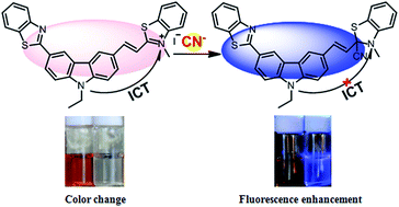
A new carbazole derivative 2-[(1E)-2-[6-(benzothiazolyl)-9-ethyl-9H-(carbazol-3-yl)]-ethenyl]-3-methyl-benzothiazolium iodide (BCB) was synthesized and its anion sensing properties were studied. BCB acts as an efficient colorimetric and ratiometric fluorescent probe used for the detection of cyanide ions in an aqueous solution even in the presence of other anions such as F, AcO, H2PO4, SO42−, SO32−, S2−, SCN, Cl, Br, I, N3, ClO4, NO2, HCO3 and CO32−. The cyanide addition to the benzothiazolium group induces an obstruction of the intramolecular charge transfer (ICT) and produces a dramatic hypsochromic shift in the absorption (152 nm) and emission (165 nm) profiles of BCB. The change in color of the solution was prominent and could be easily observed by the naked eye. The BCB probe forms a 1[thin space (1/6-em)]:[thin space (1/6-em)]1 adduct with CN with a detection limit of 0.09 μM. The probe was successfully applied for the detection of CN in natural water samples as well as in live HeLa cells.


Introduction

Among anions, cyanide (CN) is one of the most lethal poisons to living organisms. The absorption of cyanide through the lungs, gastrointestinal tract and skin, can lead to vomiting, convulsions, loss of consciousness, and ultimately death.1,2 It is known that 0.5–3.5 mg per kg of body weight is fatal for humans.3 According to the World Health Organization (WHO), the permissible level of CN in drinking water is 1.9 μM.4 Nevertheless, the increased usage of cyanide in gold mining, electroplating, tanning, polymer production and metallurgy,2 along with its necessary transportation, drastically increases the potential for contamination in the environment and increases the risk of human exposure. Therefore, the detection as well as the quantitative determination of cyanide in biological and environmental samples is of considerable importance for environmental protection and human health.

In recent years, a number of efforts has been made to develop colorimetric or fluorometric CN selective chemosensors based on versatile mechanisms, among which the reaction based probes have attracted much attention due to their unique selectivity over other anions.5 However, a majority of the reactions can only occur in an organic solvent and water soluble CN sensors are still very limited in number.6 Moreover, fluorescence based detection usually depends on the intensity change at a single wavelength, which may be influenced by variations in the sample environment. By contrast, a ratiometric method measuring the ratio of fluorescence intensities at two wavelengths provides a built-in correction for environmental effects and therefore the reliability of the measurements is substantially enhanced.7 To date, only a limited number of ratiometric fluorescence probes for cyanide detection has been reported in the literature.8 Therefore, it is of great interest to develop fluorescence chemosensors for cyanide that can show both color and fluorescence changes in an aqueous medium in a ratiometric manner.

Keeping this in mind, in the present manuscript we have designed and synthesized a new carbazole-derived (Scheme 1) intramolecular charge transfer (ICT) probe for cyanide detection in an aqueous solution. Carbazole moiety is a strong electron-donating group and thus is suitable as a donor in an intramolecular charge transfer (ICT) system. A positively-charged benzothiazolium moiety, an electron-withdrawing group, was selected as the CN reaction site, which may not only render the probe water-soluble but also manipulate an ICT process. We envisioned that there is a strong ICT process in BCB. An interaction with cyanide would interrupt the π-conjugation and block the ICT process to produce a significant ratiometric spectroscopic response. As expected, herein we observed a dramatic hypsochromic shift in the absorption and emission profiles of BCB upon reaction with cyanide, which could be utilized as a selective colorimetric and ratiometric fluorescent probe for CN detection in an aqueous solution.


image file: c5ra26587h-s1.tif
Scheme 1 The synthesis of the BCB probe.

Results and discussion

Chemosensing of CN

The anions sensing ability for BCB was investigated by UV-Vis and fluorescence spectroscopy in a DMSO–H2O (1[thin space (1/6-em)]:[thin space (1/6-em)]9, v/v, pH 7.4) HEPES buffer solution. As shown in Fig. 1a, the absorption spectrum of BCB is characterized by a higher energy band at around 320 nm due to a π → π* transition and a lower energy band at 472 nm for the intramolecular charge transfer (ICT). On addition of an increasing amount of cyanide the absorbance at 472 nm gradually decreased, but at 320 nm it gradually increased. The absorption stabilized after the amount of CN added reached 2 equivalents and a significant color change from orange-red to colorless could be observed easily (Fig. 3). The observed spectral changes suggest that the π-conjugation and ICT were both inhibited by the nucleophilic addition of CN at the benzothiazole ring carbon adjacent to the quaternary nitrogen.8e The electronic delocalization was thereby ruptured and the ICT band at 472 nm gradually disappeared accordingly. The presence of a clear isosbestic point at 385 nm indicated the clean conversion of reactant to product. The spectrophotometric titration of BCB with CN shows a linear dependence of the ratio of the absorbance at 472 nm and 320 nm (A472/A320) as a function of CN concentration, which enables the ratiometric quantification of CN. Good linearity was observed in the range of 2–20 μM (see inset of Fig. 1a).
image file: c5ra26587h-f1.tif
Fig. 1 (a) The UV-Vis and (b) fluorescence spectral changes of BCB (10 μM) in the presence of various concentrations of CN (0–20 μM). The inset in (a): ratiometric absorbance changes (A472/A320) of BCB upon the gradual addition of CN; (b): ratiometric calibration curve I424/I589 as a function of CN ion concentration.

image file: c5ra26587h-f2.tif
Fig. 2 (a) The time-dependent changes in the fluorescence spectra of BCB (1.0 × 10⁻⁵ mol L−1) treated with KCN (2.0 × 10⁻⁵ mol L−1) in DMSO[thin space (1/6-em)]:[thin space (1/6-em)]H2O (1[thin space (1/6-em)]:[thin space (1/6-em)]9, v/v, HEPES buffer, pH 7.4) and (b) the pseudo-first-order kinetic plot of the reaction of BCB (1.0 × 10⁻⁵ mol L−1) with CN (2.0 × 10⁻⁵ mol L−1).

image file: c5ra26587h-f3.tif
Fig. 3 The chromogenic response of BCB added with 10 equiv. of different anions under daylight (above) and the corresponding fluorogenic response (below) under UV light irradiation. All the images were obtained 1.5 min after the addition of the anions.

The excitation of the BCB probe at 330 nm produced a dual emission with peaks at 424 nm and 589 nm (Fig. 1b). The band at 589 nm is attributed to the ICT emission band, whereas the band at 424 nm can be ascribed to the benzothiazolyl carbazole moiety. The successive addition of CN into a solution of BCB caused a gradual quenching of the fluorescence at 589 nm and a prominent enhancement at 424 nm. It led to a bright blue emission, which is clearly visible by the naked eye under the irradiation of a hand held UV-lamp at 365 nm. A clear isoemissive point at 538 nm also indicated the formation of the BCB–CN adduct. Thus, the BCB probe can offer the fluorescent detection of cyanide at two different emission channels. There is a ca. 26.4-fold variation in the fluorescence ratio (I424/I589) from 1.42 in the absence of CN to 37.5 in the presence of 2 equivalents of CN. A good linear relationship between the intensity ratio (I424/I589) and the concentration of CN could be obtained in the range of 1.0 × 10⁻⁶ to 10 × 10−6 M (R2 = 0.98005). This ratiometric fluorescence change could be potentially useful for quantitative determination of CN. The detection limit of BCB towards CN was obtained as 0.09 μM, which is much lower than the WHO cyanide standard in drinking water (1.9 μM), indicating that BCB is a promising fluorescence ratiometric sensor for the detection of low levels of cyanide ions in water samples.

The benzothiazolyl carbazole moiety was itself fluorescent and when it was coupled with the methyl salt of 2-methylenebenzothiazole in BCB, the fluorescence was greatly reduced due to extensive π-conjugation and the ICT mechanism. The nucleophilic addition of a CN ion to BCB leads to the disruption of the π-conjugation and blockage of the ICT process and thus resulting in the recovery of the fluorescence of the benzothiazolyl carbazole moiety. As shown in Fig. 2a, two equivalents of cyanide reacted completely with BCB in ca. 2 min, indicating the high reactivity of the probe. Pseudo first-order kinetics were assumed and by monitoring the increase at 424 nm over time, the rate of the reaction was calculated from ln((FmaxFt)/Fmax) versus time (Fig. 2b), where Fmax and Ft are fluorescence intensities at time t and at end of the reaction, respectively. A rate constant of 0.027 s−1 was obtained from the slope of the straight line.9

The selectivity of the cyanide response

To examine the selectivity, BCB (10 μM) was treated with various anion species (100 μM). Among the 15 different anions tested under identical conditions, including F, AcO, H2PO4, SO42−, SO32−, S2−, SCN, Cl, Br, I, N3, ClO4, NO2, HCO3 and CO32−, only CN responded to BCB with a large blue shift and a marked color change from orange-red to colorless (Fig. 3), manifesting the good chromogenic selectivity of the probe towards CN ions in an aqueous solution. Consistent with the results of the absorption spectra, no appreciable changes in the emission spectra were observed for the other interfering anions except for the cyanide anion (Fig. 4a). Furthermore, competitive anions interaction studies show that the emission intensity of BCB + CN remained unaffected by the addition of other anions in excess (Fig. 4b). This establishes that BCB can be used to quantitatively to detect the presence of cyanide with high selectivity.
image file: c5ra26587h-f4.tif
Fig. 4 (a) The emission spectra of BCB upon the addition of different anions and (b) the fluorescence intensities of BCB at 424 nm upon the addition of CN in the presence of the interfering anions. CBCB = 10 × 10−6 mol L−1; CCN = 10 × 10−5 mol L−1; Canions = 10 × 10−5 mol L−1.

The sensing mechanism and stoichiometry

The interaction mechanism was further confirmed using a combination of 1H NMR spectroscopy and high-resolution mass spectrometry. A solution of BCB in DMSO-d6 was monitored using 1H NMR spectroscopy upon the gradual addition of KCN. As shown in Fig. 5 and Scheme 2, a significant upfield shift was observed for most of the protons of BCB upon CN addition, indicating that the CN functions as a nucleophile. The δ value for the methyl protons (Hc) adjacent to the quaternary nitrogen atom dramatically shifted upfield from 4.37 ppm to 2.97 ppm. The vinyl protons at δ 8.38 (H1) and 8.10 (H2) ppm shifted upfield towards δ 7.26 and 7.19 ppm, respectively, indicating that the electron withdrawing effect of the benzothiazolium quaternary N atom decreased. Moreover, the aromatic protons (Ha, Hb and H3) that participated in the electronic delocalization showed an upfield shift due to the removal of the electron-withdrawing effect originating from the benzothiazolium moiety. All these observations are in accordance with the nucleophilic addition of cyanide to the iminium carbon in BCB. High-resolution ESI-MS analysis also identified a peak for the BCB–CN adduct; calcd for C27H35N4O3 [BCB–CN + Na], 551.1340; found, 551.0900 (Fig. 6).
image file: c5ra26587h-f5.tif
Fig. 5 The 1H NMR spectral changes observed for BCB upon the addition of various amounts of cyanide in DMSO-d6.

image file: c5ra26587h-s2.tif
Scheme 2 A plausible mode of the interaction of cyanide with BCB.

image file: c5ra26587h-f6.tif
Fig. 6 The HRMS of product obtained by reaction between BCB and CN.

Finally, the analysis of a Job's plot for BCB, derived from fluorimetric titration, confirmed the formation of a 1[thin space (1/6-em)]:[thin space (1/6-em)]1 adduct with cyanide (Fig. 7).


image file: c5ra26587h-f7.tif
Fig. 7 The Job's plot between BCB and cyanide anion in HEPES buffer (DMSO/H2O = 1[thin space (1/6-em)]:[thin space (1/6-em)]9, v/v, pH = 7.4).

The practical applications

Because BCB showed a selective and sensitive response to CN in 90% aqueous solution, we utilized it to detect CN in tap water samples. The water sample was found to be free from cyanide and so the sample was prepared by adding known amounts of cyanide to the sample. With a given amount of CN spiked in the water samples, the recovery yields were determined using the calibration curve and are listed in Table 1. As shown in Table 1, the probe was able to measure the concentrations of spiked CN with good recovery, suggesting that the method could potentially be used for detecting CN in real samples.
Table 1 The determination of CN in water samples
Samples CN added (μM) CN found (μM) Recovery (%) RSDa (%)
a RSD: relative standard deviation, n = 5.
Tap water 1 0
Tap water 2 2 1.98 99.0% 0.81%
Tap water 3 6 6.04 100.1% 0.96%
Tap water 4 10 10.21 102.1% 0.52%
Tap water 5 14 14.16 101.1% 0.83%


The BCB probe was further examined to detect cyanide in living HeLa cells. The HeLa cells were treated with BCB (10 μM) for 30 min at 37 °C and then washed three times with phosphate-buffered saline (PBS, 10 mM, pH 7.4). The cells showed no detectable fluorescence upon incubation with only BCB (Fig. 8b). However, after treatment with KCN (20 μM) for 10 min and further incubation with BCB (10 μM) for 30 min, a bright blue fluorescence was observed (Fig. 8e). Thus, the BCB probe showed the potential to detect CN in vitro cellular systems.


image file: c5ra26587h-f8.tif
Fig. 8 The confocal laser fluorescence images of HeLa cells treated with BCB (a–c) and with both BCB and CN (d–f). (Left) The bright field image; (middle) the fluorescence image and (right) the merged image.

Experimental

Materials and instrumentation

3-Formyl-N-ethylcarbazole (99%), o-aminothiophenol (96%) and 2-methylbenzothiazole (98%) were purchased from Aladdin Reagent Co. (Shanghai, China). All other chemical reagents were of analytical grade and used as received without further purification. All the anions were supplied from their corresponding sodium or potassium salts. A stock solution of BCB was prepared in DMSO at 2.0 × 10⁻³ mol L−1. Anion stock solutions (2.0 × 10⁻³ mol L−1) were prepared by dissolving appropriate amounts of their sodium or potassium salts in water. Doubly distilled water was used throughout the experiments.

UV-Vis absorption spectra were obtained on an Agilent 8453 UV-Vis spectrophotometer (Lakewood, NJ). Fluorescence spectra were observed on a Hitachi F-7000 fluorescence spectrometer. The samples were excited at 330 nm. The excitation and emission slits were both set at 5 nm. 1H NMR and 13C NMR spectra were obtained at 600 and 400 MHz, respectively at 298 K using tetramethylsilane (TMS) as an internal standard. The following abbreviations were used to explain the multiplicities: s = singlet, d = doublet, t = triplet, q = quartet, m = multiplet, br = broad. High-resolution mass spectra were obtained on a Bruker micrOTOF-Q II mass spectrometer. Melting points were measured on a RD-II digital melting point apparatus and are uncorrected.

Synthesis of the BCB probe molecule

Compound 1 (BCB) was synthesized using the protocol shown in Scheme 1.
Synthesis of compound 3. A mixture of compound 2 (2.23 g, 10 mmol) and o-aminothiophenol (1.37 g, 11 mmol) in DMSO (20 mL) was stirred at 195 °C for 6 h under a N2 atmosphere. After completion of the reaction, it was cooled to room temperature and poured into 300 mL of water. The precipitate was filtered by vacuum filtration and chromatographed (silica, petroleum[thin space (1/6-em)]:[thin space (1/6-em)]ethyl acetate = 10[thin space (1/6-em)]:[thin space (1/6-em)]1, v/v) to give a white solid (2.65 g, 81%). Mp 142–145 °C. 1H NMR (600 MHz, DMSO-d6) δ(ppm): 8.92 (s, 1H), 8.38 (d, J = 12 Hz, 1H), 8.21 (d, J = 6 Hz, 1H), 8.13 (d, J = 12 Hz, 1H), 8.05 (d, J = 12 Hz, 1H), 7.78 (d, J = 12 Hz, 1H), 7.68 (d, J = 6 Hz, 1H), 7.54 (d, J = 6 Hz, 2H), 7.44 (t, J = 6 Hz, 1H), 7.29 (t, J = 6 Hz, 1H), 4.51 (q, J = 6 Hz, 2H), 1.36 (t, J = 6 Hz, 3H); 13C NMR (400 MHz, DMSO-d6) δ(ppm): 167.90, 153.27, 140.64, 139.66, 133.73, 125.89, 124.26, 123.35, 122.09, 121.57, 120.41, 119.04, 108.99, 36.64, 13.12; HRMS (ESI): calcd for C21H16N2S 328.1034 (M), found: 328.0948.
Synthesis of compound 4. DMF (25 mL) was placed in a 100 mL round bottom flask cooled using an ice-water bath. POCl3 (7.70 g, 50 mmol) was added dropwise to the flask. After stirring for another 30 min, the temperature of mixture was increased to room temperature and continued to stir for 1 h. To this solution, compound 3 (3.28 g, 10 mmol) in 1,2-dichloroethane (20 mL) was added in a dropwise fashion and the mixture was stirred at 90 °C for 12 h. The resulting mixture was poured into 100 mL of ice water. The solution was then brought to pH = 7 upon the addition of 1 M NaOH and extracted with CH2Cl2 (3 × 50 mL). The combined organic layer was washed with water and dried over anhydrous MgSO4. The solvent was removed by distillation and the residue material chromatographed (silica, petroleum[thin space (1/6-em)]:[thin space (1/6-em)]ethyl acetate = 5[thin space (1/6-em)]:[thin space (1/6-em)]1, v/v) to give a yellow solid (1.24 g, 35%). Mp 171–173 °C. 1H NMR (600 MHz, DMSO-d6) δ(ppm): 10.10 (s, 1H), 9.06 (s, 1H), 8.01 (s, 1H), 8.29 (d, J = 12 Hz, 1H), 8.15 (t, J = 12 Hz, 1H), 8.06 (d, J = 6 Hz, 2H), 7.86 (t, 12 Hz, 2H), 7.55 (t, J = 12 Hz, 1H), 7.45 (t, J = 12 Hz, 1H), 4.57 (q, J = 6 Hz, 2H), 1.39 (t, J = 12 Hz, 3H); 13C NMR (400 MHz, CDCl3) δ(ppm): 191.43, 144.17, 142.44, 129.33, 127.55, 126.51, 125.11, 124.71, 123.56, 123.14, 122.63, 121.63, 120.62, 109.63, 109.31, 38.31, 19.93; HRMS (ESI): calcd for C22H16N2OS 356.0983 (M), found: 357.1062.
Synthesis of compound 6. Compound 6 was synthesized according to literature methods.10 Yellow powder (1.6 g, 87%). 1H NMR (600 MHz, D2O) δ(ppm): 8.09 (d, J = 12 Hz, 1H), 8.00 (d, J = 12 Hz, 1H), 7.77 (t, J = 12 Hz, 1H), 7.70 (t, J = 12 Hz, 1H), 4.14 (s, 3H), 3.06 (s, 3H).
Synthesis of compound 1. To a stirred EtOH solution of compound 4 (3.56 g, 10 mmol) and compound 6 (2.91 g, 10 mmol) was added 1–3 drops of piperidine. The mixture was refluxed for 12 h under stirring. Then, the reaction mixture was allowed to cool to room temperature, filtered, washed thoroughly with EtOH and dried under vacuum to obtain the product as a red solid (1.6 g, 87%). 1H NMR (600 MHz, DMSO-d6) δ(ppm): 9.16 (s, 1H), 8.99 (s, 1H), 8.38 (d, J = 6 Hz, 1H), 8.33 (d, J = 12 Hz, 1H), 8.22 (d, J = 6 Hz, 1H), 8.19 (d, J = 6 Hz, 1H), 8.15 (d, J = 12 Hz, 1H), 8.13 (d, J = 6 Hz, 1H), 8.07 (d, J = 18 Hz, 1H), 8.04 (d, J = 6 Hz, 1H), 7.84 (m, 3H), 7.75 (t, J = 6 Hz, 1H), 7.53 (t, J = 6 Hz, 1H), 7.43 (t, J = 6 Hz, 1H), 4.55 (q, J = 6 Hz, 2H), 4.37 (s, 3H), 1.39 (t, J = 6 Hz, 3H); 13C NMR (400 Hz, DMSO-d6) δ(ppm): 172.27, 168.42, 154.19, 150.49, 143.26, 142.46, 142.36, 134.76, 129.95, 128.51, 127.86, 127.05, 126.65, 126.39, 125.64, 125.55, 124.52, 123.77, 123.40, 123.24, 122.83, 122.69, 120.07, 116.91, 111.24, 111.01, 110.86, 38.24, 36.65, 14.39; HRMS (ESI): calcd for C31H24N3S2 502.1406 (M), found: 502.1390.

UV-Vis and fluorescence titrations

UV-Vis and fluorescence titrations were carried out in 10 mm quartz cuvettes at 25 °C. The procedure was as follows: into a DMSO[thin space (1/6-em)]:[thin space (1/6-em)]H2O (1[thin space (1/6-em)]:[thin space (1/6-em)]9, v/v, HEPES buffer, pH 7.4) solution, containing 10 μM BCB, a CN sample was gradually titrated. At the same time, any changes in the fluorescence intensity were monitored using a fluorescence spectrometer.

Conclusions

In conclusion, we have successfully devised a novel carbazole based fluorescent probe, BCB, used for the selective detection of cyanide ions in aqueous media. Upon reaction with CN functioning as a nucleophile, BCB displays substantial dual changes in both its ratiometric emission and absorption spectra. The significant changes in color, attributed to a restricted ICT due to the formation of a stable BCB–CN adduct, could be observed directly with the naked eye. The detection limit of BCB was estimated to be 0.09 μM, which is lower than the maximum permissive level in drinking water according to the World Health Organization (WHO). Moreover, the application of BCB for imaging of CN in living cells was also successfully achieved.

Acknowledgements

This study was financially supported by Shanxi Scholarship Council of China (No. 2011-008), the Natural Science Foundation of Shanxi Province (No. 2013011040-6) and the Project Sponsored by the Scientific Research Foundation for the Returned Overseas Chinese Scholars, State Education Ministry.

Notes and references

  1. B. Vennesland, E. E. Comm, C. J. Knownles, J. Westly and F. Wissing, Cyanide in Biology, Academic Press, London, 1981 Search PubMed.
  2. C. Young, L. Tidwell and C. Anderson, Cyanide: Social, Industrial, and Economic Aspects, Minerals, Metals, and Materials Society, Warrendale, 2001 Search PubMed.
  3. W. Kaim and B. Schwederski, Bioinorganic Chemistry: Inorganic Elements in the Chemistry of Life, John Wiley & Sons Ltd., England, 1991, p. 208 Search PubMed.
  4. S. W. Merwe, F. Schoeller, W. Bolzer, R. Germonper, L. Heller, E. H. Filho, D. M. E. Formaggia, J. C. Blum, M. J. Omori and P. C. S. Mancuso, Guidelines for Drinking-Water Quality, World Health Organization, Geneva, 1996 Search PubMed.
  5. (a) S. Goswami, S. Paul and A. Manna, Dalton Trans., 2013, 42, 10682–10686 RSC; (b) Y. L. Duan and Y. S. Zheng, Talanta, 2013, 107, 332–337 CrossRef CAS PubMed; (c) N. Niamnont, A. Khumsri, A. Promchat, G. Tumcharern and M. Sukwattanasinitt, J. Hazard. Mater., 2014, 280, 458–463 CrossRef CAS PubMed; (d) M. D. Sun, S. D. Wang, Q. B. Yang, X. L. Fei, Y. X. Li and Y. P. Li, RSC Adv., 2014, 4, 8295–8299 RSC; (e) P. Zhang, B. B. Shi, X. M. You, Y. M. Zhang, Q. Lin, H. Yao and T. B. Wei, Tetrahedron, 2014, 70, 1889–1894 CrossRef CAS; (f) N. Sharma, S. I. Reja, V. Bhalla and M. Kumar, Dalton Trans., 2014, 43, 15929–15936 RSC; (g) A. B. Davis, R. E. Lambert, F. R. Fronczek, P. J. Cragg and K. J. Wallace, New J. Chem., 2014, 38, 4678–4683 RSC; (h) R. Jackson, R. P. Oda, R. K. Bhandari, S. B. Mahon, M. Brenner, G. A. Rockwood and B. A. Logue, Anal. Chem., 2014, 86, 1845–1852 CrossRef CAS PubMed; (i) Y. Zhang, D. H. Yu and G. Q. Feng, RSC Adv., 2014, 4, 14752–14757 RSC; (j) Y. B. Chen, W. Shi, Y. H. Hui, X. H. Sun, L. X. Xu, L. Feng and Z. F. Xie, Talanta, 2015, 137, 38–42 CrossRef CAS PubMed; (k) Y. K. Yue, F. J. Huo and C. X. Yin, Sens. Actuators, B, 2015, 212, 451–456 CrossRef CAS; (l) J. H. Lee, J. H. Jang, N. Velusamy, H. S. Jung, S. Bhuniya and J. S. Kim, Chem. Commun., 2015, 51, 7709–7712 RSC; (m) Y. Xu, X. Dai and B. X. Zhao, Spectrochim. Acta, Part A, 2015, 138, 164–168 CrossRef CAS PubMed; (n) Q. Li, J. H. Zhang, Y. Cai, W. J. Qu, G. Y. Gao, Q. Lin, H. Yao, Y. M. Zhang and T. B. Wei, Tetrahedron, 2015, 71, 857–862 CrossRef CAS.
  6. (a) H. J. Kim, K. C. Ko, J. H. Lee, J. Y. Lee and J. S. Kim, Chem. Commun., 2011, 47, 2886 RSC; (b) H. J. Kim, H. Lee, J. H. Lee, D. H. Choi, J. H. Jung and J. S. Kim, Chem. Commun., 2011, 47, 10918–10920 RSC; (c) X. H. Huang, X. G. Gu, G. X. Zhang and D. Q. Zhang, Chem. Commun., 2012, 48, 12195–12197 RSC; (d) J. Isaad, F. Malekc and A. E. Achariab, RSC Adv., 2013, 3, 22168–22175 RSC; (e) J. H. Lee, J. H. Jang, N. Velusamy, H. S. Jung, S. Bhuniya and J. S. Kim, Chem. Commun., 2015, 51, 7709–7712 RSC.
  7. A. Ajayaghosh, P. Carol and S. Sreejith, J. Am. Chem. Soc., 2005, 127, 14962–14963 CrossRef CAS PubMed.
  8. (a) S. Park and H. J. Kim, Sens. Actuators, B, 2012, 168, 376–380 CrossRef CAS; (b) L. Zang, D. Wei, S. Wang and S. Jiang, Tetrahedron, 2012, 68, 636–641 CrossRef CAS; (c) X. Cheng, Y. Zhou, J. Qin and Z. Li, ACS Appl. Mater. Interfaces, 2012, 4, 2133–2138 CrossRef CAS PubMed; (d) H. Li, Z. Wen, L. Jin, Y. Kam and B. Yin, Chem. Commun., 2012, 48, 11659–11661 RSC; (e) J. T. Zhang, S. L. Zhu, L. Valenzano, F. T. Luo and H. Y. Liu, RSC Adv., 2013, 3, 68–72 RSC; (f) G. L. Fu and C. H. Zhao, Tetrahedron, 2013, 69, 1700–1704 CrossRef CAS; (g) S. Goswami, S. Paul and A. Manna, Tetrahedron Lett., 2013, 54, 1785–1789 CrossRef CAS; (h) L. Wang, X. Li, J. Yang, Y. Qu and J. Hua, ACS Appl. Mater. Interfaces, 2013, 5, 1317–1326 CrossRef PubMed; (i) M. Shahid and A. Misra, Anal. Methods, 2013, 5, 434–437 RSC; (j) Y. Sun, S. W. Fan, L. Duan and R. F. Li, Sens. Actuators, B, 2013, 185, 638–643 CrossRef CAS; (k) S. Goswami, S. Paul and A. Manna, Dalton Trans., 2013, 42, 10682–10686 RSC; (l) S. Pramanik, V. Bhalla and M. Kumar, ACS Appl. Mater. Interfaces, 2014, 6, 5930–5939 CrossRef CAS PubMed; (m) W. C. Lin, S. K. Fang, J. W. Hu, H. Y. Tsai and K. Y. Chen, Anal. Chem., 2014, 86, 4648–4652 CrossRef CAS PubMed; (n) H. D. Li, T. Chen, L. Y. Jin, Y. H. Kan and B. Z. Yin, Anal. Chim. Acta, 2014, 852, 203–211 CrossRef CAS PubMed; (o) K. Y. Chen and W. C. Lin, Dyes Pigm., 2015, 123, 1–7 CrossRef CAS; (p) S. Bhardwaj and A. K. Singh, J. Hazard. Mater., 2015, 296, 54–60 CrossRef CAS PubMed; (q) R. Kumar, N. Chaudhri and M. Sankar, Dalton Trans., 2015, 44, 9149–9157 RSC; (r) N. Kaur, G. Dhaka and J. Singh, Tetrahedron Lett., 2015, 56, 1162–1165 CrossRef CAS; (s) A. K. Mahapatra, S. K. Manna, B. Pramanik, K. Maiti, S. Mondal, S. S. Ali and D. Mandal, RSC Adv., 2015, 5, 10716–10722 RSC.
  9. P. B. Pati and S. S. Zade, RSC Adv., 2013, 3, 13457–13462 RSC.
  10. X. N. Fei, Y. C. Gu, Y. Ban, Z. J. Liu and B. L. Zhang, Bioorg. Med. Chem., 2009, 17, 585–591 CrossRef CAS PubMed.

This journal is © The Royal Society of Chemistry 2016
Click here to see how this site uses Cookies. View our privacy policy here.