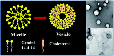
The cholesterol aided micelle to vesicle transition of a cationic gemini surfactant (14-4-14) in aqueous medium

Sonali Mondal, Animesh Pan, Sibani Das, Satya Priya Moulik and Soumen Ghosh*
Centre for Surface Science, Department of Chemistry, Jadavpur University, Kolkata-700032, India. E-mail: gsoumen70@hotmail.com; Fax: +91-33-2414-6266; Tel: +91-33-2414-6411

Received 16th December 2015 , Accepted 23rd February 2016

First published on 26th February 2016


Abstract

The cholesterol (Chol) aided micelle to vesicle transition of the cationic (14-4-14) gemini surfactant (GS) (tetramethylene-1,4-bis(dimethyltetradecylammonium bromide)) has been investigated employing spectrophotometry (visible and fluorescence), dynamic light scattering (DLS) and high resolution transmission electron microscopy (HRTEM) at different R (R = [Chol]/[GS]) values. Turbidity of the mixed solutions gradually increased with increasing R, and was highest at R = 1. There were hypsochromic spectral shifts with varied R, supporting transformation of micelles to vesicles, corroborated by DLS measurements. The vesicle sizes ranged between ∼160–240 nm. The steady state fluorescence anisotropy measurements suggested Chol induced anisotropy to the formed vesicles; they were structurally incongruent. The vesicle phase was found to be stable in the studied temperature range of 15–60 °C. The increased rotational relaxation time of the probe molecule C-153 (coumarin 153) supported enhanced rigidity of the local environment of the vesicles with an increasing proportion of Chol in the mixture.

1. Introduction

Amphiphile molecules comprising hydrophobic and hydrophilic domains may self-assemble in water. In this process, the hydrophobic parts of different molecules cluster together to minimize contacts with water whereas the hydrophilic portions orient towards water to satisfy their hydration and polarity requirements. Essentially, self-assembly formation depends on the hydrophobic, van der Waals, electrostatic, and steric interactions.1,2 The formed assemblies can be micelles, vesicles, and liquid crystals,2,3 of which vesicles are potential carriers and delivery systems for active ingredients useful in pharmaceuticals, cosmetics and medicine.1–6 They can also be used in the preparation of nano-structured materials.3,5 In this domain, prospective liquid crystalline nanocarriers (cubosomes, hexosomes, spongosomes) are also reported lately.7–11 Liposomes (lipid vesicles) are widely used nano-carriers of drugs due to their unique structure and bio-compatibility.12 However, the weak solubility of the lipids affects their stability, and the assemblies are prone to collapse into planar lamellar structures. Their stability is kinetically controlled.13 Therefore, large interest arose on nonlipid building blocks which can form stable self-assembly (e.g. supramolecular vesicles) to satisfy biological and pharmaceutical standards.14,15 There are encouraging reports on nonlipid vesicular systems (like catanionic vesicles prepared from anion–cation surfactant ion pairs),16 single surfactant molecules,17,18 bolaamphiphiles,19,20 polymersomes (derived from amphiphilic block co-polymers),21 peptosomes (from polypeptides),22 niosomes (from nonionic surfactants),23 and other chemical structures like dendrimers, amphiphilic fullerene derivatives, and recently explored room temperature ionic liquids (RTILs).24–27

Gemini surfactants (a new type of double tailed amphiphiles) are found to be prospective candidates for vesicle formation owing to their superior properties in comparison with those of conventional single tailed surfactants.28 GS has low critical micelle concentrations (CMC), better wettability, and unusual aggregation morphology.29 GS thus may have prospects in pharmaceutical, and biomedical fields. Studies on the effects of pH, temperature, additives (electrolytes, non electrolytes, solvents), co-amphiphiles, etc. on the self-assembled nanostructures of GS (cationic, anionic, and non-ionic types) have been made.29–33 Phase behaviours of GS in combination with oppositely charged conventional surfactants are also found in literature.34 Micelle to vesicle transitions of GS by the addition of co-amphiphiles,35,36 changing pH,37,38 and addition of salts are possible.39,40 For a better revelation, more systematic and planned studies are still required.

In the past years, nonionic surfactants forming “niosomes” with Chol have been promisingly promoted as potential alternatives to liposomal delivery systems.41,42 From theoretical calculations, L. F. Tasies et al.43 have proposed that cholesterol (Chol) induced micelle to vesicle transition of cationic surfactants are quite different from that without Chol. Working on cetyltrimethyammonium bromide (CTAB) they have shown that the lipid may undergo synergistic interactions with cationic surfactants. Water insoluble Chol molecules remain in the hydrophobic bilayers of the vesicular systems and make them more rigid. In this line cholesterol induced vesicle formation in aqueous solution of surface active ionic liquids (SAILs) have been employed.44 Researchers are trying to mimic the liposomes with mixed amphiphile and Chol systems for different applications.

We have herein demonstrated the influence of Chol on the micelle to vesicle transition (MVT) of the mixtures of GS and the lipid. The vesicle formation has been examined by the steady-state fluorescence anisotropy measurements of the membrane bound DPH-probe, and also from DLS, ζ-potential, and HRTEM measurements. The stability of the aggregates against temperature has been also studied. In the course of micelle to vesicle transition, the changes in the microenvironments have been probed by measuring the rotational and relaxation dynamics of C-153 (coumarin 153) from time-resolved fluorescence measurements.

2. Experimental section

2.1. Materials

N,N-Dimethyltetradecyl amine and 1,4-dibromobutane were purchased from Sigma-Aldrich (USA). At first, N,N-dimethyltetradecyl amine and 1,4-dibromobutane were refluxed in dry ethanol for 48 h, and after evaporating the solvent, the crude product was recrystallized from ethanol/acetone mixture39 for several times. The structure of the product tetramethylene-1,4-bis(dimethyltetradecylammonium bromide) (14-4-14) was confirmed by 1H NMR spectroscopy. Absence of minima in surface tension (γ) versus log[14-4-14] plot also supported the purity of the gemini. The probe coumarin 153 (C-153), pyrene (Py), 1,6-diphenyl-1,3,5-hexatriene (DPH, 98%) were purchased from Aldrich. Pyrene was re-crystallized thrice from hexane–water. Ethanol, AR grade (99.9%) was purchased from Merck (India) and preserved under desiccating condition. Cholesterol (99%) was purchased from Fluka (USA).

Characterization results of 14-4-14 (tetramethylene-1,4-bis(dimethyltetradecylammonium bromide)):

Yield 13.5 g, 19.3 mmol, 90%. 1H-NMR (CDCl3, 300 MHz): δ (ppm): 0.85–0.89 (terminal CH3, 6H), 1.25 (CH3–CH2–(CH2)10, 40H), 1.35 (CH3–CH2, 4H), 1.77 (CH3–CH2–(CH2)10–CH2, 4H), 2.1 (CH3–CH2–(CH2)12–N+Me2–CH2–CH2, 4H), 3.27 (CH2 both sides of N+Me2 gr, 8H), 3.87 (CH3 gr attached the N atom, 12H).

2.2. Protocol for solution preparation

All the experimental solutions were prepared by varying Chol concentration i.e., with different R values at a fixed GS concentration (5 mM, 35 times its CMC). The resulting solutions were sonicated for 1 h.

2.3. Turbidity measurements

Turbidities of the solutions at different R values were determined at 500 nm using a Shimadzu (Japan) UV-1601 spectrophotometer. The individual components had no absorption at the studied wavelength. Measurements were taken at [Chol]/[GS] mole ratios 0 to 2.0.

2.4. Dynamic light scattering (DLS) measurements

DLS measurements were taken in a Zetasizer nano ZS (Malvern, UK) at 90° scattering angle with a He–Ne laser (λ = 632.8 nm). The sample was allowed to equilibrate inside the DLS optical chamber for 10 min prior to taking of measurements. To determine the zeta potential (ζ) of the assemblies in solution with varying R a deep type cell was used. The data acquisition was done for at least 20 counts, and each experiment was repeated thrice. The solutions were filtered thrice through 0.45 m Millipore membrane filters.

2.5. Fluorescence measurements

Steady-state fluorescence measurements were taken in a Perkin-Elmer LS-55 (USA) fluorescence spectrophotometer attached with Peltier system PTP-1. Glass cells (1 cm path length) were used. The steady-state anisotropy (rss) values were found from the relation,45,46
 
image file: c5ra26507j-t1.tif(1)
where I and I are the emission intensities of the vertically and horizontally polarized components of the probe, respectively. In the expression of G factor the I and I are the vertically and horizontally polarized emissions, respectively resulting from horizontally polarized excitation.

2.6. Time resolved fluorescence anisotropy measurements

Time resolved fluorescence decays are collected from a TCSPC instrument by Edinburgh Instrument, U.K. (excitation wavelength 409 nm), ∼80 ps instrument response function (IRF).47 All the transients for anisotropy measurement collected at the wavelength of emission maxima and G factor calculated from longtime tail matching technique.

2.7. High resolution transmission electron microscopy (HRTEM)

TEM measurements were taken for structural characterization of Chol–GS combines at R values 0.3, 0.7, and 1 in a HRTEM, model JEOL-JEM-2100 (Japan), operating at an accelerating voltage of 200 kV. For TEM images of the vesicle solutions (10 μL) were taken on 300 mesh carbon coated copper grids, and allowed to stand for 2 min followed by staining with 0.5 wt% of uranyl acetate.

3. Results and discussion

3.1. Solution properties of 14-4-14 (GS) and mixed cholesterol–GS aggregates

3.1.1. Optical behavior. The critical micelle concentration (CMC) of 14-4-14 (GS) at 303 K is 0.167 mM.28 At this temperature, aqueous GS solution remained clear up to 100 mM. Addition of Chol in the micellar solution of [5 mM] produced initial transparency and translucency (bluish appearance) afterwards. The turbidity was assessed by measuring optical density at 500 nm with increasing R from 0 to 2. The added cholesterol was solubilized by the GS micelle forming mixed species up to R = 0.05; thereafter, transformed into vesicles with appearance of translucency and turbidity. Apart from turbidity, fluorescence anisotropy, hydrodynamic radius, and ζ-potential of the dispersions were measured in support of vesicle formation of Chol–GS in its dispersion.

The transparency, translucency and turbidity of Chol–GS mixtures at different mole ratios are depicted in Fig. 1A. Appearance of translucency started at R = 0.05 (tube 3); at R = 0.3 (tube 7), precipitation started in the samples with increasing turbidity. The photographed samples are after centrifugation (10[thin space (1/6-em)]000 rpm) and removal of precipitates from them. The OD vs. R profile is presented in Fig. 1B with identification of the vesicle formation point (tube 3) as well as the precipitation point (tube 7) showing distinct inflections. A sharp rise in OD beyond R = 1.0 was found. Even after centrifugation, fair amount of cholesterol crystals remained in the dispersions which imparted high turbidity. The MVT transition for Chol–CTAB (micellar solutions) was also studied by Cano-Sarabia et al. using the solvochromatic indicator dye pinacyanol chloride sensitive to the polarity of the medium.48 Hydrodynamic radius (Rh) dependence on R is also illustrated in Fig. 1B. The dispersion size went on increasing with a kink for sample 7 (R = 0.3), and a sharp rise after sample 10 (R = 1.0). The results supported the OD behavior of the samples. DLS measurements also supported MVT of Chol–GS combination. ζ-Potential values of the vesicular solutions at different values of R are also presented in Fig. 1C. Pure GS micelles showed ζ-potential of +28 mV. The potential rose up to +90 mV at R = 0.3, then stage wise declined. These were the combined influences of the neutral Chol and the packing of the components at the interfaces of the mixed binary species (the vesicles). The Chol induced MVT growth increased both Rh, and ζ-potential.


image file: c5ra26507j-f1.tif
Fig. 1 Representation of optical behaviors of Chol–GS mixed systems at varied R. (A) Phase behavior at R = 0.02 to 2.0; (B) dependence of optical density (O.D.) and hydrodynamic radius (Rh) with R; (C) dependence of zeta potential (ξ) of [Chol/GS] system with R values.

Steady state fluorescence anisotropy (rss) of the hydrophobic probe DPH supported assembled amphiphilic bodies (aggregates) in the mixed system.49 DPH, a membrane binding probe, was used to understand membrane rigidity.50 [Chol] modified the self-assembled systems with significant increase of rss (Fig. 2) for the fluidity of the surroundings of the DPH probe decreased. It was reported2,51 that for DPH, sphere and rod-like micelles register rss ≤ 0.14; bilayers and vesicles show rss > 0.14. At 5 mM, GS micelles produced rss = 0.075, which increased with R, and became nearly constant (∼3.5) between R = 0.5 and 1. Micelle to vesicle transition (MVT) of GS in the presence of Chol (at R = 0.05) was thus supported.


image file: c5ra26507j-f2.tif
Fig. 2 Variation of steady-state anisotropy probed by DPH of [Chol/GS] with increasing R values.

Cationic surfactants viz. CTAB conveniently form both mixed micelles and vesicles depending on Chol/CTAB ratios (R). At lower R ≤ 0.1, above the cmc of CTAB, cholesterol gets solubilized by CTAB micelles which at R = 1 reaches its solubility limit forming spontaneous bilayer.48 Different types of Chol/CTAB supramolecular assemblies might form in the dispersion. The organization was found stable with time and dilution. At R ≥ 1, insoluble Chol crystals pervaded the solution. The herein studied Chol–GS system evidenced fair similarity with Chol–CTAB combine. The MVT occurred at R = 0.05, precipitation started at R ≥ 0.3 which was though fairly lower than R ≥ 1.0 for the Chol/CTAB case. However, although not clearly known for the former, the herein studied Chol–GS at higher R showed good temperature stability, although at lower R = 0.6 (little above MVT (R = 0.05)), sigmoidal transition occurred at 27 °C. Geminis having double hydrophobic tails may form bilayer assemblies that can mimic liposomes, and have greater synergistic interaction with Chol than single tail cationic surfactants. Surface active ionic liquids (SAILs) have potential to be prospective vesicle forming representatives in association with Chol. Chol induced micelle to vesicle transition of cationic surfactants are quite different from that without Chol. It would be interesting to make a comparative study of geminis with cationic surfactants including SAILs under varied environmental conditions for their prospects in drug delivery and nano-material preparation.

3.1.2. HRTEM findings. Direct support of vesicle formation of the cholesterol–GS combines resulted from HRTEM study. The micrographs of the mixed species at R = 0.1, 0.7, and 1.0 are depicted in Fig. 3. The images evidenced (indicated) formation of spherical vesicles of average size in the range of 150–200 nm. They were comparable with those found from DLS measurements.
image file: c5ra26507j-f3.tif
Fig. 3 HRTEM images (Chol/GS) mixed vesicles (sphere, globules, etc.) at different R values (a) R = 0.1, (b) R = 0.7, and (c) R = 1.
3.1.3. Temperature effect on the vesicle system. The spontaneously formed self-assemblies are usually reversible and ordered organizations of molecules that can be affected by external stimuli. Temperature is an important stimulus for affecting their shapes and microenvironments such as micropolarity that could be ascertained using a fluorescence probe.50 The intensity ratio of the first and third vibronic peaks I1/I3 of the fluorophore pyrene was taken as a measure of the micropolarity of the vesicular system. The I1/I3 ratios at different temperatures are used to assess the phase transition temperature of the GS–Chol assemblies.52 Fig. 4A depicts the dependence of I1/I3 as a function of temperature at R = 0.5 and 1.0. The values remained almost constant in the studied temperature range of 10–60 °C (only a slight decrease was observed). The self-assembled ensembles were found fairly stable up to 60 °C. Fluorescence anisotropy of the vesicles also supported the polarity index results, and the membrane bound DPH probe also experienced almost same environment of the formed vesicles with variation of temperature at different R values (presented in Fig. 4B). The superior thermal stability of the [Chol/GS] vesicles was ascribed to the strong synergistic component interaction in the binary system. At lower R = 0.06, the rss of the Chol–GS vesicles declined with temperature with a sigmoidal (indicating morphological changes in organization) transition at 27 °C. Mixed micelles of (Chol + GS) at R = 0.02 evidenced decline in rss studied between 20–60 °C (results are obvious, and not shown to save space). The membrane of archaea bacteria is known to have superior temperature stability and thus a great potential in the field of biological applications.53,54 Herein, studied Chol–GS vesicular system having good temperature stability at higher R values has a similar prospect. Nano vesicles composed of sterols and quaternary ammonium surfactants (viz. Chol–CTAB) were reported48 to have very strong self-life and their morphology remained non-affected by rising temperature and also by dilution. They had vesicle to vesicle homogeneity irrespective of lamellarity and membrane supramolecular organization55,56 which corroborated our observations on Chol/GS vesicular system.
image file: c5ra26507j-f4.tif
Fig. 4 Representative plots for I1/I3 ratio and anisotropy of [Chol/GS] systems of different R values at different temperatures.

3.2. More exploration of microenvironment of Chol–GS assemblies

3.2.1. Steady-state fluorescence response. The absorption and emission spectra of C-153 in [Chol]/[GS] solutions at different R values are presented in Fig. 5A and B. The emission peak of C-153 (excited at 436 nm) at room temperature was at 450 nm in n-heptane medium,57,58 and 548 nm in water.59 In aqueous GS micellar solution, the emission maximum was at 530 nm indicating that the local environment in GS micellar solution was of lower polarity than water but reasonably higher than that of n-heptane. The probe molecule C-153 resided below the micelle–water interface. With increasing population of Chol both the absorption and emission maxima gradually blue shifted suggesting that the probe experienced more rigid and hydrophobic environment, a consequence of Chol imparted micelle to vesicle transformation.
image file: c5ra26507j-f5.tif
Fig. 5 (A) Absorption spectra of C-153 and (B) steady-state fluorescence emission spectra of C-153 at different R values.
3.2.2. Time resolved fluorescence study. To further investigate the local environment of the probe's residence in the amphiphilic ensembles the time dependent fluorescence anisotropy decay of C-153 in the mixed systems at different R was measured. The time resolved fluorescence anisotropy r(t) is a powerful parameter to determine the rotational restriction on the probe molecule imposed by its local environment calculated from the fluorescence decays in parallel and perpendicular directions in terms of the excitation polarization as follows:
 
image file: c5ra26507j-t2.tif(2)
where G is the grating factor, and it was determined by the use of long term tail matching technique.60t” is the time, and I and I denote horizontal and vertical emission polarizations, respectively. The orientation time correlation function is given by the relation:
 
image file: c5ra26507j-t3.tif(3)
where ri and τi,r are the individual amplitude and reorientation time constant for the ith anisotropy decay, respectively. Eqn (3) has been used for the calculation of average rotational relaxation times in self-assembled systems.

In all solutions for C-153, the decay of fluorescence anisotropy r(t) fitted well to a bi-exponential function, and the fitting parameters are given in Table 1. In aqueous solution the decay of r(t) of C-153 is single exponential with a time of ∼100 ps.61 The confinement of the probe molecules in the self-assembled organized systems made the difference. Apart from all other systems (e.g. R = 0.0 → 0.5), there was an interesting observation that the r(t) of C-153 at R = 0.7 and 1.0 did not decay to zero up to the studied t = 10 ns (Fig. 6). This result indicated that the rotational motion of the probe molecules in the vesicular solution was more hindered during the studied time window. The aggregation of the probe molecules in the bilayer region might also cause this difference. Similar results were also reported in literature, for example, hydroxypropyl cellulose–water with 40 mM γ-cyclodextrin with coumarin as the probe.62,63 Various kinds of motion of the probe molecule in the presence of aggregates may cause bi-exponential r(t) decay.45 The time constant of the overall rotational motion of the aggregates became considerably high.

Table 1 Anisotropy decay parameters of C-153 in cholesterol–GS mixtures with different R values in aqueous medium at 298 K
System (R) α1r τ1r (ns) α2r τ2r (ns) τr〉 (ns) χ2
0.0 0.42 0.27 0.58 1.97 1.26 0.95
0.3 0.47 0.29 0.53 2.16 1.28 0.92
0.5 0.48 0.25 0.52 2.45 1.39 0.91
0.7 0.50 0.25 0.50 2.61 1.43 0.92
1.0 0.50 0.29 0.50 2.81 1.55 0.91


image file: c5ra26507j-f6.tif
Fig. 6 Representative time-resolved fluorescence anisotropy decay profile of C-153 at different R values.

4. Conclusion

The aggregation behavior of the synthesized GS (14-4-14) in pure state and in the presence of Chol in aqueous medium was investigated. Chol induced transformation of GS micelles to vesicles was also studied at varied R. The MVT transition was found at R ≥ 0.05, and the vesicle formation maximized at R = 1, however, precipitation of Chol crystals occurred at R ≥ 0.3. The micelle to vesicle transition (MVT) using Chol was studied by measuring the hydrodynamic radius (Rh), ζ-potential and steady state fluorescence anisotropy. Confirmation of the formed vesicles was supported from HRTEM measurements, where the formed bilayers in the peripheral regions of the vesicles were noticed (Fig. 4). The r(t) decay of the probe C-153 showed that the average rotational relaxation time 〈τr〉 increased with R. The formed vesicles were found to have good temperature stability, and self-life supporting their pharmaceutical and cosmetic prospects. Additionally, their usefulness in the delivery of biomolecules,64 and templating prospects in nano-science and technology65 is considered prospective.

Acknowledgements

S. Mondal acknowledges UGC, Govt. of India for a Junior Research Fellowship. A. Pan thanks CSS, Department of Chemistry, Jadavpur University for granting a Research Assistantship. S. P. Moulik appreciates the support from both Indian National Science Academy and Jadavpur University for an Honorary Scientist position, and Emeritus Professorship, respectively. The authors wish to thank Dr R. K. Mitra and A. Patra, Department of Chemical, Biological & Macromolecular Sciences, S N Bose National Centre for Basic Sciences for providing the TCSPC experiments. S. M. and A. P. contribute equally to perform this work.

References

  1. Y. Moroi, Micelles, Theoretical and Applied Aspects, Plenum Press, New York, 1992 Search PubMed.
  2. D. Myers, Surfactant Science and Technology, Wiley-Interscience, A John Wiley & Sons, Inc., Publication, Hoboken, New Jersey, 2006 Search PubMed.
  3. A. Pan, S. Rakshit, S. Sahu, S. C. Bhattacharya and S. P. Moulik, Colloids Surf., A, 2015, 481, 644–654 CrossRef CAS.
  4. P. Hossel, R. Dieing, R. Norenberg, A. Pfau and R. Sander, Int. J. Cosmet. Sci., 2000, 22, 1–10 CrossRef CAS PubMed.
  5. D. D. Lasic and F. J. Martin, J. Surf. Sci. Technol., 1991, 7, 291–305 CAS.
  6. G. Liu, Q. Jin, X. Liu, L. Lv, C. Chen and J. Ji, Soft Matter, 2011, 7, 662–669 RSC.
  7. B. Angelov, A. Angelova, V. M. Garamus, R. Willumeit, R. Mutafchieva and P. Stepanek, Langmuir, 2012, 28, 16647–16655 CrossRef CAS PubMed.
  8. B. Angelov, A. Angelova, S. K. Fillippov, M. Drechsler, P. Stepanek and S. Lesieur, ACS Nano, 2014, 8, 5216–5226 CrossRef CAS PubMed.
  9. A. Angelova, B. Angelov, M. Drechsler and S. Lesieur, Drug Discovery Today, 2013, 18, 1263–1271 CrossRef CAS PubMed.
  10. B. Angelov, A. Angelova, S. Filippov, G. Karlsson, N. Terrill, S. Lesieur and P. Stepanek, Soft Matter, 2011, 7, 9714–9720 RSC.
  11. A. Angelova, B. Angelov, M. Drechsier, V. M. Garamus and S. Lesieur, Int. J. Pharm., 2013, 454, 625–632 CrossRef CAS PubMed.
  12. A. L. Ottava and H. T. Tien, J. Surf. Sci. Technol., 2000, 16, 115–148 Search PubMed.
  13. S. P. Mitra, J. Surf. Sci. Technol., 2011, 27, 15–110 CAS.
  14. P. Tanner, P. Baumann, R. Enea, O. Onaca, C. Palivan and W. Meier, Acc. Chem. Res., 2011, 44, 1039–1049 CrossRef CAS PubMed.
  15. B. N. Pandey and K. P. Mishra, J. Surf. Sci. Technol., 1997, 13, 113–122 CAS.
  16. A. J. O'Connor and A. T. Hatton, Langmuir, 1997, 13, 6931–6940 CrossRef.
  17. N. Du, R. Song, X. Zhu, W. Hou, H. Li and R. Zhang, Chem. Commun., 2014, 50, 10573–10576 RSC.
  18. S. Bhattacharya and S. N. G. Acharya, Langmuir, 2000, 16, 87–97 CrossRef CAS.
  19. J. Guilbot, T. Benvegnu, N. Legros, D. Plusquellec, J. C. Dedieu and A. Gulik, Langmuir, 2001, 17, 613–618 CrossRef CAS.
  20. Y. L. Sun, S. S. Wang, X. Han and Z. X. Chen, J. Phys. Chem. B, 2012, 116, 12372–12380 CrossRef CAS PubMed.
  21. E. Lorenceau, A. S. Utada, D. R. Link, G. Cristobal, M. Joanicot and D. A. Weitz, Langmuir, 2005, 21, 9183–9186 CrossRef CAS PubMed.
  22. H. Kukula, H. Schlaad, M. Antonietti and S. Förster, J. Am. Chem. Soc., 2002, 124, 1658–1663 CrossRef CAS PubMed.
  23. K. Kato, P. Walde, N. Koine, S. Ichikawa, T. Ishikawa, R. Nagahama, T. Ishihara, T. Tsujii, M. Shuduo, Y. Omokawa and T. Kuroiwa, Langmuir, 2008, 24, 10762–10770 CrossRef CAS PubMed.
  24. W. I. Hung, C. H. Chang, Y. H. Chang, P. S. Wu, C. B. Hung, K. C. Chang, M. C. Lai, S. C. Hsu, Y. Wei, X. R. Jia and J. M. Yeh, Langmuir, 2013, 29, 12075–12083 CrossRef CAS PubMed.
  25. S. Verma, T. Hauck, M. E. El-Khouly, P. A. Padmawar, T. Canteenwala, K. Pritzker, O. Ito and L. Y. Chiang, Langmuir, 2005, 21, 3267–3272 CrossRef CAS PubMed.
  26. H. Wang, L. Zhang, J. Wang, Z. Li and S. Zhang, Chem. Commun., 2013, 49, 5222–5224 RSC.
  27. K. S. Rao, S. So and A. Kumar, Chem. Commun., 2013, 49, 8111–8113 RSC.
  28. S. Das, B. Naskar and S. Ghosh, Soft Matter, 2014, 10, 2863–2875 RSC.
  29. S. Das, I. Mukherjee, B. K. Paul and S. Ghosh, Langmuir, 2014, 30, 12483–12493 CrossRef CAS PubMed.
  30. U. S. Siddiqui, G. Ghosh and Kabir-ud-Din, Langmuir, 2006, 22, 9874–9878 CrossRef CAS PubMed.
  31. L. Wattebled and A. Laschewsky, Langmuir, 2007, 23, 10044–10052 CrossRef CAS PubMed.
  32. G. Bai, H. Yan and R. K. Thomas, Langmuir, 2001, 17, 4501–4504 CrossRef CAS.
  33. Kabir-ud-Din, W. Fatma, S. Khatoon, Z. A. Khan and A. Z. Naqvi, J. Chem. Eng. Data, 2008, 53, 2291–2300 CrossRef CAS.
  34. M. Tian, L. Zhu, D. Yu, Y. Wang, S. Sun and Y. Wang, J. Phys. Chem. B, 2013, 117, 433–440 CrossRef CAS PubMed.
  35. T. Lu, Z. Li, J. Huang and H. Fu, Langmuir, 2008, 24, 10723–10728 CrossRef CAS PubMed.
  36. J. Lv and W. Qiao, Soft Matter, 2015, 11, 2577–2585 RSC.
  37. M. Johnsson, A. Wagenaar, M. C. A. Stuart and J. B. F. N. Engbert, Langmuir, 2003, 19, 4609–4618 CrossRef CAS.
  38. J. Lv, W. Qiao and C. Xiong, Langmuir, 2014, 30, 8258–8267 CrossRef CAS PubMed.
  39. Qi. Zhang, Z. Gao, F. Xu, S. Tai, X. Liu, S. Mo and F. Niu, Langmuir, 2012, 28, 11979–11987 CrossRef CAS PubMed.
  40. T. Lu, J. Huang, Z. Li, S. Jia and H. Fu, J. Phys. Chem. B, 2008, 112, 2909–2914 CrossRef CAS PubMed.
  41. C. Marianecci, L. Di Marzio, F. Rinaldi, C. Celia, D. Paolino, F. Alhaique, S. Esposito and M. Carafa, Adv. Colloid Interface Sci., 2014, 205, 187–206 CrossRef CAS PubMed.
  42. V. R. Yasan, S. L. Jakki, J. Natarajan and D. Kuppusamy, Drug Delivery, 2014, 4, 243–249 CrossRef PubMed.
  43. L. F. Tasies, E. M. Calvo, M. C. Sarabia, M. A. Arzo, A. Angelova, S. Lesieur, S. Ricart, J. Faraudo, N. Ventosa and J. Veciana, Langmuir, 2013, 29, 6519–6528 CrossRef PubMed.
  44. S. Mandal, J. Kuchlyan, S. Ghosh, C. Banerjee, N. Kundu, D. Banik and N. Sarkar, J. Phys. Chem. B, 2014, 118, 5913–5923 CrossRef CAS PubMed.
  45. J. R. Lakowicz, Principles of Fluorescence Spectroscopy, 3rd edn, Kluwer Academics/Plenum Publisher, New York, 2006 Search PubMed.
  46. S. Ghosh and S. Mondal, J. Surf. Sci. Technol., 2012, 28, 179–195 CAS.
  47. A. Patra, T. Q. Luong, R. K. Mitra and M. Haveinth, Phys. Chem. Chem. Phys., 2013, 15, 930–939 RSC.
  48. M. C. Sarabia, A. Angelova, N. Ventosa, S. Lesieur and J. Veciana, J. Colloid Interface Sci., 2010, 350, 10–15 CrossRef PubMed.
  49. A. Pan, P. Sil, S. Dutta, P. K. Das, S. C. Bhattacharya, A. K. Rakshit, V. K. Aswal and S. P. Moulik, J. Phys. Chem. B, 2014, 118, 3041–3052 CrossRef CAS PubMed.
  50. R. Ghosh and J. Dey, Langmuir, 2014, 30, 13516–13524 CrossRef CAS PubMed.
  51. S. Ghosh, D. Khatua and J. Dey, Langmuir, 2011, 27, 5184–5192 CrossRef CAS PubMed.
  52. J. Guilbot, T. Benvegnu, N. Legros and D. Pluspuellec, Langmuir, 2001, 17, 613–618 CrossRef CAS.
  53. E. L. Chang, Biochem. Biophys. Res. Commun., 1994, 202, 673 CrossRef CAS PubMed.
  54. M. L. Horng, J. A. Gardecki, A. Papazyan and M. Maroncelli, J. Phys. Chem., 1995, 99, 17311–17337 CrossRef CAS.
  55. M. C. Sarabia, N. Ventosa, S. Sala, C. Patino, R. Arranz and J. Veciana, Langmuir, 2008, 24, 2433–2437 CrossRef PubMed.
  56. E. Elizondo, J. Larsen, N. N. Hadjakis, L. Cabrera, T. Bjornhom, J. Veciana, D. Stamon and N. Ventosa, J. Am. Chem. Soc., 2012, 134, 1918–1921 CrossRef CAS PubMed.
  57. G. Jones II, W. R. Jackson, C. Y. Choi and W. R. Bergmark, J. Phys. Chem., 1985, 89, 294–300 CrossRef.
  58. K. Behera, P. Dahiya and S. Pandey, J. Colloid Interface Sci., 2007, 307, 235–245 CrossRef CAS PubMed.
  59. D. K. Sasmal, S. Ghosh, A. K. Das and K. Bhattacharyya, Langmuir, 2013, 29, 2289–2298 CrossRef CAS PubMed.
  60. A. Patra, T. Q. Luong, R. K. Mitra and M. Haveinth, Phys. Chem. Chem. Phys., 2014, 16, 12875–12883 RSC.
  61. S. Ghosh, C. Ghatak, C. Banerjee, S. Mandal and J. Kuchlyan, Langmuir, 2013, 29, 10066–10076 CrossRef CAS PubMed.
  62. A. Patra, P. K. Verma and R. K. Mitra, J. Phys. Chem. B, 2012, 116, 1508–1516 CrossRef CAS PubMed.
  63. D. Roy, S. K. Mondal, K. Sahu, S. Ghosh, P. Sen and K. Bhattacharyya, J. Phys. Chem. A, 2005, 109, 7359–7364 CrossRef CAS PubMed.
  64. C. Pucci, A. Scipioni and C. L. Mesa, Soft Matter, 2012, 8, 9669–9675 RSC.
  65. S. Chabba, S. Kumar, V. K. Aswal, T. S. Kang and R. K. Mahajan, Colloids Surf., A, 2015, 472, 9–20 CrossRef CAS.

This journal is © The Royal Society of Chemistry 2016
Click here to see how this site uses Cookies. View our privacy policy here.