DOI:
10.1039/C5RA26505C
(Communication)
RSC Adv., 2016,
6, 20516-20519
A large-scale on-chip droplet incubation chamber enables equal microbial culture time†
Received
15th December 2015
, Accepted 10th February 2016
First published on 11th February 2016
Abstract
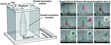
In many droplet-based screening applications, applying an equal incubation or reaction time to a large number of droplets is critical since differences in incubation time can lead to false positive or false negative hits during the selection process. Even though many droplet incubation platforms have been successfully utilized, having all droplets incubated for equal duration remains a formidable challenge. Here, we present an on-chip droplet incubation chamber that is capable of providing a constant droplet incubation time for a large number of droplets. This first-in first-out droplet incubation chamber essentially functions as a delay compartment to ensure that all droplets have equal incubation time inside the chamber before moving on to the post-incubation assay steps. The functionality of the developed chamber was tested by tracking color dye droplets flowing through the on-chip incubation chamber as well as comparing the growth of droplet-encapsulated cells of a filamentous fungus Fusarium verticillioides. The incubation time can be easily adjusted by changing the volume of the chamber or droplet collection speed. The chamber can also be easily integrated on-chip with other droplet microfluidics functional modules such as droplet generators, detectors, and sorters without any valve and tubing connection.
Introduction
Droplet microfluidics has become increasingly popular for high-throughput screening applications due to their advantages in high-speed operations, high sensitivity, small reagent volumes, and massively parallel reaction.1–4 Droplet-based high-throughput screening platforms have been utilized for screening of antibodies,5 enzymes,6 drug libraries,7 directed evolution of enzymes,8 and PCR-based analysis.9
In typical cell-based screening assays, differences in the duration of incubation, from a few to several tens of hours, are commonly required to obtain detectable differences or signals exhibiting particular phenotypes of interest. Therefore, it is imperative that an identical culture time should be applied to all droplets being screened.10 For applications where the cellular growth is the phenotype of interest, e.g. for algal library screening to identify cells that show the highest growth rate under a given condition for biofuel applications11 or drug library screening to identify compounds that inhibit the growth of target cells,7 cell-encapsulated droplets with a different incubation time could lead to false results. In most droplet-based screening systems, droplet incubation begins after the desired number of droplets is obtained in the on-chip12,13 or off-chip5,8,10,14–23 incubation platforms. In this case, the droplets that were generated first have been incubated already for a certain period of time while the droplets generated last just enter into the incubation platforms. This is problematic particularly when screening large libraries where it can take several to tens of hours to collect the desired number of droplets for cell-based screening or when culturing relatively fast-growing microorganisms, for example, E. coli. One solution to this challenge is to continuously flow droplets through a platform in a first-in first-out order so that all droplets undergo an equal incubation time. It is worth noting that continuous first-in first-out droplet incubation may not be necessary when culturing slow-growing microbial strains in droplets with relatively short droplet collection time (less than a few hours), which may not result in a significant difference in cell growth or cellular behavior.
On-chip droplet incubation is often implemented using serpentine microchannels12 or micro-scale chambers.13 However, these are typically limited to applications where the number of droplets are relatively small (∼105 droplets). Off-chip droplet incubation platforms including centrifugal tubes,14,15 tubings,8 Pasteur pipettes,20 or capillaries,22 are used for accommodating a large number of droplets (106 to 107 droplets). Some of the incubation platforms, such as microchannels/chambers, tubings and capillaries, offer some level of first-in first-out droplet flow. However, many others, such as tubes, Pasteur pipettes, vessels and syringes, do not. In addition, as more and more functions are being added to the droplet screening platforms, having a compact integrated platform with minimum tubing connections becomes important. An ideal system is to have a droplet incubation chamber large enough to hold a large number of droplets, while concurrently providing first-in first-out capability to ensure that all droplets are incubated for the same duration, as well as offering on-chip integration capabilities with other droplet processing modules.
Here, we report an on-chip integrated first-in first-out droplet incubation chamber that can provide an equal droplet incubation time for a large number of droplets. The functionality of the developed chamber was first validated by monitoring the residence time of different color dye droplets within the chamber. Next, the growth of a filamentous fungus Fusarium verticillioides, a plant pathogen causing devastating disease and food safety concerns on maize,24 in droplets was monitored and compared to confirm the equal growth of the fungus. The droplet incubation time can be easily adjusted by changing the chamber volume or droplet collection speed. This incubation chamber can also be easily integrated with other droplet manipulation modules such as droplet generation and processing modules.
Results and discussion
Design and operation principle
Fig. 1a shows the design and operation principle of the developed on-chip droplet incubation chamber that can perform first-in first-out operations for a large number of droplets. This chamber has an inlet at the top where droplets can flow into the chamber and an outlet at the center of bottom where droplets can flow out to various post-incubation droplet manipulation modules. Water-in-oil droplets float to the top of the chamber and oil sinks to bottom of the chamber due to the higher buoyancy of water-based droplets in fluorinated oil. Therefore, this top-inlet configuration allows the stacking of droplets in an orderly fashion and creates layers of droplets over time, i.e. where latter droplet layers push down the former droplet layers. The different color dye droplets (red, yellow, green, and blue droplets) in Fig. 1a illustrate droplets generated at different time points (t1–t4) flowing into the incubation chamber and creating distinct layers. Following the incubation time (t4), empty droplets (shown as colorless droplets in Fig. 1a) are generated (t5–t8), sequentially pushing color dye droplets towards the outlet of the incubator in the order of red, yellow, green, and blue droplets, all having the same incubation duration of t4. The top cone shape of the chamber was designed to prevent an abrupt change in the droplet flow by gradually reducing flow velocity, allowing for a smooth transition of flow speed and in turn minimizing the mixing of different droplet layers. In addition, the round shape around the bottom corner of the incubator was designed to minimize the dead volume.
 |
| Fig. 1 (a) Design and operating principle of the on-chip droplet incubation chamber that allows equal droplet incubation time. The red, yellow, green, and blue droplets generated at different time points (t1–t4) illustrate the first-in first-out operation scheme where all droplets undergo the same incubation duration (t4). (b) The droplet incubation chamber integrated with droplet generation (inlet) and post-incubation processing modules (outlet). | |
This droplet incubation chamber can be integrated with droplet generators and various post-incubation functional modules (mixer, splitter, detector and sorter) (Fig. 1b). Through continuously generating, incubating, and processing droplets without any interruption for a given incubation duration (t4), all droplets can go through an identical incubation time and can also be examined in a sequential order. This allows for the first droplets that move into the incubation chamber (t0) to be the first droplets that exit (t4), and the last droplets entering the chamber (t4) to be the last droplets leaving (t8). This continuous-flow operation can continue until the entire library is screened.
Validation of equal droplet incubation time resulting in equal cell growth
The droplet incubation chamber was fabricated by replicating poly(dimethylsiloxane) (PDMS) from a master mold prepared by a 3D printer (see details in ESI†). The diameter of the incubation chamber is 1 cm and the height is 4.3 cm, having a volume of 2.4 ml. Different color dye droplets (121 μm diameter, 0.9 nl) were generated using a T-junction droplet generator (see details in ESI†) at 52 Hz. As shown in Fig. 2, the color dye droplets were successfully stacked layer by layer from the top inlet to the bottom outlet, and allowed complete filling of the whole chamber within 12.5 h (2.32 × 10⁶ droplets) without any observed droplet mixing. The view from the bottom of the chamber (image inserts on upper right corners of Fig. 2) at different time points showed that each layers of the color dye droplets completely flowed out from the chamber over time. Thus, the incubation time of each color dye droplet was constant at 12.5 h, which validates the functionality of the presented incubation chamber design. This droplet incubation time can be easily modified by adjusting the volume of the chamber or collection speed of droplets. Once the droplets undergo a desired incubation time, the incubated droplets can automatically flow out from the chamber into the post-incubation processing module since they are being pushed out by the droplets continuously entering from the top side. Thus, a continuous-flow operation can be achieved without the need for any valve structure required in a stop-and-go operation.
 |
| Fig. 2 Photographs of the droplet incubation chamber showing the flow and residence of color dye droplets. This chamber enables 12.5 h of droplet incubation time, holding approximately 2.32 × 10⁶ of 121 μm diameter droplets (chamber volume: 2.4 ml). Inset images on the upper right corner show the bottom view of the incubation chamber (chamber diameter: 1 cm). | |
To further validate the capability of on-chip incubation chamber for equal incubation time, cell growth in droplets generated at different time points was monitored and compared. Single spores of F. verticillioides were encapsulated and cultured in droplets containing yeast extract–peptone–dextrose (YEPD) culture medium, and their growth was analyzed (see details in ESI†). The growth of fungal spores in droplets generated at 0, 3, and 6 hour were similar (Fig. 3a and b), indicating droplets generated at different time points were incubated for equal duration. Fig. 3c shows a single fungal spore encapsulated in a droplet at the beginning of the droplet incubation period and an elongated hyphae growing from the spore after 12.5 h of incubation. The morphology of fungal cells in the droplet culture is similar to those cultured on agar plates, and the specific growth rate, 0.16 h−1, is of the same order of magnitude as that of YEPD agar plate cultures, 0.30 h−1. In addition, throughout the droplet incubation time, no unwanted droplet merging was observed.
 |
| Fig. 3 Growth of single fungal spores in droplets after 12.5 h incubation. (a) Droplets containing fungal cells enter the chamber from the top and go through 12.5 h of incubation before exiting the chamber from the bottom. Red, yellow, green and blue boxes indicate droplets generated after 3, 6, 9, and 12.5 hours, respectively. (b) Growth of single fungal spores in droplets generated at 0, 3, and 6 hours, followed by a 12.5 h incubation period. The growth of single fungal spores are similar among droplets generated at 0, 3 and 6 h (p > 0.05). Data are presented as means ± SD (n = 10 droplets). (c) A single fungal spore encapsulated in a droplet (0 h) showing hyphae growth after 12.5 h incubation.. | |
It is known that Taylor dispersion in microfluidic channels can cause unequal incubation time of droplets due to the parabolic flow profile within the channels.25,26 In the presented chamber, the transition time of color dye droplets is in the range of 24 to 62 minutes while the droplet delay time is 12.5 h, thus, the dispersion ratio (transition time/delay time) is only 3–8%. Due to this reason, issues related to dispersion are not significant (Fig. S1 in ESI†). In addition, the selection of carrier oil (ρoil > ρaqueous) should be taken into account since certain carrier oil, for example, mineral oil, will result in the water-in-oil droplets to have lower buoyancy, making them sink to the bottom of the chamber rather than floating, in which case an inverted chamber design or alternative method may be required.
Controlling incubation time of droplets is critical for droplet-based applications, and on-chip delay-line channels have been integrated to allow incubation time from milliseconds to several hours.7,25,27 It is possible to obtain shorter incubation times by reducing chamber volume, but further analysis of Taylor dispersion is required when chamber sizes in microliter volumes or less are utilized. In addition, such shorter incubation times can be already accommodated using existing on-chip delay-line channels, thus, the chamber design presented here has its main advantages in the applications that require long culture time and a large number of droplets.
Conclusions
We designed and characterized an on-chip droplet incubation chamber that can hold a large number of droplets while ensuring an equal droplet incubation time. We validated this design by monitoring the flow of color dye droplets and the growth of fungal spores in droplets. By varying the volume of this chamber or droplet flow speed, the incubation time can be easily adjusted. This first-in first-out incubation chamber design allows for the continuous self-releasing of droplets from droplet generation to post-incubation processing without the need for any valve or tubing. Taken together, we demonstrated a droplet incubation chamber that is ideal for microfluidic droplet-based screening assays.
Acknowledgements
This work was supported through the National Science Foundation (NSF) grant #DBI-1353759 and NSF Emerging Frontiers in Research and Innovation (EFRI) grant #EFRI-1240478. The authors declare no competing financial interest.
Notes and references
- M. T. Guo, A. Rotem, J. A. Heyman and D. A. Weitz, Lab Chip, 2012, 12, 2146–2155 RSC.
- T. M. Tran, F. Lan, C. S. Thompson and A. R. Abate, J. Phys. D: Appl. Phys., 2013, 46, 114004 CrossRef.
- H. N. Joensson and H. Andersson Svahn, Angew. Chem., Int. Ed., 2012, 51, 12176–12192 CrossRef CAS PubMed.
- A. B. Theberge, F. Courtois, Y. Schaerli, M. Fischlechner, C. Abell, F. Hollfelder and W. T. S. Huck, Angew. Chem., Int. Ed., 2010, 49, 5846–5868 CrossRef CAS PubMed.
- B. El Debs, R. Utharala, I. V. Balyasnikova, A. D. Griffiths and C. A. Merten, Proc. Natl. Acad. Sci. U. S. A., 2012, 109, 11570–11575 CrossRef CAS PubMed.
- L. Granieri, J.-C. Baret, A. D. Griffiths and C. A. Merten, Chem. Biol., 2010, 17, 229–235 CrossRef CAS PubMed.
- E. Brouzes, M. Medkova, N. Savenelli, D. Marran, M. Twardowski, J. B. Hutchison, J. M. Rothberg, D. R. Link, N. Perrimon and M. L. Samuels, Proc. Natl. Acad. Sci. U. S. A., 2009, 106, 14195–14200 CrossRef CAS PubMed.
- J. J. Agresti, E. Antipov, A. R. Abate, K. Ahn, A. C. Rowat, J.-C. Baret, M. Marquez, A. M. Klibanov, A. D. Griffiths and D. A. Weitz, Proc. Natl. Acad. Sci. U. S. A., 2010, 107, 4004–4009 CrossRef CAS PubMed.
- D. Pekin, Y. Skhiri, J.-C. Baret, D. Le Corre, L. Mazutis, C. B. Salem, F. Millot, A. El Harrak, J. B. Hutchison and J. W. Larson, Lab Chip, 2011, 11, 2156–2166 RSC.
- J. Clausell-Tormos, D. Lieber, J.-C. Baret, A. El-Harrak, O. J. Miller, L. Frenz, J. Blouwolff, K. J. Humphry, S. Köster, H. Duan, C. Holtze, D. A. Weitz, A. D. Griffiths and C. A. Merten, Chem. Biol., 2008, 15, 427–437 CrossRef CAS PubMed.
- H. S. Kim, T. L. Weiss, H. R. Thapa, T. P. Devarenne and A. Han, Lab Chip, 2014, 14, 1415–1425 RSC.
- S. Koster, F. E. Angile, H. Duan, J. J. Agresti, A. Wintner, C. Schmitz, A. C. Rowat, C. A. Merten, D. Pisignano, A. D. Griffiths and D. A. Weitz, Lab Chip, 2008, 8, 1110–1115 RSC.
- S. Cho, D.-K. Kang, S. Sim, F. Geier, J.-Y. Kim, X. Niu, J. B. Edel, S.-I. Chang, R. C. Wootton and K. S. Elvira, Anal. Chem., 2013, 85, 8866–8872 CrossRef CAS PubMed.
- T. C. Scanlon, S. M. Dostal and K. E. Griswold, Biotechnol. Bioeng., 2014, 111, 232–243 CrossRef CAS PubMed.
- M. Ryckelynck, S. Baudrey, C. Rick, A. Marin, F. Coldren, E. Westhof and A. D. Griffiths, RNA, 2015, 21, 458–469 CrossRef CAS PubMed.
- L. Mazutis, J. Gilbert, W. L. Ung, D. A. Weitz, A. D. Griffiths and J. A. Heyman, Nat. Protoc., 2013, 8, 870–891 CrossRef CAS PubMed.
- V. Trivedi, A. Doshi, G. Kurup, E. Ereifej, P. Vandevord and A. S. Basu, Lab Chip, 2010, 10, 2433–2442 RSC.
- K. Churski, T. S. Kaminski, S. Jakiela, W. Kamysz, W. Baranska-Rybak, D. B. Weibel and P. Garstecki, Lab Chip, 2012, 12, 1629–1637 RSC.
- B. L. Wang, A. Ghaderi, H. Zhou, J. Agresti, D. A. Weitz, G. R. Fink and G. Stephanopoulos, Nat. Biotechnol., 2014, 32, 473–478 CrossRef CAS PubMed.
- J.-C. Baret, O. J. Miller, V. Taly, M. Ryckelynck, A. El-Harrak, L. Frenz, C. Rick, M. L. Samuels, J. B. Hutchison, J. J. Agresti, D. R. Link, D. A. Weitz and A. D. Griffiths, Lab Chip, 2009, 9, 1850–1858 RSC.
- J.-C. Baret, Y. Beck, I. Billas-Massobrio, D. Moras and A. D. Griffiths, Chem. Biol., 2010, 17, 528–536 CrossRef CAS PubMed.
- L. Mazutis, A. F. Araghi, O. J. Miller, J.-C. Baret, L. Frenz, A. Janoshazi, V. Taly, B. J. Miller, J. B. Hutchison, D. Link, A. D. Griffiths and M. Ryckelynck, Anal. Chem., 2009, 81, 4813–4821 CrossRef CAS PubMed.
- M. Najah, R. Calbrix, I. P. Mahendra-Wijaya, T. Beneyton, A. D. Griffiths and A. Drevelle, Chem. Biol., 2014, 21, 1722–1732 CrossRef CAS PubMed.
- C. P. Woloshuk and W.-B. Shim, FEMS Microbiol. Rev., 2013, 37, 94–109 CrossRef CAS PubMed.
- L. Frenz, K. Blank, E. Brouzes and A. D. Griffiths, Lab Chip, 2009, 9, 1344–1348 RSC.
- A. Ajdari, N. Bontoux and H. A. Stone, Anal. Chem., 2006, 78, 387–392 CrossRef CAS PubMed.
- H. Song and R. F. Ismagilov, J. Am. Chem. Soc., 2003, 125, 14613–14619 CrossRef CAS PubMed.
Footnote |
† Electronic supplementary information (ESI) available. See DOI: 10.1039/c5ra26505c |
|
This journal is © The Royal Society of Chemistry 2016 |
Click here to see how this site uses Cookies. View our privacy policy here.